ORIGINAL RESEARCH ARTICLE
ANALYSES OF THE DISTAL SURFACE WEAR OF CANAL IN MESIAL ROOTS OF LOWER
MOLARS AFTER THE CERVICAL PREPARATION WITHIN DIFFERENT TECHNIQUES
*
Mayse Machado Guimarães and Danilo Barral de Araújo
Health Science Institute, Federal University of Bahia, Salvador, Brazil
ARTICLE INFO ABSTRACT
Introduction Difficulty during preparation is to make the instrument widen the canal and adapt to its shape without deformation Objective quantify the wear on distal walls after Gates Glidden and Reciproc cervical preparation Method 100 molars, totaling 200 canals divided into two groups: A - Gates Glidden and B - Reciproc files. Specimens cut 2 mm cervical level so that it could be replaced in its original position in a PVC trim. Images obtained before and after cervical preparation with a stereoscopic magnifier. Images were analyzed using Image J software and measurements performed at the lowest thickness of distal dentin before and after cervical preparation. Data analyzed through Cohen's d and Student's t-test with a significance level of 5%.
Results MV roots thickness was similar at the beginning at the end of the study (d = 0.070), even with adjustment (d = 0.13). In the ML roots, the thicknesses were different at the beginning (d = 0.42) and at the end of the study (d = 0.39). However, with the adjustment, this difference disappeared (d = 0.05). Conclusion There was no statistical difference between dentin abrasion in mesial root zone risk of mandibular molars when instrumented by GG and Reciproc.
Copyright © 2019,Mayse Machado Guimarães and Danilo Barral de Araújo. This is an open access article distributed under the Creative Commons Attribution
License, which permits unrestricted use, distribution, and reproduction in any medium, provided the original work is properly cited.
INTRODUCTION
The complex anatomy of human molars requires the operator more skill and knowledge to perform endodontic treatment, especially in the presence of curvatures (Carlos Estrela, 2018). Among the variable characteristics of the dental morphology, the configuration of the root canals, which believed to be only a single tubular space, was shown as a complex system, which hinders endodontic practice, since the purpose of this treatment consists of the perfect biomechanical preparation, in the removal of existing microorganisms and in the complete hermetic sealing of this system (Mohammadi, 2015; Carlos ESTRELA, 2018; Franco de Carvalho, 2012; Guven KAYAOGLU, 2018). The presence of dentin projection added to the curvature of the lower molar canal restricts the movement of the intracanal file, acting as fulcrum and causing a greater wear in the distal wall in the middle portion of the curvature and in the mesial wall in the apical region during instrumentation of the channel (Sheila Leite, 2018). Therefore, it is necessary for the instrument to plan this region, that it is directed in the anti-furcation direction, in order not to promote
*Corresponding author: Mayse Machado Guimarães,
Health Science Institute, Federal University of Bahia, Salvador, Brazil.
perforations (Guven KAYAOGLU, 2018; Sheila Leite PINTO, 2018; Estrela, 2010). The preparation of the cervical third decreases the tension of the manual and rotational instruments during apical instrumentation by eliminating the dentin projection, and also provides greater reliability when defining the working length (Sheila Leite, 2018; Estrela C, Stephan, 2010; Weine, 1975; Abou-Rass, 1980; Kássio SOUSA1, 2018; Lim, 1987; James B.Roane, 2018; Lopes, 1999; Carlos Martín Ardila Medina, 2018). In addition, when the preparation is performed with Ni-Ti instruments, without the pre-enlargement of the cervical region with stainless steel instruments, there is a greater friction of these instruments in relation to the furca, promoting wear in the thickness of dentin to values below 0 , 5 mm (Kássio, 2018). The existence of different instrumentation techniques is justified by the anatomical variations, with the objective of obtaining a root model without alterations in the shape of the canal and the apical foramen (Lim, 1987). The cervical preparation facilitates access to the apical third, preventing formations of steps, perforations, apical "zip" (Estrela C, Stephan, 2010; Lim, 1987). The first rotating instruments developed for cervical preparation were Gates Glidden (GG), with GG # 2 tip diameter of 0.70 mm, are considered safe for instrumentation of mesial root canals of lower molars (Franco de Carvalho,
ISSN: 2230-9926
International Journal of Development Research Vol. 09, Issue, 02, pp.25908-25912, February, 2019Article History:
Received 20th November, 2018
Received in revised form 16th December, 2018
Accepted 23rd January, 2019
Published online 28th February, 2019
Key Words: Anatomy, Endodontics,
Root canal Treatment, Molars.
Citation: Mayse Machado Guimarães and Danilo Barral de Araújo. 2019. “Analyses of the distal surface wear of canal in mesial roots of lower molars
after the cervical preparation within different techniques”, International Journal of Development Research, 09, (02), 25908-25912.
2012; Kássio SOUSA1, 2018). On the other hand, because they are made of stainless steel and have limited flexibility, their use leads to scratches especially in the mesial roots of the lower molars, as excessive wear can lead to lateral perforation of the root, called "Strip Perforation". This accident may affect the prognosis of the treatment (Franco de Carvalho, 2012). During instrumentation of curved canals the wear tends to be larger towards the risk zone. With the introduction of rotating Ni-Ti instruments, the root canal system instrumentation became safer, easier and faster, becoming more efficient and producing fewer iatrogenies (Bruschi, 2017; Higuera, 2015; Bergmans, 2012). In 2008, Yared, (2008) reported a unique
instrumentation technique (Protaper® Universal) for
performing endodontic treatment with the aim of simplifying preparation and increasing safety. This study stimulated the industry to develop new instruments, predominantly reciprocating systems (Bruschi, 2017; Yared, 2008; Gavini,
2012; Kim et al., 2012). In 2010, the Reciproc® file (VDW,
Munich, Germany) was introduced. Being a unique instrument of Ni-Ti for the preparation of endodontic treatment and with a single use recommendation, it was designed to work in reciprocating movement. In this movement, the instrument rotates in one direction and then reverses direction before completing a full rotating cycle. The use of this reciprocating movement prolongs the useful life of Ni-Ti instruments, increasing resistance to clinical fatigue compared to rotating
instruments (Gavini, 2012; Lopes et al., 2013; Xi, 2018;
Bueno, 2017; Cunha et al., 2014). The Reciproc® file is a #
25.08 taper instrument, manufactured with a special Ni-Ti: M-Wire alloy which gives the instrument even more flexibility (Xi, 2018; Bueno, 2017). Based on this, they found that this alloy confers greater flexibility and resistance to cyclic fatigue compared to instruments made with the conventional Ni-Ti
alloy (Bueno et al., 2017). Allied to clinical practice, the
experimental practice of endodontic research, using extracted teeth, allows improving the knowledge of the external and internal anatomy, observing the accomplishment of the endodontic treatment protocol with greater detail and improving the calculation of the final result using the technique employed. The present study analyzed the amount of wear performed on dentin thickness in the mesial (MV and ML) roots of lower human molars after cervical preparation using two systems: Gates Glidden and Reciproc® Limas.
MATERIALS AND METHODS
The present study was approved by the Ethics and Research Committee of the UFBA Institute of Health Sciences (CAAE: 87452818.6.0000.5662). A total of 100 human mandibular, birradicular, mandibular molar roots with similar and relatively straight vestibular-palatine and mesio-distal dimensions were selected from the Biorepository of the Oral Biochemistry Laboratory of the Institute of Sciences and Health, UFBA. The mesial roots of the selected specimens were then marked one by one, 2 mm below the furca, using a millimeter ruler and permanent marker for 2.0 mm CD / DVD (PILOT Pen do Brasil / SA Indústria e Comércio, Jundiaí - SP, Brazil). Afterwards, they were fixed with transparent Orthophthalic Resin (Equifibras, Salvador, Bahia). Pieces with 32 mm of a PVC tube (Tigre / S.A., Castro - PR, Brazil) were used as mold. Bounds ready, and then a draw was held. Two groups were established. Group A: GG and Group B drills: Reciproc® file (n = 50 each). The specimens were fixed to the Elsaw precision cutter (Elquip, São Paulo, Brazil) and using a diamond disk (Buehler 111190/100493107 Ltd., Lake Bluff,
IL, USA) with constant irrigation, previously established. With the same permanent marker for CD / DVD 2.0 mm (PILOT Pen do Brasil / SA Indústria e Comércio, Jundiaí - SP, Brazil), a canal line was traced to the cementitious surface in the inner part of the root where it represents the lowest tooth thickness. After being sectioned, the images were captured with the Moticam 3+ digital camera (Motic, Canada) coupled with a Stereo Magnifying Glass (Motic K Series - Q7766, Diadema - SP, Bbrasil) at 7X magnification. These images were saved on the computer and then measured through the previously calibrated Image J® (National Institutes of Health, Bethesda, MD) program (Figure 12). The calibration of this program consists of transferring measurements in pixels to millimeters. The pixel distance used in the program was 44 for all images. Thus, the software automatically transformed the values obtained to millimeters. Then, the measurements were obtained from the internal part of the roots MV and ML towards the furca, referring to the previously demarcated line, in order to obtain the initial dentine thickness of the risk zone. Standard access to the pulp chamber was made in the teeth one by one with diamond spherical drills nº 1014 (KG Sorensen, Cotia - SP, Brazil) and Drill Endo Z drills (Dentsply Maillefer, Ballaigues, Switzerland) for posterior cervical preparation. Each specimen was individually included inside a Weld PVC Cap (Tigre / S.A., Castro - PR, Brazil) and filled with transparent natural wax (Wilson, Cotia - SP, Brazil) to stabilize them inside. A flat surface was used to facilitate the following maneuvers. The cervical preparation was performed with the Gates Glidden 32 mm drills (Dentsply Maillefer, Ballaigues, Switzerland) in increasing sequence: # 2, # 3, # 4, coupled to the contra angle and micromotor (Kavo do Brasil Ind. Com. Ltda. ., Joinville - SC, Brazil), applying slight apical pressure (Figure 18). The depth of the drills was determined by their resistance against the channel walls. The drills were replaced every 10 instrumented teeth, totaling 20 roots.
Irrigation was performed with 2 ml of distilled water. Lymph node K # 10 of 25 mm (Dentsply Maillefer, Ballaigues, Switzerland) was used for catheterization. The cervical preparations made with the Reciproc® R25, 25 mm (VDW, Munich, Germany) files were performed on the VDW Silver engine (VDW, Munich, Germany) in the "RECIPROC ALL" function (Figure 20) , initially with a length of 15 mm (Figure 17), with 3 movements of pecks and exit and incorporating a brushing action of the instrument against the walls of the canal followed by irrigation (2 ml of water) to reach the patency previously obtained with the file # 10. The Reciproc® file was replaced with each tooth, following the manufacturer's instructions. KFile No. 10 of 25 mm (Dentsply Maillefer, Ballaigues, Switzerland) was used to obtain patency and to perform catheterization. After the instrumentation, the specimens were separated and washed in the L-100 Ultrasonic Washer (Shuster, Santa Maria - RS, Brazil), activated for 180 seconds to remove excess dentin scrap produced during instrumentation. The preparation of the channels was performed by a single operator, expert in Endodontics, trained for the use of the two systems used (Gates Glidden Drills and Reciproc® Limas). After the instrumentation of all the teeth, the coronary fragment was removed and new images were obtained with the digital camera Moticam 3+ (Motic, Canada) coupled to Stereoscopic Stereoscopic Magnifying Glass (Mocha K Series - Q7766, Diadema - SP, Bbrasil) 7X. These images were saved on the computer and then measured through the Image J® program (National Institutes of Health, NHI, Bethesda, MD).
This software allowed us to obtain the measurements from the internal part of the roots MV and ML towards the furca, in the same line previously marked, in order to obtain the lowest thickness of dentin / cement after instrumentation (Figure 2 and 3). For statistical analysis, the Cohen d group was used as a measure of effect size and Student's t-test with a significance level of 5% was used. To qualify the size of the effect, the criteria of Cohen (1988) were adopted: d <0.2, no effect; d between [0.2-0.5), weak effect; d between [0.5-0.8), moderate effect and d> = 0.8, strong effect. For each root (mesio-vestibular root (MV) and mesio-lingual root (ML)) the means before and after the intervention and the differences between them in the time (adjustment) were calculated. Negative values of difference mean reduction of time values, while positive values mean increase.
RESULTS
Table 1 shows that the thicknesses were similar at the beginning (d = 0.007) of the study and at the end (d = 0.070) of the study, even with adjustment (d = 0.13). It can be observed in Table 2 that the thicknesses are different at the beginning (d = 0.42) of the study and at the end (d = 0,39) of the study. However, with the adjustment, this difference disappeared (d = 0.05).
Table 1. Mean thickness of the MV roots before and after instrumentation, mean final difference adjusted by the initial, Cohen's d values and p values
Note: measurements in millimeters (mm)
Table 2. Mean thickness of ML roots before and after instrumentation, mean final difference adjusted by initial,
Cohen's d values and p values
Note: measurements in millimeters (mm)
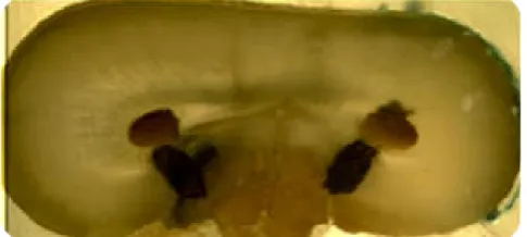
[image:3.595.314.557.57.166.2]Source: Author'sphoto
Figure 01. Smaller measures from the inner part of the root towards the Furca
[image:3.595.316.550.215.326.2]Source: Author'sphoto
Figure 02. Imageafterinstrumentationwith GG drills
Source: Author'sphoto.
Figure 03. ImageafterinstrumentationwithReciproc® file
DISCUSSION
[image:3.595.328.539.363.469.2]Many authors corroborate the need for cervical preparation in order to allow free and direct access of the instruments to the canal walls and to reduce the angle of curvature (Sheila Leite PINTO, 2018; Estrela C, Stephan, 2010; Weine, 1975; Lopes, 1999; Martin G de, 2018; Oliveira, 2014; Marco Antonio Hungaro DUARTE1, 2019). Therefore, the existence of different instrumentation techniques is justified by the anatomical variations, in order to obtain a more conservative biomechanical preparation that preserves the shape of the canal and the furcation zone (risk zone), to facilitate the access of the instruments to the apical third of roots and prevent the formation of iatrogenies (Sheila Leite, 2018; Estrela C, Stephan, 2010; Weine, 1975; Abou-Rass, 1980; Kássio SOUSA1M, 2018; Lim, 1987; James B.Roane, 2018; Lopes, 1999; Carlos Martín Ardila Medina, 2018). In addition, this relief can prevent accidents during biomechanical preparation such as: instrument fractures, root canal deviation, excessive wear on the root walls promoting weakening of the treated dental unit, foraminal transport and formation of steps (Sheila Leite, 2018; Estrela C, Stephan, 2010; Weine, 1975; Lim, 1987; Lopes, 1999; Martin, 2018; Oliveira, 2014; Marco Antonio Hungaro DUARTE1, 2018). Cervical preparations were performed on the MV roots of lower molars in order to evaluate the dentin thickness before and after instrumentation. Three instruments were used (ProTaper®, Hero Shaper and Gates Glidden Drills). The mean thicknesses of the distal dentin in the cervical third before and after the instrumentations were: 1.4 ± 0.5 mm, 1.32 ± 0.3 mm and 1.28 ± 0.31 mm for groups I, II and III, respectively. The authors concluded that GG drills performed more wear on dentin thickness. In the present study, an evaluation of the dentinal thickness was performed before and after the cervical preparations. Two instruments were used (Group A: GG and Group B: Reciproc®). The averages of the measurements obtained from the dentinal thickness of the cervical third of the MV roots before the instrumentation were: 1.297 mm for Group A and 1.299 mm for Group B.
Researchers have demonstrated that the mesial canals of the lower molars do not appear centralized in the roots and that they are closer to the furca area (Abou-Rass, 1980; Lopes, 1999). The thickness of remaining dentin found after cervical
preparation in these roots varied between 0.39 and 0.97 mm9.
These values were: 0.529 and 1.997 mm for the MV roots in Group A (Table 1) and 0.469 and 2.030 mm for the MV roots in Group B (Table 1). For ML roots, the following values were obtained after cervical preparation: 0.146 and 1.791 mm for Group A (Table 2) and 0.455 and 2.00 mm for Group B (Table 2). Different values may have been found due to the different techniques to get them.
In another study (James B.Roane, 2018), also performed on mesial roots of lower molars, they observed that wear in the risk zone was greater than wear in the safety zone. Two cervical preparation systems were used GG (# 3, # 2, # 1) and Triple Gates. There were no differences between the promoted wear between the drills tested. A mean dentin thickness of 0.6
mm was observed in both groups 12. In the present study, mean
weights found for MV channels after preparation were: 1.102 mm in Group A and 1.128 mm in Group B (Table 1). For ML canals the means obtained were: 1.028 mm for Group A and 1.167 mm for Group B (Table 2). There was no difference between groups. The Gates-Glidden # 2 drill has the diameter of the active tip equivalent to 0.70 mm and is generally considered safe for cervical preparation of mesial canals of
lower molars. However, the use of larger diameters may promote excessive wear on the dentin of the furca region, increasing the risk of perforation (Kássio SOUSA1, 2018; Marco Antonio Hungaro DUARTE1, 2018). In the present study, the sequence used for GG drills was # 2, # 3 and # 4 for cervical preparation in Group A. And although drills # 3 and # 4 were used, no drilling was observed with the use of these drills larger than GG # 2. Introduced on the market in 2010, the Reciproc® file (VDW, Munich, Germany) is designed to work on reciprocating movements. This movement consists of the instrument rotating in one direction and then reversing the direction (150º / 30º), for every three cycles of these there is a complete rotation (360º) (Gavini G, Caldeira, 2012; Lopes, 2013; Estefanía Muñoz, 2018). The use of this reciprocating motion is responsible for prolonging the useful life of Ni-Ti instruments by increasing their resistance to fatigue compared to instruments driven under continuous rotation (Gavini G,
Caldeira, 2012; Lopes et al., 2013; Xi, 2018; Bueno et al.,
2017). Moreover, this movement is still related to the preparation of biomechanical preparation more centralized within the root canals, even with the presence of curvatures. Producing in this way, more conservative preparations for the
dentinal tissues (Bruschi, 2017; Higuera, 2015; Lopes et al.,
2013; Vorster, 2018).
Conclusion
It can be concluded, within the methods used for this study, that there was no difference in wear of the "risk zone" provided by Gates Glidden and File Reciproc®.
Acknowledgement: I thank all the professors of the Oral Biochemistry Laboratory, my colleagues and all those involved in this process. I also thank the Coordination of Improvement of Higher Level Personnel for the financial support.
REFERENCES
Abou-Rass M., Frank AL., Glick DH. 1980. The Anticurvature Filing Method to Prepare the Curved Root Canal. J Am Dent Assoc. novembro de,101:792–4.
Bruschi J., Boff LB., Melo TAF de. 2017. Analysis of cutting capacity, preparation time, and apical deviation after instrumentation of artificial curved canals with the waveone ® and reciproc ® reciprocating systems. RGO - Rev Gaúcha Odontol. setembro de;65(3):191–5.
Bueno CSP, Oliveira DP de, Pelegrine RA, Fontana CE, Rocha DGP, Bueno CE da S. Fracture Incidence of WaveOne and Reciproc Files during Root Canal Preparation of up to 3
Posterior Teeth: A Prospective Clinical Study. J Endod. 1o de
maio de 2017;43(5):705–8.
Carlos Estrela, Mike R. Bueno, Fernando B. Barletta, Orlando A. Guedes2,, Olavo C. Porto1, Cyntia R.A. Estrela, Jesus Djalma Pécora. Identification of Apical and Cervical Curvature
Radius of Human Molars [Internet]. [citado 1o de novembro
de 2018]. Available: http://www.scielo.br/ scielo.php?pid= S0103-6440201500040 0351 & script= sci_arttext
Carlos Estrela, Mike Reis Bueno, Manoel Damião SOUSA-Neto, Jesus Djalma Pécora. 2018. Method for determination of root curvature radius using cone-beam computed tomography
images [Internet]. [citado 1o de novembro de 2018]. Available:
http://www.scielo.br/scie lo.php?pid=S0103-6440200800020 0005&script=sci _arttext
comparativo de la remoción de la dentina [Internet]. [citado 1 de novembro de 2018]. Available: http://scielo.sld.cu/scielo. php?script=sci_arttext&pid=S1025-02552012000400005 Coutinho-Filho T, De-Deus G, Gurgel-Filho ED, Rocha-Lima
AC, Dias KRC, Barbosa CA. Evaluation of the risk of a stripping perforation with gates-glidden drills: serial versus
crown-down sequences. Braz Oral Res. março de
2008;22(1):18–24.
Cunha RS, Junaid A, Ensinas P, Nudera W, da Silveira Bueno CE. Assessment of the Separation Incidence of Reciprocating
WaveOne Files: A Prospective Clinical Study. J Endod. 1o de
julho de 2014;40(7):922–4.
EstefaníaMuñoz, LeopoldoForner, CarmenLlena. Influence of Operator’s Experience on Root Canal Shaping Ability with a Rotary Nickel-Titanium Single-File Reciprocating Motion
System - ScienceDirect [Internet]. [citado 1o de novembro de
2018]. Available: https://www.sciencedirect.com/science/
article/pii/S0099239913007553
Estrela C., Stephan IW. 2010. Estudo comparativo do desgaste dentinário na parede distal do canal mesiovestibular do primeiro molar inferior, produzido por três técnicas de instrumentação. Rev Odontológica Bras Cent.1(1).
Franco de Carvalho EMO, Carnevalli B. Análise da alteração da curvatura, antes e após o preparo do canal radicular, pelas técnicas manual e rotatória. Rev Odontol UNESP. outubro de 2012;41(5):335–9.
Gavini G, Caldeira CL, Akisue E, Candeiro GT de M, Kawakami DAS. Resistance to Flexural Fatigue of Reciproc R25 Files under Continuous Rotation and Reciprocating Movement. J
Endod. 1o de maio de 2012;38(5):684–7.
Gluskin, A. H., Brown D. C. & Buchanan. L. S. A reconstructed
computerized tomographic comparison of Ni–Ti rotary GTTM
files versus traditional instruments in canals shaped by novice operators - Gluskin - 2001 - International Endodontic Journal -
Wiley Online Library [Internet]. [citado 1o de novembro de
2018]. Available: https://onlinelibrary.wiley.com/doi/abs/
10.1046/j.1365-2591.2001.00422.x
Guven Kayaoglu, Ilkay Peker, Mustafa Gumusok, Cigdem Sarikir, Aylin Kayadugun, Ozlem Ucok. 2018. Root and canal symmetry in the mandibular anterior teeth of patients
attending a dental clinic: CBCT study [Internet]. [citado 1o de
novembro de]. Available: http://www.scielo.br/
scielo.php?pid=S1806-8324201500010028 3&scrip t=sci_ arttext
Higuera O, Plotino G, Tocci L, Carrillo G, Gambarini G, Jaramillo DE. Cyclic Fatigue Resistance of 3 Different Nickel-Titanium Reciprocating Instruments in Artificial
Canals. J Endod. 1o de junho de 2015;41(6):913–5.
James B.Roane, Clyde L.Sabala, Manville G. Duncanson Jr. The “balanced force” concept for instrumentation of curved canals - ScienceDirect [Internet]. [citado 2 de novembro de 2018]. Available: https://www.sciencedirect.com/ science/article/pii/ S0099239985800613
Kássio SOUSA1, Carlos Vieira ANDRADE-JUNIOR2, Juliana Melo da SILVA3, Marco Antonio Hungaro DUARTE4,, Gustavo DE-DEUS5, Emmanuel João Nogueira Leal da SILVA5. Comparison of the effects of TripleGates and Gates-Glidden burs on cervical dentin thickness and root canal area by using cone beam computed tomography [Internet]. [citado
1o de novembro de 2018]. Available: http://www.scielo.br/
scielo.php?pid=S1678-77572015000200009&script=sci_arttext
Kim H.C., Kwak SW., Cheung GSP., Ko DH., Chung SM., Lee W. 2012. Cyclic Fatigue and Torsional Resistance of Two
New Nickel-Titanium Instruments Used in Reciprocation
Motion: Reciproc Versus WaveOne. J Endod. 1o de abril
de;38(4):541–4.
L. Bergmans, J.Van Cleynenbreugel, M. Beullens, M.Wevers, B. Van Meerbeek& P. Lambrechts. Smooth flexible versus active tapered shaft design using NiTi rotary instruments - Bergmans - 2002 - International Endodontic Journal - Wiley Online
Library [Internet]. [citado 1o de novembro de 2018].
Available: https://onlinelibrary.wiley.com/ doi/abs/10.1046/ j.1365-2591.2002.00574.x
Lim SS., Stock CJR. 1987. The risk of perforation in the curved canal:anticurvature filing compared with the stepback
technique. Int Endod J. 1o de janeiro de, 20(1):33–9.
Lopes HP., Elias CN., Vieira MVB., Siqueira JF., Mangelli M.,
Lopes WSP., et al., 2013. Fatigue Life of Reciproc and Mtwo
Instruments Subjected to Static and Dynamic Tests. J Endod.
1o de maio de;39(5):693–6.
Lopes HP., Siqueira Junior JF., Elias CN. 1999. Preparo químico-mecânico dos canais radiculares. In: Endodontia: biologia e técnica. p. 319–67.
Marco Antonio Hungaro DUARTE1, Ricardo Affonso
BERNARDES2, Ronald ORDINOLA-ZAPATA1, Bruno Carvalho de VASCONCELOS3, Clovis Monteiro Bramante1, Ivaldo Gomes de MORAES1. Effects of Gates-Glidden, LA Axxess and orifice shaper burs on the cervical dentin thickness and root canal area of mandibular molars [Internet].
[citado 1o de novembro de 2018]. Available: http://www.
scielo.br/scielo.php?pid=S0103-64402011000100004&script =sci_arttext
Martin G de, Azeredo RA. Análise do preparo de canais radiculares utilizando-se a diafanização [Internet]. 2018
[citado 1o de novembro de 2018]. Available: https://www.
ingentaconnect.com/content/doaj/01011774/2018/00000043/0 0000002/art00006
Mohammadi Z., Shalavi S., Jafarzadeh H. 2015. The oval shaped,
root canal: A clinical review. South Afr Dent J. junho de.,
70(5):200–4.
Oliveira MAVC de, Venâncio JF, Pereira AG, Raposo LHA, Biffi
JCG, Oliveira MAVC de, et al. Critical Instrumentation Area:
Influence of Root Canal Anatomy on the Endodontic Preparation. Braz Dent J. 2014;25(3):232–6.
Sheila Leite PINTO, Marília Fagury Videira MARCELIANO-ALVES, Renata Ximenes LINS,, Ermelindo Antônio RADETIC, Hélio Pereira LOPES. The dentin thickness remaining in the risk zone of mandibular molars after cervical
preflaring with four methods [Internet]. [citado 1o de
novembro de 2018]. Available: http://www.s cielo.br/scielo. php?pid=S1807-25772017000 100001& script=sci_arttext Vorster M, van der Vyver PJ, Paleker F. Influence of Glide Path
Preparation on the Canal Shaping Times of WaveOne Gold in
Curved Mandibular Molar Canals. J Endod. 1o de maio de
2018;44(5):853–5.
Weine FS., Kelly RF., Lio PJ. 1975. The effect of preparation procedures on original canal shape and on apical foramen
shape. J Endod. 1o de agosto de1(8):255–62.
Xi WEI, Bo HU, Haiyang PENG, Ming TANG, Jinlin SONG. The incidence of dentinal cracks during root canal preparations with reciprocating single-file and rotary-file
systems: A meta-analysis [Internet]. [citado 1o de novembro
de 2018]. Disponível em: https://www.jstage.jst .go.jp/article/ dmj/36/3/36_2016-208/_article/-char/ja/
Yared G. Canal preparation using only one Ni-Ti rotary
instrument: preliminary observations. Int Endod J. 1o de abril
de 2008;41(4):339–44.