IJPSR (2012), Vol. 3, Issue 12 (Review Article)
Received on 31 July, 2012; received in revised form 17 September, 2012; accepted 04 November, 2012
APPLICATIONS OF NOVEL VESICULAR DRUG DELIVERY SYSTEM AS OCULAR DRUG VEHICLES: A REVIEW
K.A. Modi* and P.K. Shelat
Department of Pharmaceutics & Pharmaceutical Technology, K. B. Institute of Pharmaceutical Education and Research, Kadi Sarva Vishwavidyalaya, Gandhinagar - 382023, Gujarat, India
ABSTRACT
The eye is the most easily reachable organ available for topical drug delivery of local as well as interiorly acting drugs. Ideal drug delivery system aims to provide loading and maintenance dose of drug while being within therapeutic limits at the desired site of action for the desired period of time. In the treatment of ocular disease, the site of action is very narrow and undesired side effects may cause serious injuries to eye tissues. Thus, ideal ocular drug delivery should be site specific, controlled release and non-irritating. Many drugs (more than 85%) belong to BCS class IV exhibiting poor solubility and permeability. Moreover; the precorneal losses like tear secretion, naso-lachrymal drainage and less permeability of corneal tissue causes only 1% absorption of instilled dose. These limitations lead to increased loading dose and dosing frequency to maintain minimum therapeutic level for action which ultimately causes over exposure of drug to eye and leads to serious side effects. Due to these hurdles of conventional drug delivery system, the vesicular drug delivery system is gaining more popularity day by day as they offer improved permeability and altered bioavailability of drug. These formulations mainly include liposome and niosome made up of phospholipids and non-ionic surfactants respectively. They are non-antigenic, biodegradable and non-irritating. The present article reveals the key-work done on vesicular drug delivery system as potential ocular drug delivery system and justifies the need for further research on vesicular drug delivery system as ocular vehicles.
INTRODUCTION: Eye is the most easily accessible organ for topical drug delivery for local as well as interiorly acting drugs. The eye has major two segments;
1. Anterior segment 2. Posterior segment.
Anterior structure is in front having various optical structures like cornea, pupil, aqueous humor, iris, lens
Posterior segment of eye includes sclera, choroids, retina, vitreous humor, macula and optical nerve. The cornea has relatively less permeability and hence drug penetration has always remained a critical problem. It is made up of three distinct layers, i.e. epithelium, stroma and endothelium layers, where each layer exhibits different polarity and a potential rate-limiting structure for drug permeation 1.
Keywords:
Ocular drug delivery system, Liposome,
Niosome, Chitosome
Correspondence to Author: Kushal A. Modi,
Research fellow, Department of Pharmaceutics & Pharmaceutical Technology, K. B. Institute of
Pharmaceutical Education and Research, Kadi Sarva Vishwavidyalaya, Gandhinagar - 382023, Gujarat, India
E-mail: kushal.modi@gmail.com QUICK RESPONSE CODE
IJPSR: ICV (2011)- 5.07
The ocular drug delivery has been remained highlighted issue due to its dosing problems and patient compliance. To maintain the minimum therapeutic concentration at the site of action with non-irritating characteristics is always a challenging issue in ocular drug delivery system. The uncommon barriers like tear secretion and blinking of eye causes noticeable loss of instilled dose 2.
The most common routes of drug administration for ocular disease treatment are topical, periocular and intravitreal. Among all these routes, topical route is more preferred due to its non-invasive nature and ease of administration. When drug is delivered through topical route, the absorption of drug takes place either through corneal route or non-corneal route. The corneal route involves cornea, aqueous humor and intraocular tissues where as non-corneal route involves conjunctiva, sclera and choroids.
Approximately only 5% of instilled dose reaches the intraocular tissues due to various responsible factors like pre-corneal drainage and less permeability of corneal epithelium cells. The systemic route can by pass these hurdles but the problems related this route are blood-aqueous barrier and blood-retinal barrier which lead to high loading dose to reach the target site. The high dose and dosing frequency causes unavoidable systemic side effects like stomach upset and disturbed GI motility. The endothelium cells of iris and ciliary blood vessels and non-pigmented ciliary epithelium cells exhibits tight junctional cell complex which acts as blood-aqueous barrier. This barrier plays major role in preventing entry of drugs into the aqueous humor.
Blood retinal barrier is composed of retinal capillary endothelial cells and retinal pigment epithelium (RPE) cells, which prevents the entry of drug into the retina. Frequent intravitreal dosing is required which may cause vitreous hemorrhage, retinal detachment and endophthalmitis. Another serious issue regarding loss of topically instilled drugs in conventional dosage form is from the nasal cavity. Nasal cavity exhibits large surface area and higher permeability of the nasal mucosal membrane compared to that of the cornea. Standard dropper of conventional ocular dosage form delivers around 50-75 µl per drop but large portion of this drop drains out quickly until the eye regains its
normal resident volume i.e. 7 µl 3-4. Due to this limitation, very large loss of instilled medicament occurs and hence very small fraction of dose becomes available at the site of action5. Thus, to overcome this issue, development of non-invasive, controlled release targeted drug delivery system is required which can provide drug delivery for prolonged period of time. Ideal properties of controlled ocular delivery system 6:
To provide controlled drug release at the targeted site.
Reasonable corneal penetration.
To overcome the zigzag fluctuation caused by conventional ocular drug delivery system.
Reduced dosing frequency.
To increase the corneal contact time by providing effective adhesion to corneal surface and corneal retention time, so that the released drug effectively reaches the target site.
To bypass the anatomical and physiological protective barriers like tear secretion, naso-lacrymal drainage and loss of instilled dose due to blinking of eye.
To provide comfort and compliance to the patient without blurring the vision.
limiting barriers and hence, VDDS is considered as potential drug carriers. The vesicles act as drug reservoir as well as the drug release rate and the affinity for the site of action can be altered by optimizing the formulation. VDDS generally includes liposomes and niosomes as most preferred ocular drug delivery system.
[image:3.612.26.300.426.571.2]Vesicles as Ocular Drug Delivery System (ODDS): Liposomes were first introduced as the drug carriers in 1965 8. Liposomes are made up of phospholipid bilayers surrounding an aqueous core which may or may not contain drug. It is usually of 10 nm to 10 µm or greater in diameter. They are classified as unilamellar vesicles and multilamellar vesicles (MLVs). The unilamellar vesicles are further classified into small unilamellar vesicles (SUVs), large unilamellar vesicles (LUVs) and giant unilamellar vesicles (GUVs) 9-10. Unilamellar vesicles are composed of single bilayer of phospholipids encapsulating aqueous core whereas, the multilamellar vesicles is composed of multiple phospholipids bilayers. Liposomes can entrap both hydrophilic and lipophilic drugs by partitioning them into hydrophobic domains 11-12. Figure 1 represents the basic structure of MLV and ULV.
FIGURE 1: BASIC STRUCTURE OF MLV AND ULV
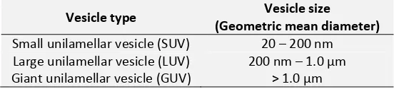
TABLE 1: SIZE OF DIFFERENT TYPES OF LIPOSOMES
Vesicle type Vesicle size (Geometric mean diameter)
Small unilamellar vesicle (SUV) 20 – 200 nm Large unilamellar vesicle (LUV) 200 nm – 1.0 µm Giant unilamellar vesicle (GUV) > 1.0 µm
Liposomes are made up of various grades of phospholipids and some other ingredients like cholesterol and lipid-conjugated hydrophilic polymers. The cholesterol acts as vesicle stabilizer by improving the firmness of bilayer membrane.
Vesicle morphology liposome depends on the various properties like surface charge, size, surface hydration and fluidity of lipid bilayers. The use of charge inducers is also well known due to their distinct applications. The cornea generally carries negative charge and hence the positively charged liposomes display better corneal permeation. Moreover, the charged liposomes exhibit less aggregation tendency as compared to neutral liposomes.
The only drawback of charged liposome is their higher uptake and rapid clearance by Reticulo-endothelial system (RES) because of their higher interaction with serum proteins. The RES clearance is also vesicle size dependent as the vesicle size below 100 nm are more prone to opsonization and hence they show more circulation time in blood 13.
The liposomes show these many of novel benefits although it has some negative points which require further improvements and optimization. The most common problem of liposome manufacturing is the susceptibility of phospholipids to oxidative degradation in air 14.
Thus, the handling and storage of liposome becomes very critical issue. Many of the researchers have enlisted requirement of nitrogen gas (N2) in ideal
[image:3.612.24.300.600.663.2]Niosomes behave in-vivo like liposomes, prolonging the circulation of entrapped drug and altering its organ distribution and metabolic stability. Encapsulation of various anti-neoplastic agents in these carrier vesicles has been shown to decrease drug induced toxic side effects, while maintaining, or in some instances, increasing the anti-tumor efficacy. Such vesicular drug carrier systems alter the plasma clearance kinetics, tissue distribution, metabolism and cellular interaction of the drug. In addition, handling and storage of niosomes requires no special conditions. In mice, the distribution of drugs incorporated in niosome vesicles has proved to be similar to that obtained after administration of drug encapsulated in liposomes. Work done in Liposomal Dosage Forms as ODDS: The liposomes were first studied as ocular drug delivery system in 1981 by Smolin et al. Authors have described preparation of Idoxuridine liposomes and have compared their therapeutic efficacy with Idoxuridine topical solution for the treatment of herpetic keratitis in the rabbit eye 18.
Schaeffer and Krohn have investigated the role of charged vesicles and size of vesicles in transcorneal permeation in rabiit eye. Authors practically demonstrated approximately four times higher corneal flux of penicillin-G loaded liposomes 19.
Singh and Mezzei have investigated role of Triamcinolone acetonide liposomes in increasing the concentration of drug at the target site. They demonstrated to achieve twice concentration of Triamcinolone acetonide when delivered in liposomal dosage form as compared to conventional drug solution.
However, liposomal formulations of Dihydrostreptomycin sulfate did not aid the corneal permeation. The justification was made that liposomes are less effective to entrap the hydrophilic drugs 20. Fitzgerald et al., have explored the effects on clearance rate of liposomes by gamma scintigraphy upon topical administration in the rabbit model. They reported that the positively charged SUVs are more effectively retained at cornea site. They justified that positively charged liposomes were being retained due to negative charge present on the corneal cells 21.
Chitosan is well known for its mucoadhesive properties and the same approach was used in liposome formulation by various researchers to improve the residence time in eye at the site of action. Chitosan is biodegradable and its byproducts are also non-toxic, thus the use of chitosan in ODDS is considered safe. Mehanna et al., have prepared chitosan coated mucoadhesive liposomes of Ciprofloxacin by reverse phase evaporation method. The prepared liposomes were further coated with chitosan of different molecular weights (chitosomes). They concluded that the liposomes coated with chitosan if high molecular weight were relatively smaller in size, which acted as a barrier to inhibit aggregation.
In vitro release studies showed chitosan coated
liposomes released drug delivery slower than uncoated liposomes due to presence of physical barrier at the outer side. Ex vivo studies demonstrated that the chitosan coated liposomes shows almost twice permeability as the chitosan aided the diffusion across the cornea cells 22.
Li et al., have determined the role of low molecular
weight chitosan coated liposomes in ocular drug delivery. Low molecular weight chitosan coated liposomes showed good influence in vivo activity. Authors claimed to achieve higher ex vivo corneal penetration across excised rabbit cornea using low molecular weight coated liposomes. The in vitro release was found to be much slower due to presence of physical barrier chitosan. The precorneal loss time was also drastically reduced due to mucoadhesive nature of chitosan. They also reported that there were no signs of adverse effects during study 23.
Zhang et al., utilized N-trimethyl chitosan chloride (TMC), a quaternary derivatives of chitosan for coating of coenzyme Q10-loaded liposomes. They reported better stability and less leakiness due to coating. They measured and reported approximately 5 times higher precorneal residence time after topical administration using gamma scintigraphy.
Habib et al., prepared and evaluated Fluconazole loaded liposomes on candidal keratitis model in rabbits. Authors aimed to enhance the antifungal action by prolonging the contact time at the site of action. They carried out comparative efficacy study of the Fluconazole solution and Fluconazole loaded liposomes. They reported to get 1.7 times faster healing with liposomal drug delivery no noticeable side effects 27.
Budai et al. prepared negatively charged ciprofloxacin loaded liposomal hydrogel for the treatment of chronic ocular infectious diseases like conjunctivitis, bacterial keratitis, which requires high drug concentration at the site of infection.
Lecithin and alpha-L-dipalmitoylphosphatidycholine (DPPC) were used as to prepare multilamellar vesicles of liposomes. Hydrogel was prepared by using Poly vinyl alcohol (PVA) and polymethacrylic acid (PMA) derivatives. The gel was evaluated for viscosity, rheological property, in vitro release and ex vivo studies. The Ciprofloxacin loaded liposomes showed better entrapment efficiency due to electrostatic charge properties and showed five time enhancement in transcorneal permeation in rabbit model 28, 29.
Felt et al., developed and evaluated oflaxacin-loaded liposomal thermo-sensitive hydrogel for transcorneal permeation of Oflaxacin. Authors reported the difference in vesicle size is arising due to difference in method of preparation. Authors used chitosan/ beta-glycerophosphate thermo-sensitive hydrogel system for fabrication of liposomes in thermo-sensitive gels and observed reduced required energy for gelation step. They also performed transcorneal permeation study using rabbit cornea and reported that Oflaxacin permeates seven times higher from the liposomal formulation compared to Oflaxacin aqueous solution due to mucoadhesive nature of the hydrogel. Researchers reported that the ocular retention time was three times higher due to mucoadhesive properties of hydrogel without any significant adverse effects 30.
Hathout et al., carried out encapsulation of Acetazolamide in liposomes by reverse phase evaporation technique (REV) for the treatment of glaucoma with topical administration.
Measurement of intraocular pressure (IOP) lowering was carried out and compared with effects produced by Acetazolamide solution. Liposomes prepared by different compositions were evaluated for entrapment efficiency, stability in vitro release and IOP lowering efficacy in rabbit model. Authors reported highest entrapment efficiency with positively charged liposomes due to ionic interaction between drug and lipid 31.
Danion et al., prepared MLV liposome loaded contact lenses for the site specific delivery of Levofloxacin in treatment of bacterial keratitis. They reported first order release kinetics up to six days. The contact lens as carrier helped to improve the retention time and continuous drug delivery 32, 33.
Mahmoud et al., prepared Chloramphenicol (CP) loaded liposomes using dimyristoylphosphatidycholine (DMPC) as lipid part by three different methods like partitioning of CP in the vesicle bilayers, entrapment of CP by normal hydration method and adsorption of CP on the vesicle surface. Those formulations were evaluated for interaction of the drug and phospholipid bilayers and for effectiveness against S. aureus. They concluded that CP located in interfacial region within the hydrophobic core of the liposomes shows better anti-bacterial activity against S. aureus up to 5 hrs 34.
Law et al., prepared Acyclovir (ACV) loaded liposomes
for topical administration and carried out in vitro corneal penetration and in vivo corneal absorption on male rabbits. They concluded that surface charge of liposomes play key role in corneal penetration and ACV absorption. They stressed on positively charged liposomes to exhibit higher drug entrapment efficiency and better drug release compared to negatively charged liposomes.
Kawakami et al., prepared liposomes of O-palmitoyl prodrug of Tilisolol to enhance the retention time of Tilisolol in the precorneal area. The administration of liposioome was carried out by topical route as well as intravitreal route in rabbit eye. Upon intravitreal application, they found higher concentration of Tilisolol as compared to free Tilisolol. By topical administration, they reported very low retention of O-palmitioyl Tilisolol in the tear fluid. Thus, to further improve this, they used 2% of sodium carmellose. Sodium carmellose acted as a reservoir for liposomes and thus retention was improved relatively 36.
Barza et al., studied the effects of liposomes size and pathological state of eye upon intravitreal elimination kinetics of carriers. They concluded that the rate of clearance of SUVs was higher than LUVs. They also observed that intraocular inflammation increases the rate of intravitreal clearance 37.
Zhang et al., prepared Tacrolimus-loaded liposomes by reverse phase evaporation method for the treatment of uveoretinitis. They further evaluated it for safety and efficacy upon intravitreal injection in rats. No retinal dysfunction was observed in the liposome-treated rats. Histo-pathological examination revealed inflammatory response in comparison to free drug. They claimed to achieve 50ng/mL up to two weeks after single administration of Tacrolimus loaded liposomes. They consluded that Tacrolimus loaded liposomes were more efficacious than the free drug 38. Cheng et al., prepared liposomal prodrug of ganciclovir (GCV) i.e. 1-O-hexadecylpropanediol-3-phospho-Ganciclovir and further injected intravitreally in rabbits. They studied its effects for antiviral treatment against herpes simplex virus type 1 (HSV-1) and human cytomegalovirus (HCMV). They concluded that intravitreal injection of 0.20 nM intravitreal concentrations was most effective without causing any side effects 39.
Fukushima et al., formulated Clodronate liposomes used to inhibit infiltration of macrophages in the conjuctiva in treatment of blepharo conjunctivitis developed in Brown Norway rats. Authors stated that Clodronate liposomes very much effective in decreasing the number of ED2-positive macrophages in the conjunctivas.
Authors suggested investigating further using liposomes larger than 550 nm to explore thorough research regarding subconjunctival clearance 40.
Work done on niosomal dosage forms as ODDS: Handjani-vila et al., first demonstrated the advantages obtained by use of vesicular systems on the skin and specifically the use of non-ionic surfactants in aqueous dispersion. They carried out a comparative toxicity study with classical formulation such as emulsions and concluded that novel vesicular system exhibits low toxicity and better permeation of actives through stratum corneum 41.
Green and Downs studied the ocular penetration of Pilocarpine using radio labeled Pilocarpine in different vehicles. They also reported that an ocular bioavailability of water soluble drug, entrapped in niosomes is due to the role of surfactant. The surfactant decreases the surface tension and hence also acts as penetration enhancers by removing the mucus layer and break functional complexes 42.
Azmin et al., prepared vesicles of Methotrexate from a nonionic surfactant, cholesterol and dicetyl phosphate and dosed to mice. They reported that intravenous dose prolonged the release of methotrexate in the blood and also improved its uptake in liver and brain. They concluded the metabolic profile of drug was also altered by the niosomes which decreses the rapid formation of 7-hydroxy methotrexate 43.
Okahata et al., prepared bilayer structure using alkyl chain length C12, C14, C16 and C18. They reported that
vesicles were lamellae and amphiphilic in nature. They concluded that the aggregates were being formed only above the phase transition temperature of surfactant
44
.
Lasic prepared bilayer vesicles by involving some physical agitation and heat. They obtained an assembly in which the hydrophobic parts of the molecule are covered by aqueous solvent and the hydrophilic head groups gets maximum contact with hydrophobic parts. He concluded that requirement of some energy is must for preparation of bilayer vesicles 45.
Saettone et al., reported non-ionic vesicles as carriers of Cyclopentolate for ocular drug delivery. They prepared vesicles by sonication polysorbate 20 and cholesterol in various concentrations. They concluded that the vesicles buffered at pH 5.5 increases the transcorneal permeation of Cyclopentolate as compared to a standard buffer solution. They also reported that vesicles remarkably increase the bioavailability Cyclopentolate at ocular site as compared to standard buffer solutions 46.
Aggarwal et al., prepared the mucoadhesive niosomes of Cyclopentolate as compared to a standard buffer solution. for ocular drug delivery system improved. Authors used chitosan and carbopol to coat niosomes of Timolol maleate prepared by reverse phase evaporation (REV) method. They carried out comparative in-vitro release study with Timolol solution and IOP lowering effect as pharmacodynamic study. They concluded that chitosan coating can significantly extend the release profile of Timolol maleate 47.
Aggarwal et al., also prepared Acetazolemide loaded niosomes by reverse phase evaporation and coated them with carbopol to enhance the bio-adhesive nature. Authors reported that sustained action was obtained for 6 hours with 33% more decrease in intraocular pressure.
The bioavailability of Acetazolemide was enhanced almost twice when dosed in niosomal dosage form as compared to free drug solution. They applied microdialysis technique to improve the regional sampling of the tissues 48.
Abdlbary and El-gendy used non ionic surfactant vesicles as carriers for the controlled ocular delivery of Gentamicin sulphate. They prepared and evaluated various nisomal formulations using different surfactants like Tween 60, Tween 80 and Brij 35 in different molar ratios and by thin film hydration
technique. They carried out photomicroscopy, transmission electron microscopy and particle size analysis for evaluation of vesicles. They reported that niosomal Gentamicin sulphate shows a high retention as compared to the free drug solution in-vitro release study. Authors also carried out ocular irritancy study and concluded that there were no signs of irritation49. CONCLUSION: The vesicular drug delivery has its distinctive advantages due to its novel structure. The strategic role of bilayer vesicles has been investigated by many researchers over the time. The critical structure of human eye and its anatomical as well as physiological barriers can be more conveniently overcome by vesicular drug delivery system. The bio-adhesive nature of phospholipids and non-irritating nature of non-ionic surfactants have laid detailed research on vesicles as ocular drug delivery system. The potential action of vesicles drug delivery system will lead to drastically reduced therapy time and excellent patient compliance.
REFERENCES:
1. Metz A: The Anatomy and Histology of the Human Eye. Nabu Press, 2010. ISBN-10: 1144060478
2. Geroski DH and Edelhauser HF: Drug delivery for posterior segment eye disease. Investigative Ophthalmology and Visual Science 2000; 40:961-964.
3. Urtti A: Challenges and obstacles of ocular pharmacokinetics and drug delivery. Advanced Drug Delivery Reviews 2006; 58:1131-1135. 4. Mitra AK: Role of transporters in ocular drug delivery system.
Pharmaceutical Research 2009; 26:1192-1196.
5. Wood RW, Lee VHE, Kreuter J and Robinson JR: Ocular disposition of poly-hexyl-2 cyano[3-14C]acrylate nanoparticles in the albino rabbit. Int.
J. Pharm1985; 23:175-183.
6. Vyas SP and Khar RK: Controlled drug delivery system – concepts and advances. Vallabh Prakashan, New Delhi, First edition 2002.
7. Biju SS, Sushama T, Mishra PR and Khar RK: Vesicular System – An overview. Indian J. Pharm Sci 2006; 68:141-153.
8. Bangham AD, Standish MM and Watkins JC: Diffusion of univalent ions across the lamellae of swollen phospholipids. Journal of Molecular Biology 1965; 13:238-252.
9. Kaur IP, Garg A, Singla AK and Aggrawal D: Vesicular systems in ocular drug delivery: an overview. International Journal of Pharmaceutics 2004; 269:1-14.
10. Jesorka A and Orwar O: Liposomes: technologies and analytical applications. Annual Review of Analytical Chemistry 2008; 1:801-832. 11. Kaur IP and Kanwar M: Ocular preparations: the formulation approach.
Drug Development and Industrial Pharmacy 2002; 28:473-493. 12. Wadhwa S, Paliwal R and Vyas SP: Nanocarriers in ocular drug delivery:
an update review. Current Pharmaceutical Design 2009; 15:2724-2750. 13. Lian T and Ho RJY: Trends and developments in liposome drug delivery
systems. Journal of Pharmaceuticals Sciences 2001; 90:667-680. 14. Florence AT and Baillie AJ: Non-ionic surfactant vesicles – alternatives to
liposomes in drug delivery. Novel Drug Delivery and its Therapeutic Application. L. F. Prescott and W. S. Nimmo, John Wiley and Sons Ltd 1989.
16. Vora B, Khopade AJ and Jain NK: Proniosome based transdermal delivery of evonorgestrel for effective contraception. J. Control Rel 1998; 54:149-165.
17. Uchegbu IF, Schätzlein A and Florence AT: Non-ionic surfactant vesicle (niosome) phase transitions. European Journal of Pharmaceutical Sciences 1996; 4:S138
18. Smolin G, Okumoto M, Feiler S and Condon D: Idoxuridine-liposome therapy for herpes simplex keratitis. American. J. Ophthalmol. 1981; 91:220-225.
19. Schaeffer HE and Krohn DL: Liposomes in topical drug delivery system. Investigative Ophthalmology and Visual Science 1982; 22:220-227. 20. Singh K and Mezzei M: Liposomal ophthalmic drug delivery system. I.
Triamcinolone acetonide. International Journal of Pharmaceutics 1983; 16:339-344.
21. Fitzgerald P, Hadgraft J and Wilson CG: A gamma scintigraphic evaluation of the precorneal residence of liposomal formulations in the rabbit. Journal of Pharmacy and Pharmacology 1987; 39:487-490. 22. Mehanna MM, Elmaradny HA and Samaha MW: Micro adhesive
liposomes as ocular delivery system: physical, microbiological and in vivo assessment. Drug Development and Industrial Pharmacy 2010; 36:108-118.
23. Li N, Zhuang C, Wang M, Sun X, Nie S, and Pan W: Liposome coated with low molecular weight chitosan and its potential use in ocular drug delivery. International Journal of Pharmaceutics 2009; 379:131-138. 24. Zhang J and Wang S: Topical use of Coenzyme Q10-loaded liposomes
coated with trimethyl chitosan: tolerance, precorneal retention and anti-cataract effect. International Journal of Pharmaceutics 2009; 372:66-75.
25. Diebold Y, Jarrin M and Saez V: Ocular drug delivery by liposome-chitosan nanoparticle complexes (LCS-NP). Biomaterials 2007; 28:1553-1564.
26. Zhang J, Guan P, Wang T, Chang D, Jiang T and Wang S: Freeze-dried liposomes as potential carriers for ocular administration of cytochrome c against selenite cataract formation. Journal of Pharmacy and Pharmacaology 2009; 61:1171-1178.
27. Habib FS, Fouad EA, Abdel-Rhaman MS and Fathalla D: Liposomes as an ocular delivery system of fluconazole: in-vitro studies. Acta Ophthalmologica 2010; 88:901-904.
28. Budai L, Hajdu M, Budai M: Gels and liposomes in optimized ocular drug delivery: studies on ciprofloxacin formulations. International Journal of Pharmaceutics 2007; 343:34-40.
29. Hosny KM: Ciprofloxacin as ocular liposomal hydrogel. AAPS PharmSciTech 2010; 11:241-246.
30. Felt O, Furrer P, Mayer JM, Plazonnet B, Buri P and Gurny R: Topical use of chitosan in ophthalmology: tolerance assessment and evaluation of precorneal retention. International Journal of Pharmaceutics 1999; 180:185-193.
31. Hathout RM, Mansour S, Mortada ND and Guinedi AS: Liposomes as an ocular delivery system for acetazolamide: in vitro and in vivo studies. AAPS PharmSciTech 2007; 8:1.
32. Danion A, Brochu H, Martin Y and Vermette P: Fabrication and characterization of contact lenses bearing surface-immobilized layers of intact liposomes. Journal of Biomedical Materials Research Part A 2007; 82:41-51.
33. Danion A, Arsenault I and Vermette P: Antibacterial activity of contact lenses bearing surface-immobilized layers of intact liposomes loaded
with levofloxacin. Journal of Pharmaceutical Sciences 2007; 9:2350-2363.
34. Mahmoud SS, Gehmam JD, Azzopardi K, Robins-Browne RM and Separovic F: Liposomal phospholipid preparations of chloramphenicol for ophthalmic applications. Journal of Pharmaceutical Sciences 2008; 97:2691-2701.
35. Law SL, Huang KJ and Chinag CH: Acyclovir-containing liposomes for potential ocular delivery corneal penetration and absorption. Journal of Controlled Release 2000; 63:135-140.
36. Kawakami S, Yamamura K, Mukai T: Sustained ocular delivery of tilisosol to rabbits after topical administration or intravitreal injection of lipophilic prodrug incorporated in liposomes. Journal of Pharmacy and Pharmacology 2001; 53:1157-1161.
37. Barza M, Stuart M and Szoka F: Effect of size and lipid composition on the pharmacokinetics of intravitreal liposomes. Investigative Ophthalmology and Visual Science 1987; 28:893-900.
38. Zhang R, He R, Qian J, Guo J, Xue K and Yuan YF: Treatment of experimental autoimmune uveoretinitis with intravitreal injection of tacrolimus (FK506) encapsulated in liposomes. Investigative Ophthalmology and Visual Science 2010; 51:3575-3582.
39. Cheng L, Hostelter KY and Chaidhawangul S: Intravitreal toxicology and duration of efficacy of a novel antiviral lipid prodrug of ganciclovir in liposome formulation. Investigative Ophthalmology and Visual Science 2000; 41:1523-1532.
40. Fukushima A, Ozaki, Ishida W, Van Rooijen N, Fukata K and Ueno H: Suppression of macrophage infiltration into the conjunctiva by clodronate liposomes in experimental immune-mediated blepharoconjuctivitis. Cell Biology International 2005; 29:277-286. 41. Handjani-Vila RM, Rlbier A. Rondot B and Vanlerberghe G: Dispersion of
lamellar phases of non-ionic lipids in cosmetic products. Int. J. Cosmetic Sci 1979; 1:303-314.
42. Green K and Downs S: Ocular penetration of pilocarpine in rabbits. Arch. Ophthalmol. 1975; 93:1165-1168.
43. Azmin MN, Florence AT, Handjani-Vila RM, Stuart JF, Vanlerberghe G and Whittaker JS: The effect of non-ionic surfactant vesicle (niosome) entrapment on the absorption and distribution of methotrexate in mice. J Pharm Pharmacol.1985; 37:237-242.
44. Okhata Y, Tanamachi S, Nagai M and Kunitake T: Synthetic bilayer membranes prepared from dialkyl amphiphiles with non-ionic and zwitterionic head groups. J. Colloid Interface Sci. 1981; 82:401-407. 45. Lasic DD: On the thermodynamic stability of liposomes. J. Colloid
interface Sci. 1990; 140:302-304.
46. Saettone MF, Perini G, Carafa M, Santucci E and Alhaique F: Non-ionic surfactant vesicles as ophthalmic carriers for cyclopentolate. A preliminary evaluation. STP Pharma Sci.1996; 6:94-98.
47. Aggarwal D and Kaur IP: Improved pharmacokinetic dynamics of timolol maleate from a mucoadhesive niosomal ophthalmic drug delivery system. Int. J. Pharm. 2005; 290:155 – 159.
48. Aggarwal D, Pal D, Mitra AK, Kaur IP: Study of the extent of ocular absorption of acetazolamide from a developed niosomal formulation by microdialysis sampling of aqueous humor. Int. J. Pharm 2007; 338:21-26.
49. Abdelbary G and El-gendy N: Niosome- Encapsulated gentamicin for ophthalmic controlled delivery. AAPS Pharm.SciTech 2008; 9:740-747.
How to cite this article:
Modi KA and Shelat PK: Applications of Novel Vesicular Drug Delivery System as Ocular Drug Vehicles: A Review. Int J Pharm Sci Res. 3(12);