INVESTIGATION
Whole Genome Sequence and Comparative
Genomics Analysis of Multi-drug Resistant
Environmental
Staphylococcus epidermidis
ST59
Zhen Xu,*,†Raju Misra,‡Dorota Jamrozy,§Gavin K. Paterson,** Ronald R. Cutler,†Mark A. Holmes,†† Saheer Gharbia,‡and Hermine V. Mkrtchyan1
*Department of Public Health, National Demonstration Center for Experimental Preventive Medicine Education, Tianjin Medical University, Tianjin, China 300070,†School of Biological and Chemical Sciences, Queen Mary University of London, London, UK E1 4NS,‡Natural History Museum, Core Research Laboratories, Molecular Biology, London, UK SW7 5BD,§The Wellcome Trust Sanger Institute, Cambridge, UK CB10 1SA,**The Royal (Dick) School of Veterinary Studies and Roslin Institute, University of Edinburgh, Easter Bush Campus, Midlothian, UK EH8 9LE, and††Department of Veterinary Medicine, University of Cambridge, Cambridge, UK CB3 0ES
ABSTRACT Staphylococcus epidermidisis a major opportunistic pathogen primarily recovered from de-vice-associated healthcare associated infections (DA-HAIs). AlthoughS. epidermidisand other coagulase-negative staphylococci (CoNS) are less virulent thanStaphylococcus aureus, these bacteria are an important reservoir of antimicrobial resistance genes and resistance-associated mobile genetic elements that can be transferred between staphylococcal species. We report a whole genome sequence of a multidrug resistant S. epidermidis(strain G6_2) representing multilocus sequence type (ST) 59 and isolated from an environ-mental sampling of a hotel room in London, UK. The genome of S. epidermidis G6_2 comprises of a 2408357 bp chromosome and six plasmids, with an average G+C content of 32%. The strain displayed a multi-drug resistance phenotype which was associated with carriage of 7 antibiotic resistance genes (blaZ,mecA, msrA,mphC, fosB, aacA-aphD, tetK) as well as resistance-conferring mutations in fusA andileS. Antibiotic resistance genes were located on plasmids and chromosome. Comparative genomic analysis revealed that antibiotic resistance gene composition found in G6_2 was partly preserved across the ST59 lineage.
KEYWORDS
Staphylococcus epidermidis antibiotic
resistance whole genome
sequence comparative
analysis
Staphylococcus epidermidisis a common human skin commensal, but also the most frequent pathogen among coagulase-negative staphylo-cocci (CoNS), causing primarily device-associated healthcare associ-ated infections (DA-HAIs). Compared with more virulentS. aureus, CoNS rarely produce toxins and less is known on whether the toxin genes contribute to strain virulence (Otto 2013a).S. epidermidisforms biofilms on medical devices and implants, from which single cells dissociate and disseminate via the bloodstream to start colonization at a different site, which might lead to sepsis, meningitis and
endocar-ditis (Beckeret al.2014). In addition,S. epidermidisand other CoNS are believed to act as a reservoir of resistance and virulence genes for
S. aureus, contributing to the evolution and emergence of successful clones of methicillin-resistantS. aureus(MRSA) (Otto 2013b).
Together withS. aureusand other CoNS,S. epidermidisaccounts for 30% of hospital associated infections (Conlanet al.2012). These nos-ocomial pathogens have developed an arsenal of strategies contributing to colonization and infection of the hosts (Beckeret al.2014), while often being resistant to multiple antibiotics. Emergence of antibiotic resistant bacteria has been mostly attributed to the healthcare-associated settings (Oliveira and Tomasz 2002). However, more recently, selec-tion of antibiotic resistance has been also associated with the com-munity which has been linked to the misuse of antibiotics (DeLeo
et al.2010). A typical example of this is the community-acquired MRSA (CA-MRSA) which, in addition to acquiring methicillin re-sistance, has gradually increased the frequency of resistance determi-nants similarly to hospital-acquired MRSA (HA-MRSA) (Chambers 2005). There is an increasing evidence that horizontal gene transfer between closely related species may contribute to this (Otto 2013a). Copyright © 2018 Xuet al.
doi:https://doi.org/10.1534/g3.118.200314
Manuscript received November 9, 2017; accepted for publication April 23, 2018; published Early Online April 30, 2018.
This is an open-access article distributed under the terms of the Creative Commons Attribution 4.0 International License (http://creativecommons.org/licenses/by/4.0/), which permits unrestricted use, distribution, and reproduction in any medium, provided the original work is properly cited.
Supplemental material available at Figshare:https://doi.org/10.25387/g3.6133946.
1Correspondence: School of Health, Sport and Biosciences, University of East
Recently, Méricet al.showed thatS. aureusandS. epidermidisshare half of the genome and while homologous recombination between the two species was rare, there was an evidence of extensive MGE sharing, in particular SCCmec, metal resistance and SaPIn1 elements (Méric
et al.2015). As a result, attention is now focusing on the multidrug-resistant coagulase-negative staphylococci and their rapid spread as opportunistic pathogens particularly in relation to patients with an immuno-compromised status (Morfin-Oteroet al.2012). Multidrug-resistant coagulase-negative staphylococci (MDR-CoNS) are primarily recovered from healthcare-associated medical devices, ambulatory pa-tients and healthy animals (Beckeret al.2014).
Molecular approaches such as pulsefield gel electrophoresis and multi-locus sequence typing have been widely used to evaluate the dissemination of resistant clones of bacteria (Miragaiaet al.2008). Recently, complete genome sequencing ofS. epidermidisstrains have been reported, however these are limited to commensal and nosoco-mial strains (Conlanet al.2012; Gillet al.2005; Zhanget al.2003). Only one study has compared whole genome sequences of fourS. epidermidis
isolated from rice seeds with that of type strain (Chaudhry and Patil 2016). To our knowledge this is thefirst whole genome based study looking at MDR-CoNS isolated from general public settings.
In this study, we present the genetic features of this multidrug resistantS. epidermidis(strain G6_2) and compare it with sixS. epidermidis
reference genomes and 133 previously published genomes of clinical
S. epidermidis.
MATERIAL AND METHODS
Isolates analyzed in this study
Between October 2012 and April 2013, we sampled different sites in three hotels in London, UK. Permission to carry out sampling was granted by the manager/owner of each hotel and the results from each hotel were reported to each manager/owner for their information. Inanimate objects in 32 hotel rooms were sampled using COPAN dry swabs (Copan Diagnostics Inc., USA). All specimens were inoculated onto Nutrient Agar (Oxoid, Basingstoke, UK) and Mannitol Salt Agar plates (Oxoid Basingstoke, UK). These cultures were incubated aero-bically at 37°for 24–72 h.
TheS. epidermidisG6_2 was recovered from one of the hotel rooms in April 2013 in London, UK. Preliminary identification was achieved by using Matrix-assisted laser desorption ionization time-flight mass-spectroscopy (Microflex LT, MALDI-TOF-MS, Bruker Daltonics, Coventry, UK) as described previously (Mkrtchyanet al.2013).
For comparative genomics analysis genomes of six S. epidermidis
reference strains were included: RP62A (Gill et al.2005), ASM1192v1), ATCC12228 (Zhang et al. 2003), ASM764v1), SEI (Davenport et al.
2014), CP009046), 949_S8 (Biswas et al. 2015), CP010942), PM221 (Savijoki et al. 2014), HG813242), and BPH 0662 (Jyh et al. 2016), NZ_LT571449) together with 129S. epidermidisgenomes derived from two previously published collections (Roachet al.2015; Tewheyet al.2014).
16S rRNA gene sequencing
Genomic DNA ofS. epidermidisG6_2 was prepared using a Qiagen DNA extraction kit (Qiagen, Crawley, UK). 16S rRNA amplification was performed as described previously (Okazaki et al.2009), PCR products were sequenced by Eurofins MWG GmBH (Ebersberg, Germany) using ABI 3730 L DNA analyzer.
Molecular characterization of S. epidermidis G6_2
Carriage of the mecAgene was determined with PCR as described previously (Hanssenet al.2004). SCCmectyping was carried out by
determination ofmecandccrcomplexes (Kondoet al.2007). Multi locus sequence tying (MLST) has been used to determine seven house-keeping genes as describe previously (Thomaset al.2006). Sequence types were determined using MLST V1.8 software (https://cge.cbs.dtu.dk/ services/MLST/).
Antibiotic susceptibility testing
The antibiotic susceptibility ofS. epidermidisG6_2 was tested against 13 antibiotics (Mast Group, Merseyside, UK) using disk diffusion meth-ods according to BSAC guidelines (J. M. Andrews and Howe 2011). This included penicillin (1 unit), amoxicillin (10mg), cefoxitin (10mg), ox-acillin (1mg), cefepime (30 mg), vancomycin (5mg), gentamicin (10mg), streptomycin (10 mg), mupirocin (20mg), erythromycin (15mg), tetracycline (10mg), fusidic acid (10mg) and chloramphen-icol (30mg). In addition, the minimum inhibitory concentration (MIC) of the isolate to oxacillin was determined using‘‘M.I.C. eval-uators’’(Oxoid Ltd., Basingstoke, UK).
Whole genome sequencing, assembly and comparative genomics
Nucleotide sequence accession numbers
Reads forS. epidermidisG6_2 were submitted to the European Bio-informatics Institute Sequence Read Archive, accession ERR387168.
Data availability
The authors state that all data necessary for confirming the conclusions presented in the article are represented fully within the article and its tables andfigures. Supplemental material available at Figshare:https:// doi.org/10.25387/g3.6133946.
RESULTS AND DISCUSSION
S. epidermidishas become a leading hospital-associated pathogen due to the increased use of medical devices (Vuonget al.2004). Treatment ofS. epidermidisinfections is challenging as the bacteria are commonly resistant to methicillin and might also display multi-drug resistance phenotype, which presents a serious public health challenge (Xuet al.
2015) .S. epidermidis, represents an important reservoir of mobilizable genes that can be horizontally transferred between staphylococci spe-cies, which has likely contributed to the development of antibiotic re-sistance inS. aureus(Otto 2013a).
S. epidermidisG6_2 was isolated from a hotel room in London, UK in 2013, and the species were determined by MALDI-TOF MS and 16S rRNA sequencing. Initial molecular analysis revealed that theS. epidermidis
G6_2 strain wasmecApositive, carrying SCCmectype IV, and rep-resented ST59.
A draft genome was assembled, comprising of 53 contigs (48.= 1kb) for the isolatedS. epidermidisG6_2 genome (Table S1; Table S2 and Figure S1). The assembly comprised of one chromosome (2408357 bp in length) and six plasmids, annotated as pG6_2_1 to pG6_2_6 (the largest, pG6_2_1, is 10570 and the smallest, pG6_2_6, is 3426 bp in length), with an average G+C content of 32.02%. It has a total (chromosome and plasmids) of 2213 predicted protein coding sequences, of which 21.5% were annotated as hypothetical proteins and 14.3% were annotated as putative functions (Table 1).
Phylogenetic relationship with other S. epidermidis isolates
A previously described collection of 129 whole genome-sequenced
S. epidermidis isolates together with 6 reference strains was used to determine the phylogenetic relationship between the G6_2 strain and
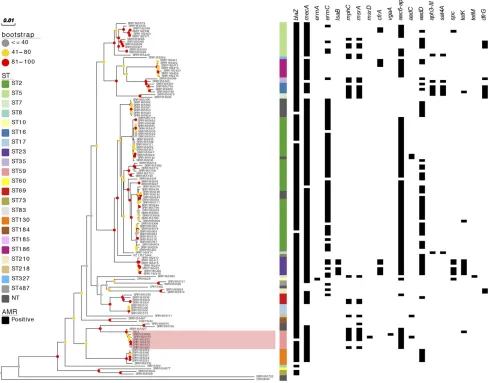
otherS. epidermidis lineages. After removal of variable sequence regions corresponsing to mobile genetic elements (MGE), recombination blocks as well as sites with more than 5% proportion of gaps, the core genome alignment contained 4262 SNP sites. Seven ST59 isolates clus-tered and formed a distinct clade withS. epidermidisG6_2 (Figure 1).
Genotypic and phenotypic characterization of antibiotic resistance
S. epidermidisG6_2, revealed 9 antibiotic resistance determinates across the chromosome and plasmids (Table 2). This included aminoglycoside resis-tance geneaac(6’)–aph(2”), beta-lactam resistance genesmecAandblaZ, fosfomycin resistance gene fosB, macrolide resistance genesmphH and
msrA(the latter also conferring resistance to lincosamide and streptogramin B) and tetracycline resistance genetet(K). This correlated with the results of antibiotic susceptibility testing as the strain was found resistant to 11 out of 13 antibiotics tested, demonstrating susceptibility to vancomycin and chlor-amphenicol only. Resistance to mupirocin and fusidic acid was associated with point mutations in chromosomally located genes,ileSandfusA, re-spectively. In addition to antimicrobial resistance genes, the G6_2 strain also carried plasmid-associatedqacCgene, which encodes the multidrug resis-tance efflux protein and mediates resistance to biocides, and a chromoso-mally-inserted copper resistance operon composed of copZ-copA-csoR
genes together with an additional copy of cobalt-zinc-cadium efflux pump geneczcD. The latter was distinct from the conserved chromosomal copy of
czcDgene, and was previously identified on a number of CoNS plasmids. The G6_2 strain carried a 47-kb composite island composed of the SCCmecIV and a SCC element that contained plasmin-sensitive surface protein genepls, spermidine N-acetyltransferase genespeGand a copper-translocating ATPase genecopA. The full sequence of this composite island was unique and did not match previously described reference genomes. However, the SCCmecIV sequence shared 99% identity with SCCmecIVa from various MRSA strains including the MRSA M1 isolated in Denmark (Larner-Svenssonet al.2013). The SCC element matched most closely the MRSA UCI62 strain representing ST5 (GenBank: CP018766). Carriage of
blaZ,tetKandqacCgenes was associated with plasmid sequences whereas other genes were inserted chromosomally. Elements carrying tetKand
qacCmatched previously reportedS. aureusplasmids. Méricet al.showed that hospital associatedS. aureusandS. epidermidisshare genes involved in pathogenicity, metal toxicity resistance and antibiotic resistance. In addi-tion they have demonstrated that high levels of recombinaaddi-tion of genes that
n Table 1 Comparative general features ofS. epidermidisG6_2 and the reference strains
Chromosomea RP62a ATCC 12228 SEI 949_S8 PM221 BPH 0662 G6_2
Length of sequences (bp) 2616530 2499279 2538314 2339868 2490012 2793003 2408357
G+C content 32.10% 32.10% 32.10% 32.00 32.10% 32.00% 32.02%
Protein coding region 2391 2419 2504 2119 2399 2699 2213
Ribosomal RNAs 4
16S 6 5 6 -b 6 5 1
23S 6 5 6 -b 6 5 1
5S 7 6 7 5 7 6 2
Transfer RNAs 59 60 58 56 59 59 60
Plasmidsc
Length of sequences (bp) P1:27310 P1:4439 P1:37688 -b P1:4439 P1:45804 P1:10570
P2:4679 P2:11152 P2:2366 P2:4909
P3:8007 P3:33094 P3:4588
P4:17261 P4:58811 P4:4576
P5:24370 P5:4271
P6:6585 P6:3426
a
Chromosome section includes: the length of the chromosome, G+C content of the chromosome, protein coding region, ribosomal RNA and transfer RNAs numbers. b
‘-’No data available in Genbankfile. Draft assembly. c
might be successful in healthcare settings contribute to proliferation of subpopulations of two species (Méricet al.2015).
Comparison of resistance determinant distribution revealed that the
S. epidermidisG6_2 strain shared a common antibiotic resistance gene composition with other ST59 isolates, suggesting that the particular combination of antibiotic resistance genes found in the G6_2 strain is preserved across the ST59 lineage (Figure 1). All ST59 isolates har-boredaac-aph,blaZandmecAgenes, and majority containedmphC
andmsrAgenes, whereastetKwas uniquely found inS. epidermidis
G6_2. The G6_2 strain also shared the qacC plasmid with other ST59 isolates as well as the SCCmec IV sequence but not full SCCmec-SCC composite island, which was not detected in any other analyzedS. epidermidisgenome.
Functional genes uniquely found in S. epidermidis G6_2 compared with reference strains
Pan-genome analysis of the G6_2 strain and sixS. epidermidisreference genomes revealed that 78 genes were unique to G6_2. After excluding genes found on plasmids, 64 chromosomally located genes were unique
to G6_2 strain. This included a number of SCCmec- and SCC-associated genes as well as some of the chromosomally inserted resistance genes such asmphC,msrA,copZ-copA-csoRoperon and the additional copy of
czcDgenes.
Comparative analysis of virulence genes
Pathogenicity ofS. epidermidishas been linked primarily with its capacity for biofilm formation. Biofilm formation occurs by initial attachment of bacteria on both biotic and abiotic surfaces, which further accumulates into multi-layered cell agglomerates. This facilitates the internalization and persistence ofS. epidermidis species in the host cells. Strains that facilitate this feature are therefore considered more virulent (Becker
[image:4.603.60.549.56.439.2]binding protein geneebh(140/140), the elastin binding protein geneebp
(135/140), thefibrinogen binding protein genessdrG(137/140) andsdrH
(138/140), serine protease genessspA(138/140) and sspB(138/140), lipase genesgeh(139/140) andlip(138/140), and the nuclease gene
nuc(138/140). The intercellular adhesion operonicaADBC, which is also associated with biofilm formation (Cramton et al.1999), was variably distributed (87/140) and absent in theS. epidermidisG6_2 strain as well as the other ST59 isolates included in this analysis. This is in agreement with previous reports of clinicalS. epidermidisST59 isolates that revealed a biofilm negative phenotype (Li et al.2009; Mendeset al.2012; Miragaiaet al.2007).
In addition to the described biofilm formation-associated virulence determinants, majority ofS. epidermidisisolates carried the hemolysin-beta genehlb(136/140), which was also present in the G6_2 strain. Less common was the delta hemolysin genehld(41/140), also detected in the G6_2 strain although absent in most other ST59 isolates.
In conclusion, this study is the first analysis of the genome of
S. epidermidisisolated from the general public environment and har-boring a cassette of resistance genes to an array of antimicrobials. The comparison of S. epidermidis G6_2 genome with clinical reference strains revealed its antibiotic resistance and virulence gene arsenal. Resistance genes were carried on both bacterial chromosome and plas-mids. We established thatS. epidermidisG6_2 harbors 12 virulence genes, and delta hemolysin genehld(41/140) is known to be detected in the G6_2 strain but absent in most other ST59 isolates. In addition, 9 antibiotic resistance determinants which are responsible for the resistance to 12 antibiotics, including streptomycin, gentamicin, penicillin, oxacillin, amoxicillin, cefoxitin, cefepime, erythromycin, fosfomycin, tetracycline, fusidic acid, mupirocin, have been identified inS. epidermidisG6_2. Additional whole genome sequence and com-parative genomics analysis are warranted to further our understanding of the origin and evaluation of multidrug resistant isolates from differ-ent ecological niches.
ACKNOWLEDGMENTS
Genome sequencing was supported by Medical Research Council (MRC) partnership grant G1001787/1.
LITERATURE CITED
Andrews, J. M., and R. A. Howe, 2011 BSAC standardized disc suscepti-bility testing method (Version 10). J. Antimicrob. Chemother. 66: 2726– 2757.https://doi.org/10.1093/jac/dkr359
Andrews, S., 2011 pp. 175–176 inFastQC A Quality Control Tool for High Throughput Sequence Data. Babraham Institute, Cambridge, UK. Bankevich, A., S. Nurk, D. Antipov, A. A. Gurevich, M. Dvorkinet al.,
2012 SPAdes: a new genome assembly algorithm and its applications to single-cell sequencing. J. Comput. Biol. 19: 455–477.https://doi.org/ 10.1089/cmb.2012.0021
Becker, K., C. Heilmann, and G. Peters, 2014 Coagulase-negative staphylococci. Clin. Microbiol. Rev. 27: 870–926.https://doi.org/10.1128/CMR.00109-13
Biswas, R. K., M. M. Kock, T. Adelowotan, W. Strasheim, T. G. Mahomed et al., 2015 Draft genome sequences offive clinical methicillin-resistant Staphylococcus aureusisolates and a methicillin-resistantStaphylococcus epidermidisisolate. Genome Announc. 3: e00836–e15.https://doi.org/ 10.1128/genomeA.00836-15
Chambers, H. F., 2005 Community-associated MRSA–resistance and vir-ulence converge. N. Engl. J. Med. 352: 1485–1487.https://doi.org/ 10.1056/NEJMe058023
Chaudhry, V., and P. B. Patil, 2016 Genomic investigation reveals evolution and lifestyle adaptation of endophyticStaphylococcus epidermidis. Sci. Rep. 6: 19263.https://doi.org/10.1038/srep19263
Chen, L., D. Zheng, L. Bo, Y. Jian, and J. Qi, 2016 VFDB 2016: hierarchical and refined dataset for big data analysis—10 years on. Nucleic Acids Res. 44: D694–D697.https://doi.org/10.1093/nar/gkv1239
Conlan, S., H. H. Kong, and J. A. Segre, 2012 Species-level analysis of DNA sequence data from the NIH human microbiome project. PLoS One 7: e47075.https://doi.org/10.1371/journal.pone.0047075
Cramton, S. E., C. Gerke, N. F. Schnell, W. W. Nichols, and F. Gotz, 1999 The intercellular adhesion (Ica) locus is present inStaphylococcus aureusand is required for biofilm formation. Infect. Immun. 67: 5427–5433. Croucher, N. J., A. J. Page, T. R. Connor, A. J. Delaney, J. A. Keaneet al.,
2015 Rapid phylogenetic analysis of large samples of recombinant bacterial whole genome sequences using gubbins. Nucleic Acids Res. 43: e15.https://doi.org/10.1093/nar/gku1196
Davenport, K. W., H. E. Daligault, T. D. Minogue, K. A. Bishoplilly, S. M. Broomallet al., 2014 Complete genome assembly of Staphylo-coccus epidermidisAmMS 205. Genome Announc. 2: e01059–e14.https:// doi.org/10.1128/genomeA.01059-14
n Table 2 Genotypic and phenotypic characterization of antibiotic resistance inS. epidermidisG6_2
Product Gene name
Accession number
(Identity %) Location Function Class of antibiotic Antibiotics
Aminoglycoside-modifying enzymes
aac(6’)-aph(2’’) M13771 (100) plasmid Aminoglycoside resistance
Aminoglycoside Gentamycin streptomycin
b-lactamase blaZ AJ302698 (100) plasmid Beta-lactam
resistance
Beta-lactam Penicillin oxacillin Amoxcillin cefepime cefoxitin Penicillin-binding
protein 2a
mecA AB505628 (100) Chromosome Beta-lactam
resistance Fosfomycin
resistance protein
fosA ACHE01000077 (100) Chromosome Fosfomycin
resistance
Phosphonic Fosfomycin
Macrophage
scavenger receptors
msr(A) X52085 (98.98) plasmid Macrolide, Lincosamide and Streptogramin B resistance
Microlide Erythromycin
Inactivating enzymes mph(C) AF167161 (100) plasmid Macrolide
resistance Tetracycline efflux
pump
tet(K) U38428 (99.93) plasmid Tetracycline resistance
Tetracycline Tetracycline
Isoleucyl RNA synthetase
ileS — — Fusidic acid
resistance
Fusidic acid Fusidic acid
Elongation factor G fusA — — Monoxycarbolic
resistance
DeLeo, F. R., M. Otto, B. N. Kreiswirth, and H. F. Chambers,
2010 Community-associated meticillin-resistantStaphylococcus aureus. Lancet 375: 1557–1568.https://doi.org/10.1016/S0140-6736(09)61999-1
Gill, S. R., D. E. Fouts, G. L. Archer, E. F. Mongodin, R. T. Deboyet al., 2005 Insights on evolution of virulence and resistance from the com-plete genome analysis of an early methicillin-resistantStaphylococcus aureusstrain and a biofilm-producing methicillin-resistant Staphylococ-cus epidermidisstrain. J. Bacteriol. 187: 2426–2438.https://doi.org/ 10.1128/JB.187.7.2426-2438.2005
Gupta, S. K., B. R. Padmanabhan, S. M. Diene, R. Lopez-Rojas, M. Kempf et al., 2014 ARG-ANNOT, a new bioinformatic tool to discover anti-biotic resistance genes in bacterial genomes. Antimicrob Agents Ch. 58: 212–220.https://doi.org/10.1128/AAC.01310-13
Hanssen, A., G. Kjeldsen, and J. U. Ericson Sollid, 2004 Local variants of staphylococcal cassette chromosomemecin sporadic methicillin-resistant Staphylococcus aureusand methicillin-resistant coagulase-negative staphylococci: evidence of horizontal gene transfer? Antimicrob Agents Ch. 48: 285–296.https://doi.org/10.1128/AAC.48.1.285-296.2004
Haubold, B., F. Klötzl, and P. Pfaffelhuber, 2015 Andi: fast and accurate estimation of evolutionary distances between closely related genomes. Bioinformatics 31: 1169–1175.https://doi.org/10.1093/bioinformatics/ btu815
Inouye, M., H. Dashnow, L. A. Raven, M. B. Schultz, B. J. Popeet al., 2014 SRST2: rapid genomic surveillance for public health and hospital microbiology labs. Genome Med. 6: 90.https://doi.org/ 10.1186/s13073-014-0090-6
Jyh, L., I. R. Monk, S. J. Pidot, S. Singh, C. Kylet al., 2016 Functional analysis of thefirst complete genome sequence of a multidrug resistant sequence Type 2Staphylococcus epidermidis. Microb. Genom. 2: e000077.
https://doi.org/10.1099/mgen.0.000077
Kondo, Y., T. Ito, X. X. Ma, S. Watanabe, B. N. Kreiswirthet al., 2007 Combination of multiplex PCRs for staphylococcal cassette chromosomemectype assignment: rapid identification system formec, ccr, and major differences in junkyard regions. Antimicrob Agents Ch. 51: 264–274.https://doi.org/10.1128/AAC.00165-06
Larnersvensson, H., P. Worning, M. D. Bartels, H. L. Hestbjerg, K. Boyeet al., 2013 Complete genome sequence ofStaphylococcus aureusstrain M1, a unique t024–ST8-IVa danish methicillin-resistantS. aureusclone. Ge-nome Announc. 1: S69–S82.
Li, M., X. Wang, Q. Gao, and Y. Lu, 2009 Molecular characterization of Staphylococcus epidermidisstrains isolated from a teaching hospital in Shanghai, China. J. Med. Microbiol. 58: 456–461.https://doi.org/10.1099/ jmm.0.007567-0
Mendes, R. E., L. M. Deshpande, A. J. Costello, and D. J. Farrell, 2012 Molecular epidemiology ofStaphylococcus epidermidisclinical isolates from U.S. hospitals. Antimicrob Agents Ch. 56: 4656–4661.
https://doi.org/10.1128/AAC.00279-12
Méric, G., M. Miragaia, M. de Been, K. Yahara, B. Pascoeet al.,
2015 Ecological overlap and horizontal gene transfer inStaphylococcus aureusandStaphylococcus epidermidis. Genome Biol. Evol. 7: 1313–1328.
https://doi.org/10.1093/gbe/evv066
Miragaia, M., J. A. Carriço, J. C. Thomas, I. Couto, M. C. Enrightet al., 2008 Comparison of molecular typing methods for characterization of Staphylococcus epidermidis: proposal for clone definition. J. Clin. Micro-biol. 46: 118–129.https://doi.org/10.1128/JCM.01685-07
Miragaia, M., J. C. Thomas, I. Couto, M. C. Enright, and H. de Lencastre, 2007 Inferring a population structure forStaphylococcus epidermidis from multilocus sequence typing Data. J. Bacteriol. 189: 2540–2552.
https://doi.org/10.1128/JB.01484-06
Mkrtchyan, H. V., C. A. Russell, N. Wang, and R. R. Cutler, 2013 Could public restrooms be an environment for bacterial resistomes? PLoS One 8: e54223.https://doi.org/10.1371/journal.pone.0054223
Morfin-Otero, R., M. A. Martínez-Vázquez, D. López, E. Rodríguez-Noriega, and E. Garza-González, 2012 Isolation of rare coagulase-negative iso-lates in immunocompromised patients:Staphylococcus gallinarum, Staphylococcus pettenkoferiandStaphylococcus pasteuri. Ann. Clin. Lab. Sci. 42: 182–185.
Okazaki, M., K. Ohkusu, H. Hata, H. Ohnishi, K. Sugaharaet al., 2009 Mycobacterium Kyorinensesp. nov., a novel, slow-growing species, related to mycobacterium celatum, isolated from human clinical speci-mens. IJSEM 59: 1336–1341.https://doi.org/10.1099/ijs.0.000760-0
Oliveira, D. C., and A. L. H. Tomasz, 2002 Secrets of success of a human pathogen: molecular evolution of pandemic clones of meticillin-resistant Staphylococcus aureus. Lancet Infect. Dis. 2: 180–189.https://doi.org/ 10.1016/S1473-3099(02)00227-X
Ondov, B. D., N. H. Bergman, and A. M. Phillippy, 2011 Interactive metagenomic visualization in a web browser. BMC Bioinformatics 12: 385.https://doi.org/10.1186/1471-2105-12-385
Otto, M., 2013a Coagulase-negative staphylococci as reservoirs of genes facilitating MRSA infection: staphylococcal commensal species such as Staphylococcus epidermidisare being recognized as important sources of genes promoting MRSA colonization and virulence. BioEssays 35: 4–11.
https://doi.org/10.1002/bies.201200112
Otto, M., 2013b Staphylococcal infections: mechanisms of biofilm matura-tion and detachment as critical determinants of pathogenicity. Annu. Rev. Med. 64: 175–188.https://doi.org/10.1146/annurev-med-042711-140023
Page, A. J., C. A. Cummins, M. Hunt, V. K. Wong, S. Reuteret al., 2015 Roary: rapid large-scale prokaryote pan genome analysis. Bioin-formatics 31: 3691–3693.https://doi.org/10.1093/bioinformatics/btv421
Quail, M. A., I. Kozarewa, F. Smith, A. Scally, P. J. Stephenset al., 2008 A large genome center’s improvements to the illumina sequencing system. Nat. Methods 5: 1005–1010.https://doi.org/10.1038/nmeth.1270
Roach, D. J., J. N. Burton, C. Lee, B. Stackhouse, S. M. Butler-Wuet al., 2015 A year of infection in the intensive care unit: prospective whole genome sequencing of bacterial clinical isolates reveals cryptic transmis-sions and novel microbiota. PLoS Genet. 11: e1005413 (erratum: PLoS Genet. 13: e1006724).https://doi.org/10.1371/journal.pgen.1005413
Savijoki, K., A. Iivanainen, P. Siljamäki, P. K. Laine, L. Paulinet al., 2014 Genomics and proteomics provide new insight into the com-mensal and pathogenic lifestyles of bovine- and human-associated Staphylococcus epidermidisstrains. J. Proteome Res. 13: 3748–3762.
https://doi.org/10.1021/pr500322d
Seemann, T., 2014 Prokka: rapid prokaryotic genome annotation. Bioin-formatics 30: 2068–2069.https://doi.org/10.1093/bioinformatics/btu153
Stamatakis, A., 2014 RAxML Version 8: A tool for phylogenetic analysis and post-analysis of large phylogenies. Bioinformatics 30: 1312–1313.
https://doi.org/10.1093/bioinformatics/btu033
Tewhey, R., B. Gu, T. Kelesidis, C. Charlton, A. Bobenchiket al., 2014 Mechanisms of linezolid resistance among coagulase-negative Staphylococci determined by whole-genome sequencing. MBio 5: e00894–e14.https://doi.org/10.1128/mBio.00894-14
Thomas, J. C., M. R. Vargas, M. Miragaia, S. J. Peacock, G. L. Archeret al., 2006 Improved multilocus sequence typing scheme forStaphylococcus epidermidis. J Clin Microl. 45: 616–619.https://doi.org/10.1128/JCM.01934-06
Vuong, C., S. Kocianova, Y. Yao, A. B. Carmody, and M. Otto, 2004 Increased colonization of indwelling medical devices by quorum-sensing mutants ofStaphylococcus epidermidisin vivo. J. Infect. Dis. 190: 1498–1505.https:// doi.org/10.1086/424487
Wood, D. E., and S. L. Salzberg, 2014 Kraken: ultrafast metagenomic sequence classification using exact alignments. Genome Biol. 15: R46.
https://doi.org/10.1186/gb-2014-15-3-r46
Xu, Z., V. M. Hermine, and R. R. Cutler, 2015 Antibiotic resistance and mecAcharacterization of coagulase-negative staphylococci isolated from three hotels in London, UK. Front. Microbiol. 9: 947.
Zhang, H., S. Gao, M. J. Lercher, S. Hu, and W. Chen, 2012 EvolView, an online tool for visualizing, annotating and managing phylogenetic trees. Nucleic Acids Res. 40: W569–W572.https://doi.org/10.1093/nar/ gks576
Zhang, Y., S. Ren, H. Li, Y. Wang, G. Fuet al., 2003 Genome-based analysis of virulence genes in a non-biofilm-formingStaphylococcus epidermidisstrain (ATCC 12228). Mol. Microbiol. 49: 1577–1593.
https://doi.org/10.1046/j.1365-2958.2003.03671.x