Journal of Chemical and Pharmaceutical Research
__________________________________________________
ISSN No: 0975-7384 CODEN(USA): JCPRC5
J. Chem. Pharm. Res., 2011, 3(3):52-63
Effect of Lovastatin on Lipoprotein Lipid Peroxidation and
Antioxidant Status in Inflammation Induced Hyperlipidemic Rats
Samir Chhetria, Amir Khanb*, N S Rathoree, Fouzia Ishaqc, Abhay S Chandela and Deepti Malhotra
a
Dept. of Biotechnology, HNB Garhwal University, Srinagar, Uttarakhand, India
b
Dept. of Biotechnology and Biomedical Science, DIBNS, Dehradun, Uttarakhand, India
c
Dept. of Environmental Science, Gurukul Kangri University, Haridwar, Uttarakhand, India
d
Dept. of Biotechnology, Shri Guru Ram Rai (P.G) College Dehradun India
e
Dept. of Biotechnology, Graphic Era University, Dehradun India
__________________________________________________________________________________
ABSTRACT
Cardiovascular diseases (CVD) are the major contributor to the global burden of disease. Coronary heart disease, cerebrovascular disease, hypertension, artery disease, rheumatic heart disease, congenital heart disease and failure of heart came under the category of CVD. Epidemiological studies have suggested a link between atherosclerosis, infection and inflammation. Atherosclerosis is a multifaceted disease process with several different well defined risks factors, such as hypercholesterolemia, hypertension and diabetes. In this study we investigate the efficacy of hypolipidemic, anti-atherogenic and antioxidant agent Lovastatin by analyzing all the parameters in plasma, Total lipid, TC, TG, VLDL-C, LDL-C, non-HDL-C, MDA and Hepatic TG, TC as well as hepatic antioxidant enzymes (Catalase, Superoxide dismutase, Glutathione peroxidase and Glutathione reductase). All the plasma lipids parameters as well as hepatic antioxidant enzymes level were significantly increased/decreased in inflammation induced hyperlipidemic (IIH-C) rats. After 4-weeks administration of Lovastatin significantly restore the above altered parameters. In conclusion, Lovastatin may be useful in the prevention and treatment of inflammation induced hyperlipidemia, CVD and atherosclerosis.
Keywords: CVD, Hypercholesterolemia, Hypolipidemic, Inflammation, Atherosclerosis
Lovastatin and Hepatic antioxidant enzymes.
__________________________________________________________________________________
INTRODUCTION
EXPERIMENTAL SECTION
Chemicals:-1- Chloro 2, 4-Dinitrobenzene was purchased from Central drug house, Pvt. Ltd.
(India). All other chemicals used for this study were of analytical grade and obtained from HIMEDIA (India), Sisco (India), Ashirwad (India), Sigma-Aldrich (USA), Miles (USA), Acros (USA) and Lovastatin drug was supplied as a gift from Saimira Innoform Pvt. Ltd. Chennai, India.
Estimation: Fractionation of Plasma lipoprotein such as LDL[21], HDL and its fractions-HDL2, HDL3[22], Plasma FRAP[23], determination of triglyceride and total cholesterol in liver homogenate[24], activities of antioxidant enzymes such as Catalase[25], Superoxide dismutase[26], Glutathione peroxidase [27] and Glutathione reductase [28] in liver homogenate were measured by following known procedures.
Experimental Design: The experimental study was approved by the Dolphin Institute of
Biomedical and Natural Sciences, Dehradun, Uttarakhand, where the study was conducted. Healthy male albino rats, weighing about 150-180 g were purchased from Indian Veterinary Research Institute (IVRI), Bareilly (India), were maintained to animal house environmental condition prior to the experiment. For the present study, animals were divided into following 3 groups:
Normal Control (NC); six rats were given 1.0 ml saline/rat/day through gastric intubation for 4 weeks, inflammation induced hyperlipidemic Control (IIH-C) rats; six rats in this group were administered 1.0 ml saline/rat/day through gastric intubation for 4 weeks, Inflammation induced hyperlipidemic Lovastatin treated rats (IIH-LT); six rats in this group were given 1.0 mg Lovastatin/rat/day through gastric intubation for 4 weeks.
Diet/Drug Administration:-The rats were given pelleted rat chow. Maintenance and treatment
of all the animals was done in accordance with the principles of Institutional Animal Ethics Committee constituted as per the directions of the Committee for the Purpose of Control and Supervision of Experiments on Animals (CPCSEA), India. Six rats in IIH-LT group were given 1.0 mg Lovastatin/rat/day, through gastric intubation for 4 weeks.
Induction of Inflammation: - Inflammation was induced in IIH-C and IIH-LT group by the
subcutaneous injection of turpentine (0.5ml/rat) in the dorsolumbar region and left for five hours.
Collection of Blood and Plasma: For the estimation of different parameters, overnight fasted
rats in each group were anaesthetized and blood drawn from cardiac puncture, and were collected in heparinised tube. Plasma was separated from blood by centrifugation at 2500 rpm for 30 min.
Total cholesterol and triglyceride estimation in liver homogenate:- Liver were excised and
chilled in ice cold saline. Weight of all liver was taken only after drying the tissue. The volume of each homogenate was recorded and centrifuged at 1000 rpm for 10 min at 40C. After centrifugation, a portion of each homogenate from liver thus obtained was used for the estimation of total cholesterol and triglyceride content in it.
Statical evaluation: This was done by employing two-tailed student t-test as describe by Bennet
RESULT
Effects of Lovastatin on average body weight in each group of rats:-Table-1 depicts the
[image:4.595.211.403.226.324.2]average body weight (g) of N-C, IIH-C, IIH-LT was 165g, 170g and 176g, whereas, the average body weight of N-C, IIH-C, IIH-LT rats showed a significant gain of 34%, 13% and 42% respectively after 4 weeks of treatment. These results demonstrate that in inflammation induced hyperlipidemic lovastatin treated (IIH-LT) rats the gain in body weight after 4 weeks were significantly higher than N-C rats.
Table-1 Average body weight in each group of rats before and after 4 weeks of Lovastatin treatment
Average body weight/rat (g)
Group Before treatment After Treatment
N-C 165.52±2.13* 223.25±12.23 (+34.87%)
IIH-C 170.53±4.11* 194.05±9.45 (+13.79%)b
IIH-LT 176.23±4.72* 250.44±11.15 (+42.10%)a
*Values are mean ± SD from 6 rats in each group. N-C normal Control; IIH-C inflammation induced hyperlipidemic control rats; IIH-LT fed 1mg Lovastatin/rats/day for 4 weeks, Significantly different from N-C at
bp<0.001. Significantly different from IIH-C at ap<0.001
[image:4.595.84.539.384.603.2]
Fig.1 Impact of Lovastatin on, plasma Total lipid (TL), Triglycerides (TG) and Total Cholesterol (TC) in inflammation induced hyperlipidemic rats after 4 weeks of treatment.
*
Values are mean (mg/dl) ± SD from pooled plasma of 6 rats in each group. N-C, normal control; IIH-C, Inflammation induced hyperlipidemic control rats; IIH-LT fed 1 mg Lovastatin/rat/day for 4 weeks. Significantly
different from N-C at ap < 0.001, Significantly different from IIH-C at ap<0.001 and bp<0.05.
Effects of Lovastatin on plasma total lipid (TL), triglycerides (TG) and total cholesterol (TC) in inflammation induced hyperlipidemic rats after 4 weeks of treatment: As seen in
Fig 1, all the plasma lipids parameters were significantly increased in Inflammation induced hyperlipidemic (IIH-C) rats, when compared to N-C values. Total lipids (TL), triglycerides (TG) and total cholesterol (TC) significantly increased from 380, 52, and 79 mg/dl in N-C to 483, 102,
a
a
a b
a a
0 100 200 300 400 500 600
TL TG TC
mg/dl
Parameters
and 151 mg/dl, respectively, in IIH-C group. After 4 weeks of Lovastatin treatment, levels of TL, TG, and TC were significantly decreased by 6 %, 33 %, and 42 %, respectively, when compared to corresponding N-C values. These results demonstrate that 4-week treatment of inflammation induced hyperlipidemic rats with 1.0 mg Lovastatin mediated a significant reduction in above lipid parameters.
Effects of Lovastatin on plasma lipoprotein fraction and the ratios of LDL-C/HDL-C and HDL-C/TC: As seen in Fig 2, plasma VLDL-C, LDL-C and non-HDL-cholesterol
(non-HDL-C) levels were significantly increased from 9 mg/dl, 52 mg/dl and 63 mg/dl in N-C to 20 mg/dl (112%), 110 mg/dl (111 %) and 125 mg/dl (98 %) respectively in IIH-C. After 4 weeks of Lovastatin treatment, both VLDL-C, LDL-C and non-HDL-C levels showed a significant reduction 39 %, 49 % and 44 %, respectively, in IIH-LT. Whereas HDL-C, HDL2-C and HDL3 -C levels were decreased from 19, 6 and 10 mg/dl in IIH--C to 16 mg/dl (15 %), 4 mg/dl (34 %) and 9 mg/dl (5 %), respectively, in IIH-C values. After 4 weeks of Lovastatin treatment (IIH-LT) HDL-C, HDL2-C and HDL3-C levels showed a significant increase of 54 %, 122 % and 40 %, respectively, when compared to corresponding values in IIH-C. These results demonstrate that Lovastatin is effective in reducing VLDL-C and LDL-C levels. On the other hand, in comparison to IIH-C values, treatment of Inflammation induced hyperlipidemic rats with Lovastatin mediated a significantly higher increase in HDL-C, HDL2-C and HDL3-C concentration.
[image:5.595.84.531.353.599.2]
Fig 2 Impacts of Lovastatin on plasma VLDL-C, LDL-C, HDL-C, HDL2-C, HDL3-C and Non-HDL-C in inflammation induced hyperlipidemic rats after 4 weeks of treatment
Values are mean (mg/dl) ± SD from pooled plasma of 6 rats in each group, N-C, normal control; IIH-C, Inflammation induced hyperlipidemic control rats; IIH-LT fed 1 mg Lovastatin/rat/day for 4 weeks, Significantly
different from N-C at ap<0.001 and bp<0.02, Significantly different from IIH-C at ap<0.001.
On the other hand, LDL-C/HDL-C and HDL-C/TC ratios were calculated from the data presented in Table 2 and 3. LDL-C/HDL-C ratio was significantly increased from 2.73 in N-C to 6.83 (150 %) in IIH-C group, when compared to ratio in N-C. After 4 weeks of treatment, the increase in LDL-C/HDL-C ratio was significantly prevented and decreased to 2.22 in IIH-LT, which is close to normal control value. HDL-C/TC ratio was significantly decreased from 0.240 in N-C to 0.106 (55 %) in IIH-C group, as seen in Table 2. Lovastatin treatment to these rats
a
a
a
a b
a
a
a
a
a a
a
0 20 40 60 80 100 120 140
VLDL-C LDL-C HDL-C HDL2-C HDL3-C Non-HDL-C
mg/dl
Parameters
significantly prevented the increase in HDL-C/TC ratios and fully restored them to a ratio value similar to N-C.
Table 2 Impacts of Lovastatin on the ratio of LDL-C/HDL-C, HDL-C/TC, in inflammation induced hyperlipidemic rats after 4 weeks of treatment.
Parameters N-C IIH-C IIH-LT
LDL-C/HDL-C 2.73±0.023** 6.83±0.043 **
(+150.18 %)a
2.22±0.011** (-67.49 %)a
HDL-C/TC 0.240±0.027** 0.106±0.018** (-55.83 %)b
0.285±0.019** (+62.80 %)a
For the calculation of ratio, data have been taken from fig 1 and 2.
Values are mean (mg/dl) ± SD from pooled plasma of 6 rats in each group, N-C, normal control; IIH-C, Inflammation induced hyperlipidemic control rats; IIH-LT fed 1 mg Lovastatin/rat/day for 4 weeks, Significantly
different from N-C at ap<0.001, Significantly different from IIH-C at ap<0.001.
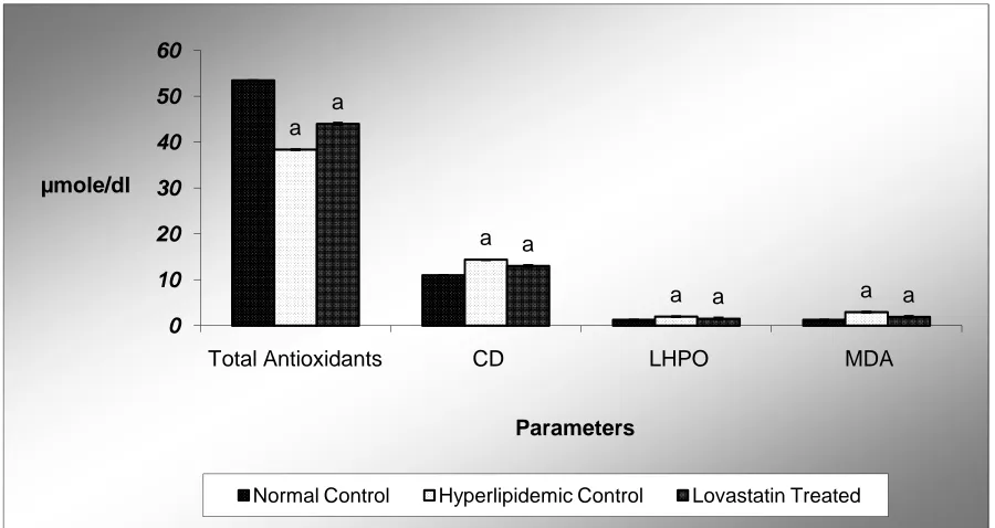
Fig.3 Impact of Lovastatin on plasma total antioxidant, Conjugated diene (CD), Lipid hydroperoxide (LHPO) and Malondialdehyde (MDA) contents in inflammation induced hyperlipidemic rats after 4 weeks of
treatment
Values are mean (µmole/dl) ± SD from pooled plasma of 6 rats in each group. N-C normal Control; IIH-C inflammation induced hyperlipidemic control rats; IIH-LT feed 1mg Lovastatin /rats/day for 4 weeks. Significantly
different from N-C at a p< 0.001.Significantly different from IIH-C at ap<0.001.
Effects of Lovastatin on plasma total antioxidants and lipid peroxidation products:-Fig-3
depicts the antioxidant impact of lovastatin on plasma concentrations of total antioxidants, conjugated diene (CD), lipid hydroperoxide (LHPO) and malondialdehyde (MDA) in inflammation induced hyperlipidemic rats. In IIH-C rats, plasma total antioxidants level was reduced from a control value of 53 to 38 (27%) µmole/dl. Treatment of IIH-LT rats with Lovastatin for 4 weeks resulted in a significant increase of total antioxidants levels by 13 % when compared to IIH-C value. The oxidative stress induced in IIH-C rats significantly enhanced plasma lipid peroxidation products, such as conjugated diene, lipid hydroperoxide and MDA. Formation of conjugated diene, lipid hydroperoxide and MDA in plasma was increased from 10.97, 1.26 and 1.29 in N-C to 14.38 (31 %), 1.98 (57 %) and 2.96 (129 %) µmole/dl,
a
a
a a
a
a
a a
0 10 20 30 40 50 60
Total Antioxidants CD LHPO MDA
µmole/dl
Parameters
respectively, in IIH-C. After Lovastatin treatment, in IIH-LT, a significant decrease of 9 %, 22 %and 35 % was seen in the formation of conjugated diene, lipid hydroperoxide and MDA, respectively, when compared to corresponding values in IIH-C rats. These results demonstrate that in IIH-C rats, due to increase in oxidative stress, total antioxidants level was decreased, whereas, concentration of plasma conjugated diene, lipid hydroperoxide and MDA were significantly increased. Tocotrienols treatment significantly restored the total antioxidants level and blocked the increase in plasma conjugated diene, lipid hydroperoxide and MDA to a level close to corresponding normal values.
Lovastatin effect on Triglycerides (TG), Total Cholesterol (TC) and various Lipid peroxidation products in the Liver homogenate:-As seen in Table 3, hepatic levels of
[image:7.595.151.457.440.578.2]triglyceride (TG) and total cholesterol (TC) were significantly increased in inflammation induced hyperlipidemic control rats (IIH-C) by 38 % and 113 % respectively, when compared to corresponding values in N-C. Feeding of lovastatin inflammation induced rats for 4 weeks was associated with a significant decline in liver TG and TC levels by 9 % and 29% respectively, in IIH-LT. On the other hand, formation of conjugated diene, lipid hydroperoxide and MDA in liver of inflammation induced hyperlipidemic (IIH-C) rats was significantly increased by 47%, 34 % and 42 %, respectively. Feeding of lovastatin to IIH-LT rats for 4 weeks, was associated with a significant decline in the formation of liver conjugated diene, lipid hydroperoxide and MDA by 26 %, 21 % and 20 % respectively, when compared to corresponding values in IIH-C group. These results demonstrate that increased levels of TG, TC, conjugated diene, lipid hydroperoxide and MDA in liver of inflammation induced hyperlipidemic rats were significantly reduced after 4 weeks of lovastatin treatment.
Table 3 impact of Lovastatin on triglycerides, total cholesterol and various lipid peroxidation products in the Liver homogenate after 4 weeks treatment of inflammation induced hyperlipidemic rats.
Parameter NC IIH-C IIH-LT
Triglycerides* 0.493±0.001* 0.685±0.006 *
(+38.94%)a
0.623±0.003* (-9.05%)a
Total cholesterol* 1.34±0.034 2.86±0.018 (+113.43%)a
2.02±0.011 (-29.37%)a
Conjugated diene** 4.97±0.020* 7.32±0.020 *
(+47.28%)a
5.35±0.005* (-26.91%)a
Lipid Hydroperoxide** 0.952±0.001 1.280±0.002 (+34.45%)a
1.002±0.018 (-21.71%)a
MDA** 2.25±0.013 3.20±0.083 (+42.22%)a
2.55±0.011 (-20.31%)d
*Values are mean mg/100mg protein ± SD from homogenate of pooled liver of 6 rats in each group. ** Values are
mean µmole/dl ± SD from homogenate of pooled Liver of 6 rats in each group. N-C normal control; IIH-C inflammation induced hyperlipidemic control rats; IIH-LT feed 1 mg of Lovastatin/rats/day for 4 weeks, significantly different from N-C at a p<0.001 and bp<0.001, significantly different from IIH-C at ap<0.001.
Effects of Lovastatin on the various antioxidant enzymes activities in the liver homogenate:- As seen in Table 4, Catalase (CAT) activity in liver was significantly decreased
(IIH-LT) rats resulted in a significant increase in hepatic SOD activity by 22 %, respectively from normal value. In inflammation induced rats, Glutathione peroxidase (Gpx) activity in liver was significantly increased from a value of 48 units in N-C to 61 (27 %) units, in IIH-C rats. As evident, after 4 weeks of treatment with lovastatin, Gpx activity in liver was significantly decreased by 32 %, when compared to corresponding tissue values in IIH-C group. On the other hand, in smoke exposed rats, the enzymatic activities of hepatic Glutathione reductase (Gred) was decreased significantly by 37 %, when compared to corresponding values of N-C rats. Feeding of lovastatin to inflammation induced hyperlipidemic rats significantly blocked the decrease in hepatic Gred activities and increased them to a similar value of 36 %, Compared to corresponding values of Gred activities in N-C. Administration of lovastatin to smoke exposed rats significantly prevented the decrease in Gred activity and increased to a level, which is similar to normal value. In summary, hepatic CAT, SOD, Gpx and Gred enzymes, which constitute a mutually supportive team of defense against ROS, are significantly decreased in inflammation, induced hyperlipidemic rats. However, lovastatin treatment to inflammation induced hyperlipidemic rats substantially quenches these free radicals (ROS), thus positively normalizing the above enzyme levels.
Table 4 Impact of Lovastatin on Liver Catalase, Superoxide dismutase, Glutathione peroxidase and Glutathione reductase activities in inflammation induced hyperlipidemic rats after 4 weeks of treatment †One
unit(U/mg protein) of enzyme activity is defined as the µmoles of H2O2 decomposed/min/mg protein. ††One
unit (U/mg protein) of enzyme activity is defined as the amount of enzyme required to inhibit O.D. at 560 nm of chromogen production by 50%in one minute. βOne unit (U/mg protein) of enzyme activity is defined as
nmole oxidized Glutathione formed /min/mg homogenate protein. ‡One unit (U/mg protein) of enzyme activity is defined as nmole NADPH oxidized/min/mg PMS protein.
Group Catalase† Superoxide dismutase††
Glutathione peroxidaseβ
Glutathione Reductase‡ N-C 3.70±0.123* 0.755±0.003 48.49±1.02* 8.45±0.198 IIH-C 2.30±0.201*
(-37.83 %)a
0.552±0.002 (-26.88%)a
61.56±1.41* (+27.08%)a
5.26±0.231 (-37.75%) a IIH-LT 3.20±0.013*
(+39.13 %)a
0.665±0.005 (+22.47 %)a
41.38±1.74* (-32.78%)a
7.18±0.199 (+36.50%)a
*Values are mean ± SD from homogenate or PMS fraction of pooled liver of 6 rats in each group, N-C, normal
control; IIH-C inflammation induced hyperlipidemic control rats and IIH-LT fed 1 mg Lovastatin/rats/day for 4 weeks. Significantly different from N-C at ap<0.001. Significantly different from IIH-C at ap<0.001.
DISCUSSION
Lovastatin for 28 days significantly prevented the turpentine induced adverse effects and ameliorated the levels of all the evaluated parameters. Our results strongly suggest that the alleviation of inflammatory conditions is due to potent lipid lowering and free radical scavenging properties of Lovastatin and, thus, can be useful in the therapy of systemic inflammatory process which might induce atherosclerosis. Based on these findings, the antiinflammatory potential of Lovastatin looks promising and more comprehensive studies should be undertaken to determine their actual mode of action. In conclusion, considering the strong hypolipidemic/atheroprotective and antioxidant, and possibly anti-inflammatory actions of Lovastatin, intake of Lovastatin may be useful in the prevention and treatment of infection/inflammation induced hyperlipidemia and atherosclerosis.
REFERENCES
[1] CD Mathers; A Lopez; C Stein; D MaFat; C Rao; M Inoue. In Disease Control Priorities Project Working 2001 Paper 18. Bethesda, MD.
[2] CJ Murray; AD Lopez. Global Comparative Assessments in the Health Sector Disease Burden, Expenditures, and Intervention Packages Geneva., 1994, WHO.
[3] The World Health Report. Reducing Risks, Promoting Healthy Life. Geneva., 2002, WHO. [4] EA Enas; S Yusuf; JL Mehta. Am. J. Cardiol., 1992. 70(9): 945-9
[5] KR Feingold; I Staprans; RA Memon. J. Lipid Res., 1992a, 33:1765–76.
[6] NJ Todd; JT Whicher; C Westacott; A Gilbert. Clin. Chim. Acta., 1990, 189:47–54.
[7] TS Rao; JL Currie; AF Shaffer; PC Isakson. . J. Pharmacol. Exp. Ther., 1994, 269:917–925. [8] BJ Van Lenten; SY Hama; FC de Beer; DM Stafforini; TM McIntyre; SM Prescott; BN La Du; AM Fogelman; M Navab. J. Clin. Invest., 1995, 96:2758–2767.
[9] VG Cabana; JN Siegel; SM Sabesin. . J. Lipid Res., 1989, 30:39–49. [10] JM McCord. Clin. Biochem., 1993, 26:351—7.
[11] P Gregorevic; GS Lynch; D Williams. Eur. J. Appl. Physiol., 2001, 86:24—7. [12]CL Fattman; LM Schaefer; TD Oury. Free Radic. Biol. Med.,2003, 35:236—56.
[13] M Kanter; O Coskun; F Armutcu; Y Uz Hulya; G Kizilay. Tohoku J. Exp. Med., 2005, 206. 155-162.
[14]O Coskun; F Armutcu; M Kanter; GM Kuzey. J. Appl. Toxicol., 2005, 25: 8–12 [15]V Vimal; T Devaki. Journal of Ethnopharmacology., 2004, 90: 151–154. [16]JA Berliner; JW Heinecke. . Free Radic. Biol. Med., 1996. 20:707–727. [17]D Steinberg.. J. Biol. Chem., 1997, 272: 20963–6.
[18]B Frei. Crit. Rev. Food Sci. Nutr., 1995, 35: 83-98.
[19]JT Willerson; PM Ridker. Inflammation as a cardiovascular risk factor. Circulation., 2004, 109:II2–II10.
[20]MS Elkind. Inflammation, atherosclerosis, and stroke. Neurologist., 2006, 12:140 –148. [21]H Wieland; D Seidel. J. Lipid Research., 1989, 24: 904-909.
[22]W Patsch; SA Brown; JD Morrisett; AM Jr Gotto; JR Patsch. . Clin. Chem., 1989, 35: 265-270.
[23]IFF Benzie; JJ Strain. The FRAP assay. Analytical Biochem., 1996, 239: 70-76. [24]J Folch; N Lees; SG Stanley. . J. Biol. Chem., 1957, 226: 497-509.
[25]AK Sinha. Anal. Biochem ., 1972, 47: 389-394.
[26]P Kakkar; B Das; PN Viswanathan. Indian J. Biochem. Biophys ., 1984, 21: 130-132. [27]DG Hafeman; RA Sunde; WG Hoekstra. J. Nutr., 1974, 104: 580-587.
[28]I Carlberg; EB Mannervik. J. Biol. Chem ., 1975. 250: 4475-4480.
[30]W Khovidhunkit; RA Memon; KR Feingold; C Grunfeld. J. Infect. Dis., 2000, 181(Suppl3):S462–72.
[31]W Khovidhunkit; MS Kim; RA Memon. J. Lipid Res., 2004, 45:1169–96.
[32]MA Navarro; R Carpintero, S Acın. Immune-regulation of the apolipoprotein A-I/C-III/A-IV gene cluster in experimental inflammation. Cytokine., 2005, 31: 52-63.
[33]CD Gardner; SP Fortmann; RM Krauss. JAMA., 1996, 276:875–81.
[34]BA Griffin; DJ Freeman; GW Tait; J Thomson; MJ Caslake; CJ Packard; J Shepherd. Role of plasma triglyceride in the regulation of plasma low density lipoprotein (LDL) subfractions: relative contribution of small, dense LDL to coronary heart disease risk. Atherosclerosis., 1994, 106:241–253.
[35]B Lamarche; A Tchernof; S Moorjani. Small, dense low-density lipoprotein particles as a predictor of the risk of ischemic heart disease in men. Circulation., 1997, 95: 69 -75.
[36]H Drexel; W Franz; RK Amann; C Neuenschwander; A Luethy; S Khan; F Follath. Am. J.
Cardiol., 1992, 70: 436-440.
[37]M Minhajuddin; J Iqbal; ZH Beg. Current Adv. Atheroscler. Res., 1999, 2: 120-128.
[38]AA Qureshi; BA Bradlow; L Brace; J Manganello; DM Peterson; BC Pearce; JJK Wright; A Gapor; CE Elson. Response of hypercholesterolemia subjects to administration of tocotrienols. Lipids., 1995, 30: 1171-1177.
[39]AA Qureshi; N Qureshi; JJK Wright. Am J Clin Nut., 1991, 53: 1021S-6S.
[40]RA Parker; BC Pearce; RW Clark; DA Godan; JJK Wright. J. Biol. Che., 1993. 268: 11230-11238.
[41]A Theriault; Q Wang; A Gapor; K Adeli. Arterioscler. Thromb. Vasc. Biol., 1999a, 19: 704-712.
[42]C Gabay; I Kushner. N. Engl. J. Med., 1999, 340: 448–454.
[43]JL Mehta; TGP Saldeen; K Rand. J. Am. Coll. Cardio., 1998, 31:1217–25.
[44]F Nielsen; BB Mikkelsen; JB Nielsen; HR Andersen; P Grandjean. Clin. Chem., 1997, 43:1209-14.
[45]HH Draper; M Hadley. Malondialdehyde determination as index of lipid peroxidation. Methods Enzymol., 1990, 186:421—31.
[46]HR Andersen; JB Nielsen; F Nielsen; P Grandjean. J.Clin. Chem., 1997, 43: 562—8.S [47]M Inoue; IM Arias; JL Boyer; N Fausto; WB Jakoby; DA Schachter; DA Shafritz. The liver: biology and pathobiology. Raven Press: New York, USA., 1994, 443-59.