Open Access
Research article
Altered mRNA expression of genes related to nerve cell activity in
the fracture callus of older rats: A randomized, controlled,
microarray study
Martha H Meyer, Wiguins Etienne and Ralph A Meyer Jr*
Address: Orthopaedic Research Laboratory, Department of Orthopaedic Surgery, Carolinas Medical Center, P.O. Box 32861, Charlotte, NC 28232-2861 USA
Email: Martha H Meyer - [email protected]; Wiguins Etienne - [email protected]; Ralph A Meyer* - [email protected] * Corresponding author
Abstract
Background: The time required for radiographic union following femoral fracture increases with age in both humans and rats for unknown reasons. Since abnormalities in fracture innervation will slow skeletal healing, we explored whether abnormal mRNA expression of genes related to nerve cell activity in the older rats was associated with the slowing of skeletal repair.
Methods: Simple, transverse, mid-shaft, femoral fractures with intramedullary rod fixation were induced in anaesthetized female Sprague-Dawley rats at 6, 26, and 52 weeks of age. At 0, 0.4, 1, 2, 4, and 6 weeks after fracture, a bony segment, one-third the length of the femur, centered on the fracture site, including the external callus, cortical bone, and marrow elements, was harvested. cRNA was prepared and hybridized to 54 Affymetrix U34A microarrays (3/age/time point).
Results: The mRNA levels of 62 genes related to neural function were affected by fracture. Of the total, 38 genes were altered by fracture to a similar extent at the three ages. In contrast, eight neural genes showed prolonged down-regulation in the older rats compared to the more rapid return to pre-fracture levels in younger rats. Seven genes were up-regulated by fracture more in the younger rats than in the older rats, while nine genes were up-regulated more in the older rats than in the younger.
Conclusions: mRNA of 24 nerve-related genes responded differently to fracture in older rats compared to young rats. This differential expression may reflect altered cell function at the fracture site that may be causally related to the slowing of fracture healing with age or may be an effect of the delayed healing.
Background
Bone formation to bridge the fracture gap following skel-etal fracture slows with age in both humans [1-6] and rats [7-9]. While young, 6-week-old rats reach radiographic union by 4 weeks after femoral fracture, adult, 26-week-old rats require 10 weeks, and 26-week-older, 52-week-26-week-old rats
need in excess of 6 months [7]. Despite this increased time to radiographic union with age, there was no increase in the time of expression of Indian hedgehog or any of the bone morphogenetic proteins in the fracture callus for adult rats [10] or for older rats [11,12]. Radiographic union for adult and older rats occurred well after the time Published: 03 August 2004
BMC Musculoskeletal Disorders 2004, 5:24 doi:10.1186/1471-2474-5-24
Received: 25 March 2004 Accepted: 03 August 2004
This article is available from: http://www.biomedcentral.com/1471-2474/5/24
© 2004 Meyer et al; licensee BioMed Central Ltd.
of expression of these skeletally active cytokines [10,11]. Except for markers of osteoblast activity and bone matrix formation, few genes remain up-regulated during the time period when bone forms to bridge the fracture gap [10-12].
These earlier studies done with RT-PCR revealed a paucity of data for genes differentially expressed by age. We had hypothesized that bone formation to bridge the fracture gap would be under a negative-feedback control system. Thus, the genes which stimulate bone formation should be up-regulated in adult or older rats to attempt to accel-erate their slower progression of bony healing. This was not observed in adult [10] or older [11,12] rats. Either bone formation to bridge the fracture gap is not subject to negative-feedback control, or the genes up-regulated to control this bone formation are not those normally thought of as being involved in skeletal homeostasis. This suggested the need for a wider search for genes active dur-ing the fracture reparative process.
In this project, mRNA gene expression was measured by DNA microarray technology at various time points after fracture for young, adult, and older rats. The goal was to identify genes whose expression following fracture was altered by age. Such genes may either show reduced expression, if the age-related slowing of healing is caused by inadequate expression levels, or they may show enhanced expression, in an attempt to stimulate some poorly responding pathway. Among the genes which were differentially expressed at the fracture site with age were genes related to nerve cell activity.
In this study, we explored whether abnormal mRNA expression of genes related to nerve cell activity was asso-ciated with the slowing of skeletal repair in older rats. Abnormalities in the innervation of the fracture site will slow skeletal healing clinically [13-15] and experimen-tally [16-18].
Methods
Rats
Intact female Sprague-Dawley rats (Harlan Sprague-Daw-ley, Inc., Indianapolis, IN) were purchased at one or six months of age and housed in our vivarium in pairs until they were the proper age for experimentation. The rats were fed Teklad Rodent Diet [W] (#8604, Harlan Teklad, Madison, WI) and tap water ad libitum. The work was done in an AAALAC-accredited vivarium under protocols approved by our Institutional Animal Care and Use Committee.
Surgery
Intact female Sprague-Dawley rats at 6 (young), 26 (adult) or 52 (older) weeks of age, weighing 154 ± 11 g (mean ±
SD), 281 ± 25 g, and 330 ± 30 g respectively, were anaes-thetized with an intraperitoneal injection of ketamine and xylazine (30 mg and 5 mg/kg body weight respectively) as described earlier [7,11]. The left knee was shaved, scrubbed with Betadine Solution (Purdue Frederick, Stamford, CT), and draped with sterile sheets. A medial incision was made at the knee, the patella was deflected laterally and a 1.0 mm hole was drilled into the inter-condylar notch. An intramedullary rod (1.0 mm diameter, stainless steel, type 304V, O-SWGX-400, Small Parts, Miami Lakes, FL) was placed retrograde into the left femur [11]. The incision was closed with wound clips. A closed simple transverse mid-diaphyseal femoral fracture was induced with a Bonnarens and Einhorn device [19]. Ran-domly selected rats from among those scheduled for sur-gery were used for 0 time no-fracture sham controls. Rats were euthanized at 0, 0.4, 1, 2, 4, and 6 weeks after frac-ture for a total of 6 time points at each of the 3 ages. Six rats per time point per age group were selected for micro-array analysis (2 rats/micro-array). Radiographs were made at fracture, at 1 week after fracture, and at euthanasia. The femora were rapidly harvested, and one third of the fem-oral length, centered on the fracture site, was collected. This contained the fracture callus with associated cortical bone and marrow and was frozen in liquid nitrogen and stored at -75 C.
RNA Sample Preparation and Microarray Processing
G2500A (Agilent Technologies, Palo Alto, CA). Three arrays from three independent samples (six rats) were done for each age at each time point.
Data Analysis
The Rat U34A GeneChip Microarray has probe sets for over 8,700 rat genes. Most probe sets have 20 different probes for the same gene on each array with 20 additional mismatch controls. The data were analyzed with Affyme-trix Microarray Suite 5.0 and AffymeAffyme-trix Data Mining Tool 3.0 software. Microarray Suite was used to scale the mRNA expression (signal value) of all genes to an average of 500 for each array. For each gene, the software reported a sig-nal value and a Present/Margisig-nal/Absent call. This latter algorithm was a statistical comparison of the variation among the several probe sets for each gene compared to the noise level and gave a call for each gene as Present, Marginal, or Absent. The program then compared the sig-nal value of each gene in the fractured samples against the signal value of the same gene in the unfractured control sample. The difference between the two signal levels, rela-tive to the variability between the multiple probes for each gene, yielded a probability of change due to chance alone. Genes with p less than 0.005 were judged significantly dif-ferent from the same gene in the unfractured sample. This more conservative p value was employed to minimize false positive responses.
The Data Mining Tool was used for cluster analysis with the Self Organizing Map (SOM) algorithm. The data were clustered on the signal values between 20 and 20,000 with the maximum/minimum ratio of at least 3.0 and the max-imum – minmax-imum difference of at least 100. One hun-dred clusters were specified.
Nerve-related genes were identified by searches for nerve-related names in the gene descriptions of each gene on the microarray. This association was confirmed by a review of the information for that gene in the NetAffx web site http:/ /www.affymetrix.com and in the PubMed database http:/ /www.ncbi.nlm.nih.gov/entrez/query.fcgi. GenBank accession numbers and names are shown for each gene. Each graph shows the average ± SEM of the three microar-rays that were done for each time point for each age. Sig-nificant changes in gene expression were demonstrated by t test and linear regression [20].
This report conforms to the MIAME standards of MGED http://www.mged.org. A copy of the full microarray data set has been deposited in the NCBI Gene Expression Omnibus http://www.ncbi.nlm.nih.gov/geo/ as series GSE594.
Results
Radiology
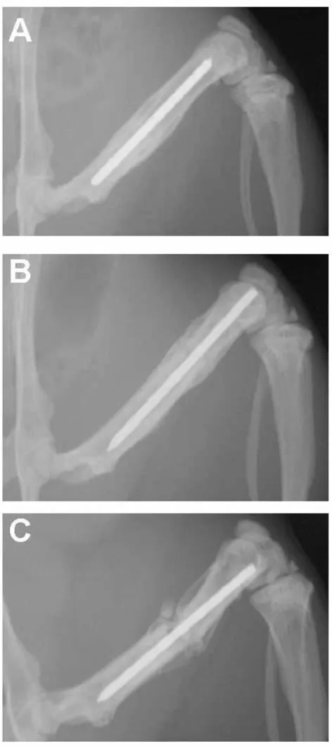
In all young rats, bone bridged the fracture gap by four weeks after surgery. By six weeks after fracture, remodeling was beginning to obscure the fracture site (Fig. 1). In con-trast, bone bridging in the adult rats progressed more slowly. The adult rats did have a vigorous periosteal reac-tion at the site of the fracture and were approaching radi-ographic union by six weeks after surgery (Fig. 1). In the older, one-year-old rats, bridging of the fracture gap by bone progressed the slowest. They had a minimal perio-steal reaction at six weeks after surgery (Fig. 1).
General results
On each array, on average, 5,200 genes were scored as absent, and 3,300 as present. Of these, 1,159 were signif-icantly up-regulated and 928 were signifsignif-icantly down-reg-ulated at two weeks after fracture in the adult rats of the first series (see Additional File 1). Up-regulated genes included cytokines and matrix genes for both cartilage and bone. Down-regulated genes included genes related to blood cell synthesis and mitochondrial function.
SOM clusters identified genes up- or down-regulated by fracture. Most genes affected by fracture followed the same time course at all three ages. These genes showed approximately the same peak expression level and regressed to baseline at about the same time point at all three ages. Among the genes affected by fracture were a number of genes associated with nerve cells. These were selected for more intense analysis.
Similar responses at all three ages
Up-regulated nerve-related genes are shown in Table 1. Two examples are shown in the upper two graphs in Fig-ure 2. Both of these genes were significantly up-regulated from the 0 time control (P < 0.001 by t test for 9 samples (3 ages × 3 replicates) of 0 time vs. 0.4 week (Fig. 2, top graph) or vs. 0 time vs. 2 week (Fig. 2, middle graph)). Other nerve-related genes were down-regulated by frac-ture at all three ages (Table 2). These regained near normal activity by six weeks after fracture. An example is shown in the bottom graph of Figure 2. This gene (TAG-1) had a sig-nificant down-regulation after fracture (P < 0.001 by t test for 9 samples of 0 time vs. 0.4 week), followed by a signif-icant increase at 6 weeks after fracture compared to 0.4 week after fracture (P < 0.001 by t test for 9 samples).
Defects in the older rats
Table 3, and three examples of these genes are shown in Figure 3. All three of these genes had a significantly decreased mRNA expression levels at 1 week after fracture compared to 0 time control (P < 0.001 by t test for 9 sam-ples (3 ages × 3 replicates)). At 4 and 6 weeks after frac-ture, the young rats showed faster recovery in mRNA expression than did the older rats for the three genes in Fig. 3 (P < 0.01 by t tests for 4 and 6 week young vs. 4 and 6 week old).
In the second type of defect, other genes were up-regu-lated by fracture, but the response was weaker in the older rats. These genes are shown in Table 4. Three examples are shown in Figure 4. The broad peaks of the genes in Figure 4 permitted the t test to demonstrate a significantly higher expression level in the young rats at 1 and 2 weeks after fracture in comparison to the same time points of older rats. These comparisons for the three genes in Figure 4 were significant at P < 0.001 (top graph, AF034963), P < 0.02 (middle graph, AB005541) and P < 0.01 (bottom graph, U09357) for 6 samples per age group (2 time points pooled for 3 replicates).
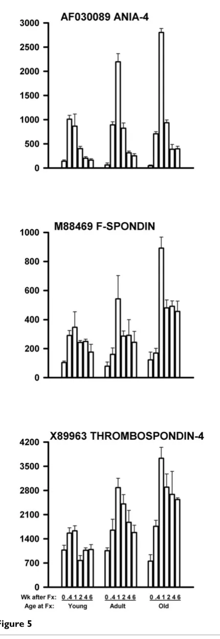
In the third type of defect, genes were also up-regulated by fracture. However, the response was stronger in the older rats than in the younger rats. These genes are shown in Table 5, and three examples are shown in Figure 5. The peak values for these three genes significantly increased with age by linear regression (P < 0.001 (top graph, AF030089), P = 0.01 (middle graph, M88469), and P < 0.001 (bottom graph, X89963) for 9 data points (3 ages × 3 replicates)).
Present/Marginal/Absent calls
For each gene for each array, the Microarray Suite software reported a statistical decision as to whether the mRNA was Present, Marginal, or Absent. We have reviewed these calls for the genes shown in Figures 2,3,4,5. For Figure 2, the Present/Marginal/Absent calls were: Fig. 2 Top: 53/1/0; Fig. 2 Middle: 39/2/13; and Fig. 2 Bottom: 0/0/54. For Fig-ure 3, the calls were: Fig. 3 Top: 45/2/7; Fig. 3 Middle: 7/ 0/47; and Fig. 3 Bottom: 32/6/16. For Figure 4, the calls were: Fig. 4 Top: 0/0/54; Fig. 4 Middle: 0/0/54; and Fig. 4 Bottom: 51/0/3. For Figure 5, the calls were: Fig. 5 Top: 45/1/8; Fig. 5 Middle: 52/0/2; and Fig. 5 Bottom: 54/0/0.
Discussion
In this study, as in our earlier work [7,10-12], the time required to reach radiographic union after femoral frac-ture increased with age in the female rat. This slowing of fracture repair with age is associated with changes in the mRNA expression of specific genes within the healing fracture site [10-12]. To study this further, microarray technology was used to identify additional genes whose mRNA expression was affected by skeletal fracture. Radiographs
Figure 1
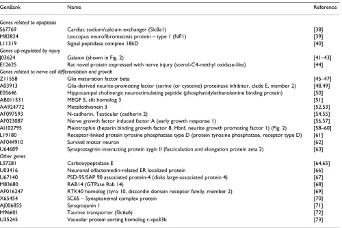
Table 1: Nerve-related genes up-regulated by fracture at all three ages
GenBank Name Reference
Genes related to apoptosis
S67769 Cardiac sodium/calcium exchanger (Slc8a1) [38] M82824 Leucopus neurofibromatosis protein – type 1 (NF1) [39] L11319 Signal peptidase complex 18kD [40]
Genes up-regulated by injury
J03624 Galanin (shown in Fig. 2) [41–43] E12625 Rat novel protein expressed with nerve injury (sterol-C4-methyl oxidase-like) [44]
Genes related to nerve cell differentiation and growth
Z11558 Glia maturation factor beta [45–47] A03913 Glia-derived neurite-promoting factor (serine (or cysteine) proteinase inhibitor, clade E, member 2) [48,49] E05646 Hippocampal cholinergic neurostimulating peptide (phosphatidylethanolamine binding protein) [50] AB011531 MEGF 5, slit homolog 3 [51] AA924772 Metallothionein 3 [52,53] AF097593 N-cadherin, Testicular (cadherin 2) [54,55] AF023087 Nerve growth factor induced factor A (early growth response 1) [56,57] AI102795 Pleiotrophin (heparin binding growth factor 8, Hbnf, neurite growth promoting factor 1) (Fig. 2) [58–60] L19180 Receptor-linked protein tyrosine phosphatase type D (protein tyrosine phosphatase, receptor type D) [61] AF044910 Survival motor neuron [62] U64689 Synaptotagmin interacting protein zygin II (fasciculation and elongation protein zeta 2) [63]
Other genes
L07281 Carboxypeptidase E [64,65] U03416 Neuronal olfactomedin-related ER localized protein [66] U67140 PSD-95/SAP 90 associated protein-4 (disks large-associated protein 4) [67] M83680 RAB14 (GTPase Rab 14) [68] AF016247 RTK40 homolog (tyro 10, discordin domain receptor family, member 2) [69] X65454 SC65 – Synaptonemal complex protein [70] AJ006855 Synaptojanin 1 [71] M96601 Taurine transporter (Slc6a6) [72] U35245 Vacuolar protein sorting homolog r-vps33b [73] Alternative names are shown in parentheses. References to physiological roles are shown to the right.
Table 2: Nerve-related genes down-regulated by fracture at all three ages
Genbank Name Reference
Genes associated with the growth cone of nerve fibers
M31725 Axonal glycoprotein TAG-1 (Contactin 2) (shown in Fig. 2) [74] S82649 Neuronal activity-regulated pentraxin (Narp) [75] X59149 Neural cell adhesion molecule L1 [76–78]
Genes associated with synaptic signaling pathways
L08496 GABA-A receptor delta subunit [79] S68944 Na+/Cl--dependent neurotransmitter transporter [80]
S76742 Neurotransmitter transporter rB21a (X transporter protein 3) [81] U39549 Synaptogyrin (synaptogyrin 1) [82] D28512 Synaptotagmin III (synaptotagmin 3) [83] AF007758 Synuclein 1 (synuclein alpha) [84]
Other genes
[image:5.612.58.573.501.698.2]More than two-thirds of the detectable genes on the rat U34A microarray have a change in mRNA expression level following fracture [21]. Most of these genes were not known to participate in the healing process of bone before the advent of microarray technology [21,22]. This reflects changes in both the types of cells at the fracture site as well as changes in the activity of the existing cells. Among the cells affected by fracture are nerve fibers. Protein and mRNA of genes related to neuronal functioning are found in intact bone and in the fracture callus [23-29]. Since proper innervation of the fracture site is needed for fracture repair clinically [13-15] and experimentally [16-18], this led to the hypothesis that the age-related slowing of fracture repair may be related to the abnormal nerve cell activity at the fracture site.
To evaluate this hypothesis, nerve-related genes were stud-ied from among the genes present on the Affymetrix Rat U34A microarray. Genes were identified for which the mRNA response to femoral fracture was changed in the older rats compared to the young rats. Three types of change with age were found:
1. The mRNA expression levels of the genes shown in Table 3 and Figure 3 were decreased by fracture. While gene expression in the young rats was approaching pre-fracture levels by six weeks after pre-fracture, gene expression showed minimal return to normal in older rats. Genes in this category were all related to signaling molecules or to signal receptors (references shown in Table 3).
2. Other nerve-related genes had strong up-regulation after fracture in young rats but only mild up-regulation in older rats. These are shown in Table 4 and Figure 4. This partial loss of function with age was observed in genes associated with nerve cell differentiation or cell cycle or Figure 2
mRNA levels of three nerve-related genes affected by frac-ture in young, adult, and older rats
Figure 2
genes related to synaptic structure (references cited in Table 4).
[image:7.612.53.565.99.214.2]3. A third set of genes was increased in mRNA expression by fracture, but the increase was greater in the older rats. These are shown in Table 5 and Figure 5. Many of these
Table 3: Nerve-related genes with prolonged down-regulation after fracture in older rats
[image:7.612.59.553.282.409.2]GenBank Name Reference L09119 C kinase substrate calmodulin-binding protein (neurogranin) (shown in Fig. 3) [89] S77867 G coupled protein receptor UHR-1 (G protein coupled receptor 10) [90] AF109405 GABA-B receptor 2 (G protein-coupled receptor 51) [91] U08290 Neuronatin alpha (neuronatin) [92] M15880 Neuropeptide Y (shown in Fig. 3) [93,94] S56508 Fatty acid coenzyme A ligase, long chain 6 [95] AF041083 RoBo-1 (Slc11a1) (shown in Fig. 3) [55,96,97] X86789 Sensory neuron synuclein (gamma-synuclein) [98,99] Data presented as in Table 1. All genes are related to signals and signal receptors.
Table 4: Nerve-related genes with a greater up-regulation in younger rats
GenBank Name Reference
Genes related to nerve cell differentiation and the cell cycle
X86003 Neuron-derived orphan receptor-2 [100] AB005540 PCTAIRE 2 [101] AB005541 PCTAIRE 3 (shown in Fig. 4) [102] U09357 Protein tyrosine phosphatase, receptor-type, Z polypeptide 1 (shown in Fig. 4) [103]
Genes related to the synapse and sensory perception
[image:7.612.54.587.477.638.2]AI179632 Proton gated cation channel DRASIC [104] AF034863 Synaptic scaffolding molecule (S-SCAM, activin receptor interacting protein 1) (shown in Fig. 4) [105,106] S56141 Sodium-dependent neurotransmitter transporter [107] Data presented as in Table 1.
Table 5: Nerve-related genes with a greater up-regulation in older rats
GenBank Name Reference
Genes related to signals and signal transduction
X74832 Acetylcholine receptor alpha subunit (cholinergic receptor, nicotinic, alpha polypeptide 1) [108] M16112 Brain type II calcium/calmodulin dependent protein kinase beta subunit [109]
Genes related to cell adhesion
M88469 F-spondin (shown in Fig. 5) [110] AF060879 Neurocan (chondroitin sulfate proteoglycan 3) [111,112] U16845 Neurotrimin [113] M96375 Non-processed neurexin 1-beta (neurexin 1) [114] X89963 TSP-4 (thrombospondin-4) (shown in Fig. 5) [115–117]
Other genes
genes were related to cell adhesion or to cell signal or sig-nal transduction (references cited in Table 5).
All three classes of genes showed altered expression in the older rats compared to young rats. We hypothesize that bone fracture may physically disrupt nerve fibers in bone. A sub-population of these skeletal nerve fibers may regrow into the fracture site or regain function at a slower rate in older rats. This may account for the failure to recover from low mRNA values for the first group (pro-longed down-regulation) or the failure to up-regulate mRNA expression adequately after fracture in the older rats in the second group (diminished up-regulation). Other genes in the third group with increased levels of mRNA after fracture in the older rats may represent attempts to stimulate nerve regrowth or other processes that are not responding. This may represent negative-feed-back-induced up-regulation caused by effector cell resist-ance. Taken together, these changes in nerve cell function with age may contribute to the slowing of fracture repair in older rats. It must be pointed out that the associations noted here do not necessarily reflect cause and effect. It is also possible that the delayed re-innervation of the fracture site is an effect of the delayed healing in the older rats and not a cause of the delayed healing.
Experimental studies have been done to detect the role of innervation on fracture healing. Studies of sectioning the sciatic nerve in concert with tibial fracture have been reported to speed fracture healing [30-33]. However, sec-tioning both femoral and sciatic nerves inhibits fracture healing [18]. Aro et al. [16] have reported mechanorecep-tors (Pacinian corpuscles) in the periostium of the rat fib-ula, which, if removed, lead to non-union [17]. Direct application of nerve growth factor to the fracture site increases healing in the rat rib [34].
In humans, abnormal bone healing is also associated with lack of nerve activity at the fracture site. Nagano et al. [13] have noted scaphoid nonunion in the wrists of patients with neuroarthropathy from a long-standing nerve palsy. Santavirta et al. [14] have found a lack of peripheral inner-vation at the fracture site of noninfected fractures with delayed union or nonunion of diaphyseal bones. Nord-Figure 3
mRNA levels of three nerve-related genes with more pro-longed down-regulated after fracture in the older rats Figure 3
strom et al. [15] have found a lack of stromal innervation associated with delayed union or pseudoarthrosis in spondylolysis.
Humans [1-6] show a slowing of fracture healing with increasing age as do rats [7-9]. The cause of the slowing of fracture healing with age is not well understood. The fem-ora of young rats regain normal biomechanical properties by 4 weeks after fracture, while adults take 12 weeks, and older rats require in excess of 6 months [7]. This model presents an opportunity to elucidate novel genes important to this healing process. The slowing could reflect a loss of function as some processes essential for the rapid healing of fractures in young animals are inhib-ited with age. Alternatively, the slowing of skeletal repair with age may be caused by partial resistance of the healing process to stimulation in adult or older individuals. Such resistance should result in enhanced stimulation by regu-latory systems to attempt to evoke a healing response. Both patterns were seen among the genes studied in this report. These genes are candidates for further study.
These changes with age are not limited to genes related to neuronal activity. We have also noted similar changes in genes related to mitochondrial activity [35]. It is likely that the age-related changes in fracture repair are caused by failure of several metabolic pathways. Methods, such as DNA microarrays, which sample many different biological pathways will be useful in defining these novel, multi-faceted defects.
The specificity of these changes is seen in the majority of the nerve-related genes for which the expression pattern following fracture was unaffected by age. These transcripts had similar increases or decreases following fracture in the young, adult, and older rats. These uniform responses suggest that most metabolic patterns were unaffected by age. Nerve-related genes similarly up-regulated by femoral fracture at all three ages were broadly related to differenti-ation and growth of nerve cells, to known up-reguldifferenti-ation following nerve injury, or to association with apoptosis (references cited in Table 1). Some of these genes were slower to return to baseline values in older rats, such as galanin and TAG-1. In contrast, nerve-related genes similarly down-regulated by femoral fracture at all three Figure 4
mRNA levels of three genes related to nerve function and up-regulated more in young rats than in older rats following femoral fracture
Figure 4
ages were broadly related to the nerve growth cone or to synaptic signaling pathways (references cited in Table 2).
In this study gene expression was measured by quantifica-tion of the mRNA level for each gene with microarray technology. It must be kept in mind that there are other control systems which influence the protein synthetic rate and also protein degradation. Protein synthesis will be low in the absence of mRNA for that gene, but elevated mRNA levels are not a guarantee that protein levels will also be elevated for that gene. Changes noted at the mRNA level will need to be confirmed at the protein and struc-tural levels. Assignment of the genes studied herein as nerve-related is made on the basis of currently available information. Other cell types in the fracture callus may also express these genes. Histological studies will permit the association of these genes with specific cell types within the fracture callus. These experiments are now in progress.
We have compared mRNA gene expression by microarray to that measured by reverse transcription – polymerase chain reaction (RT-PCR)[36]. Good correlation was found between the two methods if the transcripts were judged mostly present, the signal level did not approach the upper limit of the detector, and the probe sets or PCR primers were from the same region of the gene [36]. Some other genes, even though most samples were judged absent, also gave good correlation between the two meth-ods. These latter genes were at the upper range of the absent calls and had good precision between samples [36]. The genes reported herein have the marked variation in mRNA levels that have been reported previously in frac-ture samples [10,11] with large changes in expression after fracture which return to the prefracture levels as healing progresses. The finding here of moderate signal levels, good precision among the three samples for each time point at each age, and a strong response to fracture indicate the ability of this technology to report changes in mRNA levels for these genes.
Conclusions
[image:10.612.59.284.82.735.2]In summary, most genes respond to bone fracture with altered mRNA gene expression, including genes related to neuronal functioning. However, a number of these genes Figure 5
mRNA levels of three genes related to nerve function and up-regulated more in older rats than in younger rats follow-ing femoral fracture
Figure 5
responded to fracture differently in older rats than in young rats. Such differential expression with age may reflect altered cell functioning at the fracture site that may be related to the slowing of fracture healing in older rats.
Competing interests
None declared.
Authors' contributions
MM, WE, and RM participated in the surgery and the sam-ple collection. MM and WE prepared and hybridized the samples and analyzed the data. RM prepared the manu-script. All authors read and approved the final manuscript.
Additional material
Acknowledgements
This study was supported in part by grants from the North Carolina Bio-technology Center and the Orthopaedic Trauma Association. We thank Drs. Scott Porter, James Fiechtl and Daniel Heiner for their surgical assist-ance. We thank Ms. Virginia Mooney, Mr. Michael Steuerwald, and Ms. Jes-sica Heath for their technical assistance and Ms. Carolyn Ayers for her secretarial assistance. A preliminary report of this study has appeared [37].
References
1. Bronk JT, Ilstrup D, Melton J, Bolander ME: Age and gender effect fracture repair in the humerus. Trans Orthop Res Soc 1997,
22:258 (Abstract).
2. Skak SV, Jensen TT: Femoral shaft fracture in 265 children. Log-normal correlation with age of speed of healing.Acta Orthop Scand 1988, 59:704-707.
3. Nieminen S, Nurmi M, Satokari K: Healing of femoral neck frac-tures; influence of fracture reduction and age. Ann Chir Gynaecol 1981, 70:26-31.
4. Nilsson BE, Edwards P: Age and fracture healing: A statistical analysis of 418 cases of tibial shaft fractures.Geriatrics 1969,
24:112-117.
5. Hee HT, Wong HP, Low YP, Myers L: Predictors of outcome of floating knee injuries in adults: 89 patients followed for 2–12 years.Acta Orthop Scand 2001, 72:385-394.
6. Kawai T, Murakami S, Hiranuma H, Sakuda M: Radiographic changes during bone healing after mandibular fractures.Br J Oral Maxillofac Surg 1997, 35:312-318.
7. Meyer RA Jr, Tsahakis PJ, Martin DF, Banks DM, Harrow ME, Kiebzak GM: Age and ovariectomy impair both the normalization of mechanical properties and the accretion of mineral by the fracture callus in rats.J Orthop Res 2001, 19:428-435.
8. Ekeland A, Engesæter LB, Langeland N: Influence of age on mechanical properties of healing fractures and intact bones in rats.Acta Orthop Scand 1982, 53:527-534.
9. Bronk JT, Urabe K, Liang T, Bolander ME: The effect of age on fracture repair in a rat femur model.Trans Orthop Res Soc 1996,
21:200 (Abstract).
10. Desai BJ, Meyer MH, Porter S, Kellam JF, Meyer RA Jr: The effect of age on gene expression in adult and juvenile rats following femoral fracture.J Orthop Trauma 2003, 17:689-698.
11. Meyer RA Jr, Meyer MH, Tenholder M, Wondracek S, Wasserman R, Garges P: Gene expression in older rats with delayed union of femoral fractures.J Bone Joint Surg [Am] 2003, 85A:1243-1254. 12. Meyer RA Jr, Meyer MH, Phieffer LS, Banks DM: Delayed union of
femoral fractures in older rats: Decreased gene expression.
BMC Musculoskelet Disord 2001, 2:2.
13. Nagano J, Tada K, Masatomi T, Horibe S: Arthropathy of the wrist in leprosy–what changes are caused by long-standing periph-eral nerve palsy?Arch Orthop Trauma Surg 1989, 108:210-217. 14. Santavirta S, Konttinen YT, Nordstrom D, Makela A, Sorsa T,
Hukkanen M, Rokkanen P: Immunologic studies of nonunited fractures.Acta Orthop Scand 1992, 63:579-586.
15. Nordstrom D, Santavirta S, Seitsalo S, Hukkanen M, Polak JM, Nord-sletten L, Konttinen YT: Symptomatic lumbar spondylolysis. Neuroimmunologic studies.Spine 1994, 19:2752-2758. 16. Aro H, Eerola E, Aho AJ: Osteolysis after rigid fixation. The
pos-sible role of periosteal neural mechanoreceptors in bone remodeling.Clin Orthop 1982, 166:292-300.
17. Aro H, Eerola E, Aho AJ: Development of nonunions in the rat fibula after removal of periosteal neural mechanoreceptors.
Clin Orthop 1985, 199:292-299.
18. Madsen JE, Hukkanen M, Aune AK, Basran I, Moller JF, Polak JM, Nor-dsletten L: Fracture healing and callus innervation after peripheral nerve resection in rats. Clin Orthop 1998,
351:230-240.
19. Bonnarens F, Einhorn TA: Production of a standard closed frac-ture in laboratory animal bone.J Orthop Res 1984, 2:97-101. 20. Zar JH: Biostatistical Analysis 2nd edition. Englewood Cliffs:
Prentice-Hall, Inc.; 1984.
21. Meyer RA Jr, Desai BR, Meyer MH: Age alters mRNA gene expression in the fracture callus of rats by microarray analysis.Trans Orthop Res Soc 2003, 28:489 (Abstract).
22. Hadjiargyrou M, Lombardo F, Zhao S, Ahrens W, Joo J, Ahn H, Jur-man M, White DW, Rubin CT: Transcriptional profiling of bone regeneration. Insight into the molecular complexity of wound repair.J Biol Chem 2002, 277:30177-30182.
23. Dickson GClingen-Vance H: Neuropeptide immunoreactivity in repairing I.M. nailed rat femoral osteotomies.Program, Interna-tional Society for Fracture Repair 2002:51 (Abstract).
24. Grills BL, Schuijers JA: Immunohistochemical localization of nerve growth factor in fractured and unfractured rat bone.
Acta Orthop Scand 1998, 69:415-419.
25. Hattorl A, Tsujimoto M, Hayashi K, Kohno M: Bone morphoge-netic protein-2 is markedly synergistic with tumor necrosis factor in stimulating the production of nerve growth factor in fibroblasts.Biochem Mol Biol Int 1996, 38:1095-1101.
26. Rusanen M, Korkala O, Gronblad M, Partanen S, Nederstrom A: Evo-lution of substance P immunofluorescent nerves in callus tis-sue during fracture healing.J Trauma 1987, 12:1340-1343. 27. Hukkanen M, Konttinen YT, Santavirta S, Paavolainen P, Gu XH,
Ter-enghi G, Polak JM: Rapid proliferation of calcitonin gene-related peptide-immunoreactive nerves during healing of rat tibial fracture suggests neural involvement in bone growth and remodelling.Neuroscience 1993, 54:969-979.
28. Li J, Ahmad T, Spetea M, Ahmed M, Kreicbergs A: Bone reinnerva-tion after fracture: a study in the rat.J Bone Miner Res 2001,
16:1505-1510.
29. Hukkanen M, Konttinen YT, Santavirta S, Nordsletten L, Madsen JE, Almaas R, Oestreicher AB, Rootwelt T, Polak JM: Effect of sciatic nerve section on neural ingrowth into the rat tibial fracture callus.Clin Orthop 1995, 311:247-257.
30. Frymoyer JW, Pope MH: Fracture healing in the sciatically den-ervated rat.J Trauma 1977, 17:355-361.
Additional File 1
Additional File 1. Response of adult rats at two weeks after fracture. This spreadsheet is a listing of all genes altered by fracture in the first adult rat sample at two weeks after fracture compared to the first adult no-fracture sample. The data are sorted on the probability value for the comparison of each gene between the two arrays. For each array, the signal (mRNA tran-script level), detection (present, absent, or marginal) and detection p-value are given. In addition, for the two-week sample, the change (increase, marginal increase, marginal decrease or decrease) and change p-value are given for the comparison of the two-week sample to the no-fracture sample. These two arrays are archived in the GEO repository http://www.ncbi.nlm.nih.gov/geo/ as samples GSM9028 (no-fracture sample) and GSM9031 (2-week sample).
Click here for file
31. Aro H, Eerola E, Aho AJ, Penttinen R: Healing of experimental fractures in the denervated limbs of the rat.Clin Orthop 1981,
155:211-217.
32. Aro H: Effect of nerve injury on fracture healing. Callus for-mation studied in the rat.Acta Orthop Scand 1985, 56:233-237. 33. Nordsletten L, Madsen JE, Almaas R, Rootwelt T, Halse J, Konttinen
YT, Hukkanen M, Santavirta S: The neuronal regulation of frac-ture healing. Effects of sciatic nerve resection in rat tibia.Acta Orthop Scand 1994, 65:299-304.
34. Grills BL, Schuijers JA, Ward AR: Topical application of nerve growth factor improves fracture healing in rats.J Orthop Res
1997, 15:235-242.
35. Meyer RA, Meyer MH: The effect of age on the expression of mitochondrial genes during the healing of femoral fractures in the rat.J Bone Miner Res 2003, 18:S258 (Abstract).
36. Etienne W, Meyer MH, Peppers J, Meyer RA Jr: Comparison of mRNA gene expression by RT-PCR and DNA microarray.
BioTechniques 2004, 36:618-626.
37. Meyer MH, Etienne W, Meyer RA: The effect of age on the expression of nerve-related genes during the healing of fem-oral fractures in the rat. J Bone Miner Res 2003, 18:S258 (Abstract).
38. Diaz-Horta O, Kamagate A, Herchuelz A, Van Eylen F: Na/Ca exchanger overexpression induces endoplasmic reticulum-related apoptosis and caspase-12 activation in insulin-releas-ing BRIN-BD11 cells.Diabetes 2002, 51:1815-1824.
39. Feldkamp MM, Gutmann DH, Guha A: Neurofibromatosis type 1: Piecing the puzzle together.Can J Neurol Sci 1998, 25:181-191. 40. Signs SA, Jacquet R: Induction of ethanol dependence increases
signal peptidase mRNA levels in rat brain. Mol Cell Biochem
1994, 139:21-26.
41. Su Y, Ganea D, Peng X, Jonakait GM: Galanin down-regulates microglial tumor necrosis factor-alpha production by a post-transcriptional mechanism.J Neuroimmunol 2003, 134:52-60. 42. Wynick D, Thompson SW, McMahon SB: The role of galanin as a
multi-functional neuropeptide in the nervous system.Curr Opin Pharmacol 2001, 1:73-77.
43. Bacon A, Holmes FE, Small CJ, Ghatei M, Mahoney S, Bloom S, Wyn-ick D: Transgenic over-expression of galanin in injured pri-mary sensory neurons.Neuroreport 2002, 13:2129-2132. 44. Uwabe K, Gahara Y, Yamada H, Miyake T, Kitamura T:
Identifica-tion and characterizaIdentifica-tion of a novel gene (neurorep 1) expressed in nerve cells and up-regulated after axotomy.
Neuroscience 1997, 80:501-509.
45. Zaheer A, Lim R: In vitro inhibition of MAP kinase (ERK1/ ERK2) activity by phosphorylated glia maturation factor (GMF).Biochemistry 1996, 35:6283-6288.
46. Bourgeois F, Guimiot F, Mas C, Bulfone A, Levacher B, Moalic JM, Simonneau M: Identification and isolation of a full-length clone of mouse GMFB (Gmfb), a putative intracellular kinase reg-ulator, differentially expressed in telencephalon.Cytogenet Cell Genet 2001, 92:304-309.
47. Kaplan R, Zaheer A, Jaye M, Lim R: Molecular cloning and expres-sion of biologically active human glia maturation factor-beta.
J Neurochem 1991, 57:483-490.
48. Carter RE, Cerosaletti KM, Burkin DJ, Fournier RE, Jones C, Green-berg BD, Citron BA, Festoff BW: The gene for the serpin thrombin inhibitor (PI7), protease nexin I, is located on human chromosome 2q33-q35 and on syntenic regions in the mouse and sheep genomes.Genomics 1995, 27:196-199. 49. Crisp RJ, Knauer MF, Knauer DJ: Protease nexin 1 is a potent
uri-nary plasminogen activator inhibitor in the presence of col-lagen type IV.J Biol Chem 2002, 277:47285-47291.
50. Moore C, Perry AC, Love S, Hall L: Sequence analysis and immu-nolocalisation of phosphatidylethanolamine binding protein (PBP) in human brain tissue. Brain Res Mol Brain Res 1996,
37:74-78.
51. Itoh A, Miyabayashi T, Ohno M, Sakano S: Cloning and expressions of three mammalian homologues of Drosophila slit suggest possible roles for Slit in the formation and maintenance of the nervous system.Brain Res Mol Brain Res 1998, 62:175-186. 52. Chung RS, Vickers JC, Chuah MI, Eckhardt BL, West AK:
Metal-lothionein-III inhibits initial neurite formation in developing neurons as well as postinjury, regenerative neurite sprouting.Exp Neurol 2002, 178:1-12.
53. Palmiter RD, Findley SD, Whitmore TE, Durnam DM: MT-III, a brain-specific member of the metallothionein gene family.
Proc Natl Acad Sci USA 1992, 89:6333-6337.
54. Tran NL, Adams DG, Vaillancourt RR, Heimark RL: Signal trans-duction from N-cadherin increases Bcl-2. Regulation of the phosphatidylinositol 3-kinase/Akt pathway by homophilic adhesion and actin cytoskeletal organization.J Biol Chem 2002,
277:32905-32914.
55. Rhee J, Mahfooz NS, Arregui C, Lilien J, Balsamo J, VanBerkum MF:
Activation of the repulsive receptor Roundabout inhibits N-cadherin-mediated cell adhesion.Nat Cell Biol 2002, 4:798-805. 56. Janssen-Timmen U, Lemaire P, Mattei MG, Revelant O, Charnay P:
Structure, chromosome mapping and regulation of the mouse zincfinger gene Krox-24; evidence for a common reg-ulatory pathway for immediate early serum-response genes.
Gene 1989, 80:325-336.
57. Sukhatme VP, Cao X, Chang LC, Tsai-Morris CH, Stamenkovich D, Ferreira PCP, Cohen DR, Edwards SA, Shows TB, Curran T, Le Beau MM, Adamson ED: A zinc finger-encoding gene coregulated with c-fos during growth and differentiation, and after cellu-lar depocellu-larization.Cell 1988, 53:37-43.
58. Tare RS, Oreffo RO, Sato K, Rauvala H, Clarke NM, Roach HI:
Effects of targeted overexpression of pleiotrophin on postna-tal bone development. Biochem Biophys Res Commun 2002,
298:324-332.
59. Kretschmer PJ, Fairhurst JL, Decker MM, Chan CP, Gluzman Y, Boh-len P, Kovesdi I: Cloning, characterization and developmental regulation of two members of a novel human gene family of neurite outgrowth-promoting proteins. Growth Factors 1991,
5:99-114.
60. Pavlov I, Voikar V, Kaksonen M, Lauri SE, Hienola A, Taira T, Rauvala H: Role of heparin-binding growth-associated molecule (HB-GAM) in hippocampal LTP and spatial learning revealed by studies on overexpressing and knockout mice.Mol Cell Neurosci
2002, 20:330-342.
61. Walton KM, Martell KJ, Kwak SP, Dixon JE, Largent BL: A novel receptor-type protein tyrosine phosphatase is expressed during neurogenesis in the olfactory neuroepithelium.Neuron
1993, 11:387-400.
62. La Bella V, Cisterni C, Salaun D, Pettmann B: Survival motor neu-ron (SMN) protein in rat is expressed as different molecular forms and is developmentally regulated.Eur J Neurosci 1998,
10:2913-2923.
63. Bloom L, Horvitz HR: The Caenorhabditis elegans gene unc-76 and its human homologs define a new gene family involved in axonal outgrowth and fasciculation.Proc Natl Acad Sci USA
1997, 94:3414-3419.
64. Manser E, Fernandez D, Loo L, Goh PY, Monfries C, Hall C, Lim L:
Human carboxypeptidase E. Isolation and characterization of the cDNA, sequence conservation, expression and processing in vitro.Biochem J 1990, 267:517-525.
65. Cool DR, Normant E, Shen F, Chen HC, Pannell L, Zhang Y, Loh YP:
Carboxypeptidase E is a regulated secretory pathway sort-ing receptor: Genetic obliteration leads to endocrine disor-ders in Cpe(fat) mice.Cell 1997, 88:73-83.
66. Danielson PE, Forss-Petter S, Battenberg EL, deLecea L, Bloom FE, Sutcliffe JG: Four structurally distinct neuron-specific olfacto-medin-related glycoproteins produced by differential pro-moter utilization and alternative mRNA splicing from a single gene.J Neurosci Res 1994, 38:468-478.
67. Takeuchi M, Hata Y, Hirao K, Toyoda A, Irie M, Takai Y: SAPAPs. A family of PSD-95/SAP90-associated proteins localized at postsynaptic density.J Biol Chem 1997, 272:11943-11951. 68. Elferink LA, Anzai K, Scheller RH: rab15, a novel low molecular
weight GTP-binding protein specifically expressed in rat brain.J Biol Chem 1992, 267:5768-5775.
69. Lai C, Lemke G: Structure and expression of the Tyro 10 receptor tyrosine kinase.Oncogene 1994, 9:877-883.
70. Chen Q, Pearlman RE, Moens PB: Isolation and characterization of a cDNA encoding a synaptonemal complex protein. Bio-chem Cell Biol 1992, 70:1030-1038.
72. Smith KE, Borden LA, Wang CH, Hartig PR, Branchek TA, Weinshank RL: Cloning and expression of a high affinity taurine trans-porter from rat brain.Mol Pharmacol 1992, 42:563-569. 73. Pevsner J, Hsu SC, Hyde PS, Scheller RH: Mammalian homologues
of yeast vacuolar protein sorting (vps) genes implicated in Golgi-to-lysosome trafficking.Gene 1996, 183:7-14.
74. Furley AJ, Morton SB, Manalo D, Karagogeos D, Dodd J, Jessell TM:
The axonal glycoprotein TAG-1 is an immunoglobulin super-family member with neurite outgrowth-promoting activity.
Cell 1990, 61:157-170.
75. Tsui CC, Copeland NG, Gilbert DJ, Jenkins NA, Barnes C, Worley PF:
Narp, a novel member of the pentraxin family, promotes neurite outgrowth and is dynamically regulated by neuronal activity.J Neurosci 1996, 16:2463-2478.
76. Senner V, Kismann E, Puttmann S, Hoess N, Baur I, Paulus W: L1 expressed by glioma cells promotes adhesion but not migration.Glia 2002, 38:146-154.
77. Hlavin ML, Lemmon V: Molecular structure and functional test-ing of human L1CAM: An interspecies comparison.Genomics
1991, 11:416-423.
78. Kamiguchi H, Lemmon V: A neuronal form of the cell adhesion molecule L1 contains a tyrosine-based signal required for sorting to the axonal growth cone. J Neurosci 1998,
18:3749-3756.
79. Bianchi MT, MacDonald RL: Slow phases of GABA(A) receptor desensitization: Structural determinants and possible rele-vance for synaptic function.J Physiol 2002, 544:3-18.
80. el Mestikawy S, Giros B, Pohl M, Hamon M, Kingsmore SF, Seldin MF, Caron MG: Characterization of an atypical member of the Na+/Cl(-)-dependent transporter family: Chromosomal localization and distribution in GABAergic and glutamater-gic neurons in the rat brain.J Neurochem 1994, 62:445-455. 81. Smith KE, Fried SG, Durkin MM, Gustafson EL, Borden LA, Branchek
TA, Weinshank RL: Molecular cloning of an orphan trans-porter. A new member of the neurotransmitter transporter family.FEBS Lett 1995, 357:86-92.
82. Stenius K, Janz R, Sudhof TC, Jahn R: Structure of synaptogyrin (p29) defines novel synaptic vesicle protein.J Cell Biol 1995,
131:1801-1809.
83. Li C, Ullrich B, Zhang JZ, Anderson RG, Brose N, Sudhof TC:
Ca(2+)-dependent and -independent activities of neural and non-neural synaptotagmins.Nature 1995, 375:594-599. 84. Ribeiro CS, Carneiro K, Ross CA, Menezes JR, Engelender S:
Synphi-lin-1 is developmentally localized to synaptic terminals, and its association with synaptic vesicles is modulated by alpha-synuclein.J Biol Chem 2002, 277:23927-23933.
85. Kim J, Jones BW, Zock C, Chen Z, Wang H, Goodman CS, Anderson DJ: Isolation and characterization of mammalian homologs of the Drosophila gene glial cells missing.Proc Natl Acad Sci USA
1998, 95:12364-12369.
86. Oppenheim RW, Houenou LJ, Johnson JE, Lin LF, Li L, Lo AC, New-some AL, Prevette DM, Wang S: Developing motor neurons res-cued from programmed and axotomy induced cell death by GDNF.Nature 1995, 373:344-346.
87. Sangameswaran L, Hempstead J, Morgan JI: Molecular cloning of a neuron-specific transcript and its regulation during normal and aberrant cerebellar development.Proc Natl Acad Sci USA
1989, 86:5651-5655.
88. Kawasaki M, Uchida S, Monkawa T, Miyawaki A, Mikoshiba K, Marumo F, Sasaki S: Cloning and expression of a protein kinase C-regulated chloride channel abundantly expressed in rat brain neuronal cells.Neuron 1994, 12:597-604.
89. Wu J, Li J, Huang KP, Huang FL: Attenuation of protein kinase C and cAMP-dependent protein kinase signal transduction in the neurogranin knockout mouse. J Biol Chem 2002,
277:19498-19505.
90. Welch SK, O'Hara BF, Kilduff TS, Heller HC: Sequence and tissue distribution of a candidate G-coupled receptor cloned from rat hypothalamus. Biochem Biophys Res Commun 1995,
209:606-613.
91. Martin SC, Russek SJ, Farb DH: Molecular identification of the human GABABR2: Cell surface expression and coupling to adenylyl cyclase in the absence of GABABR1.Mol Cell Neurosci
1999, 13:180-191.
92. Dou D, Joseph R: Cloning of human neuronatin gene and its localization to chromosome 20q11.2-12: The deduced pro-tein is a novel "proteolipid".Brain Res 1996, 723:8-22.
93. Pedrazzini T, Seydoux J, Kunstner P, Aubert JF, Grouzmann E, Beer-mann F, Brunner HR: Cardiovascular response, feeding behav-ior and locomotor activity in mice lacking the NPY Y1 receptor.Nat Med 1998, 4:722-726.
94. Kitlinska J, Lee EW, Movafagh S, Pons J, Zukowska Z: Neuropeptide Y-induced angiogenesis in aging.Peptides 2002, 23:71-77. 95. Fujino T, Yamamoto T: Cloning and functional expression of a
novel long-chain acyl-CoA synthetase expressed in brain.J Biochem (Tokyo) 1992, 111:197-203.
96. Dickson BJ: Molecular mechanisms of axon guidance.Science
2002, 298:1959-1964.
97. Noel LS, Champion BR, Holley CL, Simmons CJ, Morris DC, Payne JA, Lean JM, Chambers TJ, Zaman G, Lanyon LE, Suva LJ, Miller LR:
RoBo-1, a novel member of the urokinase plasminogen acti-vator receptor/CD59/Ly-6/snake toxin family selectively expressed in rat bone and growth plate cartilage.J Biol Chem
1998, 273:3878-3883.
98. Alimova-Kost MV, Ninkina NN, Imreh S, Gnuchev NV, Adu J, Davies AM, Buchman VL: Genomic structure and chromosomal local-ization of the mouse persyn gene.Genomics 1999, 56:224-227. 99. Pan ZZ, Bruening W, Giasson BI, Lee VM, Godwin AK:
Gamma-synuclein promotes cancer cell survival and inhibits stress-and chemotherapy drug-induced apoptosis by modulating MAPK pathways.J Biol Chem 2002, 277:35050-35060.
100. Petropoulos I, Part D, Ochoa A, Zakin MM, Lamas E: NOR-2 (neu-ron-derived orphan receptor), a brain zinc finger protein, is highly induced during liver regeneration. FEBS Lett 1995,
372:273-278.
101. Hirose T, Tamaru T, Okumura N, Nagai K, Okada M: PCTAIRE 2, a Cdc2-related serine/threonine kinase, is predominantly expressed in terminally differentiated neurons.Eur J Biochem
1997, 249:481-488.
102. Okuda T, Cleveland JL, Downing JR: 1 and PCTAIRE-3, two members of a novel cdc2/CDC28-related protein kinase gene family.Oncogene 1992, 7:2249-2258.
103. Milev P, Friedlander DR, Sakurai T, Karthikeyan L, Flad M, Margolis RK, Grumet M, Margolis RU: Interactions of the chondroitin sul-fate proteoglycan phosphacan, the extracellular domain of a receptor-type protein tyrosine phosphatase, with neurons, glia, and neural cell adhesion molecules. J Cell Biol 1994,
127:1703-1715.
104. Waldmann R, Bassilana F, de Weille J, Champigny G, Heurteaux C, Lazdunski M: Molecular cloning of a non-inactivating proton-gated Na+ channel specific for sensory neurons.J Biol Chem
1997, 272:20975-20978.
105. Hirao K, Hata Y, Ide N, Takeuchi M, Irie M, Yao I, Deguchi M, Toyoda A, Sudhof TC, Takai Y: A novel multiple PDZ domain-contain-ing molecule interactdomain-contain-ing with N-methyl-D-aspartate recep-tors and neuronal cell adhesion proteins.J Biol Chem 1998,
273:21105-21110.
106. Shoji H, Tsuchida K, Kishi H, Yamakawa N, Matsuzaki T, Liu Z, Naka-mura T, Sugino H: Identification and characterization of a PDZ protein that interacts with activin type II receptors. J Biol Chem 2000, 275:5485-5492.
107. Uhl GR, Kitayama S, Gregor P, Nanthakumar E, Persico A, Shimada S:
Neurotransmitter transporter family cDNAs in a rat mid-brain library: 'orphan transporters' suggest sizable struc-tural variations.Brain Res Mol Brain Res 1992, 16:353-359. 108. Witzemann V, Stein E, Barg B, Konno T, Koenen M, Kues W, Criado
M, Hofmann M, Sakmann B: Primary structure and functional expression of the alpha-, beta-, gamma-, delta- and epsilon-subunits of the acetylcholine receptor from rat muscle.Eur J Biochem 1990, 194:437-448.
109. Lund LM, McQuarrie IG: Calcium/calmodulin-dependent pro-tein kinase IIbeta isoform is expressed in motor neurons dur-ing axon outgrowth and is part of slow axonal transport.J Neurosci Res 2002, 67:720-728.
110. Klar A, Baldassare M, Jessell TM: F-spondin: A gene expressed at high levels in the floor plate encodes a secreted protein that promotes neural cell adhesion and neurite extension. Cell
1992, 69:95-110.
Publish with BioMed Central and every scientist can read your work free of charge "BioMed Central will be the most significant development for disseminating the results of biomedical researc h in our lifetime."
Sir Paul Nurse, Cancer Research UK
Your research papers will be:
available free of charge to the entire biomedical community
peer reviewed and published immediately upon acceptance
cited in PubMed and archived on PubMed Central
yours — you keep the copyright
Submit your manuscript here:
http://www.biomedcentral.com/info/publishing_adv.asp
BioMedcentral
neurocan binds to the neural cell adhesion molecules Ng-CAM/L1/NILE and N-CAM, and inhibits neuronal adhesion and neurite outgrowth.J Cell Biol 1994, 125:669-680.
112. Qi ML, Wakabayashi Y, Enomoto M, Shinomiya K: Changes in neu-rocan expression in the distal spinal cord stump following complete cord transection: A comparison between infant and adult rats.Neurosci Res 2003, 45:181-188.
113. Chen S, Gil O, Ren YQ, Zanazzi G, Salzer JL, Hillman DE: Neurot-rimin expression during cerebellar development suggests roles in axon fasciculation and synaptogenesis. J Neurocytol
2001, 30:927-937.
114. Rowen L, Young J, Birditt B, Kaur A, Madan A, Philipps DL, Qin S, Minx P, Wilson RK, Hood L, Graveley BR: Analysis of the human neurexin genes: Alternative splicing and the generation of protein diversity.Genomics 2002, 79:587-597.
115. Arber S, Caroni P: Thrombospondin-4, an extracellular matrix protein expressed in the developing and adult nervous sys-tem promotes neurite outgrowth. J Cell Biol 1995,
131:1083-1094.
116. Tucker RP, Adams JC, Lawler J: Thrombospondin-4 is expressed by early osteogenic tissues in the chick embryo.Dev Dyn 1995,
203:477-490.
117. Lawler J, McHenry K, Duquette M, Derick L: Characterization of human thrombospondin-4.J Biol Chem 1995, 270:2809-2814. 118. Burgess HA, Reiner O: Cleavage of doublecortin-like kinase by
calpain releases an active kinase fragment from a microtu-bule anchorage domain.J Biol Chem 2001, 276:36397-36403. 119. Sakai H, Nakashima S, Yoshimura S, Nishimura Y, Sakai N, Nozawa Y:
Molecular cloning of a cDNA encoding a serine protease homologous to complement C1s precursor from rat C6 glial cells and its expression during glial differentiation.Gene 1998,
209:87-94.
Pre-publication history
The pre-publication history for this paper can be accessed here: