R E S E A R C H A R T I C L E
Open Access
Quantitative analysis of the reversibility of knee
flexion contractures with time: an experimental
study using the rat model
Guy Trudel
1,2*, Hans K Uhthoff
2,3, Louis Goudreau
2,4and Odette Laneuville
5Abstract
Background:Knee flexion contractures prevent the full extension of the knee joint and cause disability. The etiology is not well defined. Extended periods of immobilization of joints lead to contractures difficult to completely reverse by rehabilitation treatments. Recovery of the complete range of motion without intervention has not been studied but is of importance to optimize clinical management. This study was designed to quantify the spontaneous reversibility of knee flexion contractures over time.
Methods:Knee flexion contractures of increasing severities were induced by internally fixing one knee of 250 adult male rats for 6 increasing durations. The contractures were followed for four different durations of spontaneous recovery up to 48 weeks (24 groups, target n = 10 per group). The angle of knee of extension at a standardized torque was measured. Contralateral knees constituted controls.
Results:Full reversibility characterized by knee extension similar to controls was only measured in the lowest severity group where 4 weeks of spontaneous recovery reversed early-onset contractures. Spontaneous recovery of 2, 4 and 8 weeks caused partial gain of knee extension in longer-lasting contractures (P≤0.05; all 4 comparisons). Extending the durations of spontaneous recovery failed to further improve knee extension (P > 0.05, all 12 comparisons). No reversal occurred in the highest severity group (32 week; P > 0.05).
Conclusions:Reversibility of knee flexion contractures was dependent on their severity. Full spontaneous recovery was limited to the least severe contractures. While contractures initially improved, a plateau was reached beyond which additional durations of spontaneous recovery led to no additional gain of knee extension. These results support our view that without treatment, permanent losses in knee mobility must be anticipated in immobility-induced contractures.
Keywords: Contracture, Knee joint, Biomechanics, Reversibility, Temporal study
Background
A contracture limits the passive range of motion of a joint and is caused by multiple factors that include joint immobility [1]. Contractures are prevalent clinically as a consequence of casting, joint arthroplasty, sports in-juries, bed rest and others [2-6]. Once established, joint contractures can limit function and performance [1].
Just how effective is spontaneous recovery in returning a contractured joint to normal range of motion? The re-covery potential of joint contracture has been the focus of little research, restricted to animal experimentation of which very few studies reported quantitative data mea-sured over time [7]. In addition, published reports are controversial; some investigations in a rabbit model pro-posed that contractures left untreated may be fully re-versible [8,9]; this view was recently echoed [10]. Other studies suggest the contrary: only contractures of recent onset have the potential to recover without treatment in the rat, rabbit and horse [11-15]. The potential for re-versibility of joint contractures needs to be established. * Correspondence:[email protected]
1
Department of Medicine, Faculty of Medicine, University of Ottawa, 451 Smyth Rd., Ottawa, ON K1H 8M5, Canada
2
Bone and Joint Research Laboratory, Faculty of Medicine, University of Ottawa, 451 Smyth Rd., Ottawa, ON K1H 8M5, Canada
Full list of author information is available at the end of the article
Determining the reversibility of joint contractures in an animal model can have clinical relevance for patient care and for resource utilization. If contractures are largely reversible, treatment is not justified. If largely irreversible, delays in diagnosis or treatment may be costly since cur-rently, there is no effective medical treatment to reverse or cure long-lasting joint contractures. For these reasons, a comprehensive reversibility experiment using a rat mo-del and including quantitative measures of ROM over a time course would provide the rationale to improve the management of joint contractures.
In the current study, knee joint flexion contractures of various severities underwent incremental durations of spontaneous recovery in a rat model. The range of knee extension was measured using an automated goniom-eter. Our objectives were 1) to determine whether knee joint contractures are fully reversible during recovery without treatment and 2) to determine the duration of spontaneous recovery that produces maximal partial re-versibility of joint contractures. Our hypotheses were that 1) while early-onset knee flexion contractures may be reversible, long-lasting contractures do not fully re-verse spontaneously and 2) while initial partial reversibil-ity may occur, extending the duration of recovery does not lead to further gains in knee extension.
This study provides evidence-based guidelines on the timing of intervention to manage joint contractures of various severities by accurately predicting their natural course.
Methods
This project was approved by the University of Ottawa Animal Care Committee. Rat knee contractures of vari-ous severities were produced by extra-articular fixation of one knee of 250 adult male Sprague–Dawley rats, each weighing 325 g, aiming for a final sample size of 40 each for six durations: 1, 2, 4, 8, 16 or 32 weeks [7]. Briefly, under general anesthesia and alternating right and left legs, a Delrin® plate was surgically fixed with screws to the proximal femur and distal tibia. This surgi-cal internal fixation spanned the knee joint, away from all intra-articular knee structures while achieving rigid fixation in 45° of flexion [7]. Preoperatively the rats re-ceived slow-release buprenorphine and ketamine. Bupiva-caine hydrochloride was applied transdermally at closure. Rats had access to food ad libitum and their activity was unrestricted in cages. Leg pain beyond 4 days post-op was treated with gabapentin s/c for 7 days. Nonsteroidal anti-inflammatory medications were avoided. Wound infec-tions were treated with amoxicillin trihydrate/clavulanate potassium orally twice daily for 7 days; fluoroquinolones were avoided. More animals were operated in experimen-tal groups of greater severity in anticipation of attrition.
Animals with surgical failures or requiring euthanasia ahead of endpoint were replaced.
At the end of the fixation period, the plate and screws were removed; also, any fibrous tissue covering the plate was divided at the proximal femur and distal tibia. The procedure created knee flexion contractures of various levels of severity depending on the duration of fixation. The rats were assigned to 1 of 4 durations of spontan-eous recovery (approximately 10 per duration; Figure 1). One group was killed immediately after plate removal and represented the contracture with no recovery. All groups had one duration of recovery equal to that of fixation (Figure 1). For the first 5 durations of fixation (1, 2, 4, 8 and 16 weeks) one group had a duration of re-covery double the duration of fixation (Figure 1). Finally, for the first 4 durations of fixation (1, 2, 4 and 8 weeks), one group had a duration of recovery four times that of the duration of fixation (Figure 1). We could not double the duration of recovery of knees surgically fixed for 32 weeks or quadruple the duration of recovery in ani-mals fixed for 16 and 32 weeks owing to the 2-year life expectancy of the Sprague–Dawley rat. Therefore, for these 2 durations, spontaneous recovery period equal to half the duration of fixation was studied (Figure 1).
The knee contractures were not complicated by factors such as intra-articular trauma or change in neurological status. The joints received no physical intervention after fixation removal: the data correspond to spontaneous or untreated recovery of knee contracture.
Knee images were analyzed by the same person, blin-ded to the experimental set-up of the animal. Images were opened with ImageJ version 1.45 s (National Insti-tutes of Health, Bethesda, Maryland) and calibrated. The two arms of the femorotibial angle were drawn using the angle tool. The femoral line was drawn from the lateral condyle to the middle of the femur clamp (aligned with the femoral diaphysis) (Figure 2). The tibial line went from the lateral femoral condyle to the lateral malleolus (Figure 2). The femoro-tibial angle corresponded to the
maximal angle of knee extension reached at the preset torque.
Statistical analyses
[image:3.595.59.539.88.551.2]angle of extension than contralateral knees. AP value of ≤0.05 was interpreted as statistically significant: contrac-tures had not fully reversed. Second, to detect the dur-ation of recovery that improved contracture: the mean angle of extension of the experimental knees after each duration of recovery was compared to that at the previous duration of recovery using univariate analyses (recov-ery 1 vs 0, 2 vs 1, and 3 vs 2). A P value of≤0.05 after Bonferroni correction (given the same rats were in-cluded in two different analyses) was interpreted as sta-tistically significant: a plateau had not been achieved and this additional duration of recovery improved the contracture. Third, we tested for a change with time in the contralateral knee using an ANOVA for each con-tracture severity; a P value of ≤0.05 was interpreted as statistically significant. A confidence level of 95% was used on all analyses.
Results
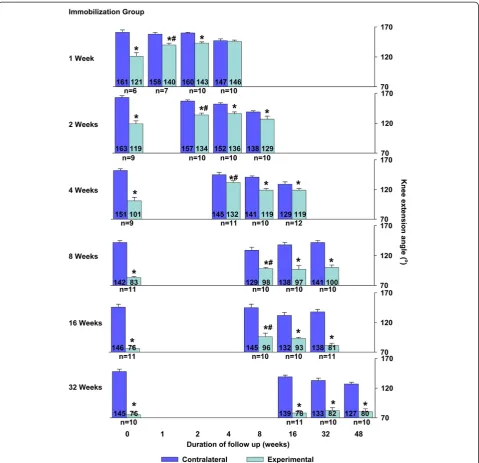
Thirteen rats required local wound care, of which 12 re-ceived antibiotics; all 13 were treated and included. At endpoint, data for 12 animals were not analyzed for per-sistent fibrous adhesions, leg fracture during testing, ex-tension angle over 195° or images not recorded. Final number of experimental animals was 238; the distri-bution per group, fixation and recovery durations are
shown in Figure 1. The animal model created
contractures of various severities with maximal knee ex-tension reaching 121° (101–140), 119° (96–133), 101° (77–136), 83° (75–93), 76° (60–103) and 76° (59–94) after 1, 2, 4, 8, 16 and 32 weeks of internal fixation, respectively (P = .003 for first comparison and <0.001 for the remaining 5 comparisons to contralateral knees; Figure 1). Full reversal occurred after 4 weeks of recovery in 1-week-old contracture. At all other durations of recovery, the mean angle of knee extension was smaller than con-tralateral knees (Figure 1). Thus, except for 1-week-old contractures, a full spontaneous reversal of the knee con-tracture was not observed in any joint concon-tracture.
Durations of recovery that produced partial reversal of knee contracture were identified. Recovery for 2, 4, 8 and 8 weeks led to partial reversal of the contracture caused by 2, 4, 8 and 16 weeks of fixation, respectively (respective p-values of: .052, <.001, .048 and .009; Figure 1). The ex-tent of partial gain was 15°, 31°, 15° and 20° for initial con-tractures of 44°, 40°, 59° and 70° respectively (Figure 1). These constituted a plateau after which additional dura-tions of recovery added no significant gain in knee ex-tension compared with the previous recovery duration (P> 0.05 for all 8 comparisons; Figure 1). Specifically, doubling the duration of recovery added no knee exten-sion compared to the duration of recovery equal to the duration of fixation in all applicable groups (P> 0.05 for all 4 comparisons; Figure 1), and quadrupling the duration of recovery, when feasible, added no gain in knee exten-sion compared to recovery double the duration of fixation (P> 0.05 for all 3 comparisons; Figure 1).
Recovery durations of 16, 32 or 48 weeks after 32 weeks of surgical fixation did not change the mean an-gles of knee extension (P> 0.05 for all 3 comparisons; Figure 1).
Knees contralateral to the experimental knees showed a decrease in angle of extension over the duration of re-covery for groups with unilateral fixation durations of 1, 2, 4 and 32 weeks (respective p-values of: .002, <.001, p < .001 and p = .005; Figure 1).
Discussion
Prolonged immobility of healthy joints resulted in con-tractures in the rat model. We quantified the spontan-eous reversibility of knee joint contractures of various severities in 24 situations over 80 weeks. The data con-stitute convincing evidence for dose–response relation-ships not only between severity of the knee contracture and duration of internal fixation but also between sever-ity of the contractures and potential for reversibilsever-ity with spontaneous recovery (Figure 3).
[image:4.595.59.289.89.258.2]Spontaneous recovery of rat knee contractures for 4 weeks allowed full reversal of 1-week-old contractures. At all other contracture severities, knee flexion contrac-tures were not fully reversible no matter the duration of Figure 2Arthrometer used to measure angle of extension.The
recovery. These results confirm the first hypothesis that only recent-onset knee flexion contractures fully reverse spontaneously. This study confirmed reports of incom-plete reversal of joint contractures in various animal mo-dels [11-16] and improved upon previous investigations by the broad range of 18 clinically relevant durations of spontaneous recovery, from adult to geriatric age, by stan-dardized mechanical testing, and by sufficient sample size for statistical testing. These data should help resolve the controversy regarding the potential for full spontaneous recovery of joint contractures secondary to immobility [8,10].
Within a specific time window — recovery for 2–8 weeks, some knee joint contractures were partially re-versible. The extent of partial reversal was modest, an average gain of 20° of knee extension. This corresponded to an average 41% of the contractures (Figure 1). These constituted plateaus in the spontaneous recovery; exten-ding the duration of recovery never led to a significant improvement of the contractures, which confirms the sec-ond hypothesis. Once a joint contracture is diagnosed, the current study implies that simple observation is not an ap-propriate option. Our data predict a poor prognosis, with recovery of only 20° (or 41%) of the contracture. Therefore intervention may be necessary to regain knee extension beyond natural recovery.
This study also measured decreased knee extension over time in the rat knees contralateral to a contractured knee (Figure 1). In this animal model, at least three fac-tors can contribute: both surgical procedures involved general anesthesia and postoperative recovery (approxi-mately 1 week each). The temporary general hypomobi-lity may have contributed to the loss of extension in
contralateral knees. Secondly, an index contracture in the experimental leg may limit extension in contralateral knees to smoothen the gait pattern; patients with osteo-arthritis and a unilateral knee flexion contracture lacked knee extension of the contralateral knee [17]. Thirdly, aging remains a controversial contributor to decreased range of motion in diarthrodial joints [18-24]. In this study, using the contralateral knee joint for comparison controlled for the postoperative general hypomobility and for aging.
This animal model studied the simple immobility-induced joint contractures. No articular trauma, osteo-chondral damage or hemarthrosis accompanied the im-mobilization. No neurological injury altered the limb muscle tone (increased or decreased tone with an upper or a lower motor neuron injury, respectively) Import-antly, no treatment was provided. This study produced normative data on the effects of joint immobility. The ef-fects of other variables (partial mobility, articular trauma, change in neurological status, various treatments) need to be studied separately. Similarly, distinguishing the tissue limiting the knee joint, articular or muscular, can be ob-tained by comparing range of extension before and after myotomies [25].
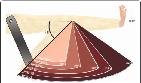
[image:5.595.56.291.89.228.2]The range of extension of the knee is a key variable in the biomechanical gait assessment. Full knee extension reaching 180 degrees is necessary for normal walking in humans. The magnitude of the knee flexion contracture proportionately disturbs the gait pattern. In humans, knee joint contractures required more quadriceps force for stability, caused a shorter stride length, increased oxy-gen consumption and negatively impacted balance and risk of falling [26-28]. For some patients, the immobility of normal joints is temporary (e.g., brace, cast, intensive care stay), and eventually they will actively use their joints again. The current study in the rat model shows that a statistically significant lack of knee extension re-mained despite long periods of spontaneous recovery similar to patients with fixed knee flexion contractures (Figure 3). The quantitative results measured over a time course of unassisted recovery in the current study may apply to patients with knee flexion contracture caused by prolonged immobility: after 2 weeks of immobility, their joints may develop a contracture that will not be fully reversible without treatment [3].
In the current study, 2 weeks of knee joint fixation had already caused contractures, not completely revers-ible without intervention. Early surveillance and inter-vention may decrease the risk of contractures [29]. A survey found that, of patients spending 2 weeks or more in an intensive care unit, 26% never had their joint mo-tion documented [30]. Similar to the current experimen-tal results, the development of contractures was linked to the duration of immobility: patients staying 8 weeks Figure 3Range of extension after spontaneous recovery of
or longer in intensive care had an adjusted odds ratio of 7.1 to 1 of presenting a joint contracture compared to patients staying for 2–3 weeks [3]. The current study confirmed that the longer a joint contracture remains undiagnosed or untreated, the more severe and irrevers-ible the structural changes [31].
The animal model differed from clinical practice in that a brace or a cast causes less rigid immobility than the extraarticular fixation. Clinically, spasticity or mus-culoskeletal lesions often accompanies joint contractures in stroke, spinal cord injury, peripheral nerve injuries, trauma [2,6]. Finally, in clinical practice some form of treatment may be instituted when a contracture is diagnosed.
Study limitations
First, the quadruped gait of the rat permitted long-term tolerance of the knee fixation in flexion because they never walk with their knee in full 180° of extension. However, functionally, a knee extension deficit in the rat is different than in humans [27]. Whereas the basic mechanical data from this study may apply to other dia-rthrodial joints, the functional impact of contractures varies from joint to joint. Second, we did not study the effect of recovery beyond a fourfold duration of fixation, because we had already reached the life expectancy of some animals. Whether extended periods of recovery would have been beneficial is unlikely, since doubling or quadrupling the duration of recovery did not lead to any significant gain in knee extension. Finally, short dura-tions of recovery after long periods of fixation may have shown that the plateau in partial reversal was achieved earlier than after 8 weeks of recovery.
Conclusions
In this animal model knee flexion contractures of 2-week onset or longer did not fully recover when untreated. Spontaneous recovery initially allowed modest, partial range of knee extension before a plateau was reached; extending the duration of spontaneous recovery was an ineffective intervention. In the absence of treatment, per-manent losses in knee extension must be anticipated.
Competing interests
The authors declare that they have no competing interests.
Authors’contributions
All authors were involved in drafting the article or revising it critically for important intellectual content, and all authors approved the final version to be published. GT had full access to all of the data in the study and takes full responsibility for the integrity of data analysis. Study conception and design: GT, HKU, LG. Acquisition of data: GT, HKU. Analysis and interpretation of data: GT, HKU, OL, LG. Drafting the article or revising it critically: GT, HKU, OL, LG.
Acknowledgements
This research was performed in the Bone and Joint Research Laboratory at the University of Ottawa and supported by Canadian Institute of Health Research grant MOP 97831. None of the authors has a conflict of interest in
regards to this research. The authors would like to acknowledge Joao Tomas and Tony Zandbelt for the arthrometer, Ying Nie for the surgeries with Sophie C. Bérubé, Li Dong and Hakim Louati, veterinarian staff, Khaoula Louati for image processing, Elizabeth Coletta for data analysis and graphs, Tim Ramsay for statistical review and Gloria Baker for editing the manuscript.
Author details
1
Department of Medicine, Faculty of Medicine, University of Ottawa, 451 Smyth Rd., Ottawa, ON K1H 8M5, Canada.2Bone and Joint Research
Laboratory, Faculty of Medicine, University of Ottawa, 451 Smyth Rd., Ottawa, ON K1H 8M5, Canada.3Division of Orthopedic Surgery, Faculty of Medicine,
University of Ottawa, 451 Smyth Rd., Ottawa, ON K1H 8M5, Canada.
4Biomedical Engineering, The Ottawa Hospital Rehabilitation Centre, 505
Smyth Road, Ottawa, ON K1H 8M2, Canada.5Department of Biology, Faculty of Science, University of Ottawa, 30 Marie Curie, Ottawa, ON K1N 6N5, Canada.
Received: 26 May 2014 Accepted: 29 September 2014 Published: 7 October 2014
References
1. Dudek N, Trudel G:Joint Contractures.InEssentials of Physical Medicine and Rehabilitation: Musculoskeletal Disorders, Pain, and Rehabilitation.2nd edition. Edited by Frontera WR, Silver JK, Rizzo TD Jr. Philadelphia: Saunders; 2008:651–655. Chapter 117.
2. Bushby K, Finkel R, Birnkrant DJ, Case LE, Clemens PR, Cripe L, Kaul A, Kinnett K, McDonald C, Pandya S, Poysky J, Shapiro F, Tomezsko J, Constantin C:Diagnosis and management of Duchenne muscular dystrophy, part 2: implementation of multidisciplinary care.Lancet Neurol
2010,9:177–189.
3. Clavet H, Hébert PC, Fergusson DA, Doucette S, Trudel G:Joint contractures following prolonged stay in the intensive care unit.CMAJ
2008,178:691–697.
4. Fox P, Richardson J, McInnes B, Tait D, Bedard M:Effectiveness of a bed positioning program for treating older adults with knee contractures who are institutionalized.Phys Ther2000,80:363–372.
5. Huang T, Blackwell SJ, Lewis SR:Ten years of experience in managing patients with burn contractures of axilla, elbow, wrist and knee joints. Plast Reconstr Surg1978,61:70–76.
6. Singer BJ, Dunne JW, Singer KP, Jegasothy GM, Allison GT:Non-surgical management of ankle contracture following acquired brain injury.Disabil Rehabil2004,26:335–345.
7. Trudel G, Uhthoff HK, Brown M:Extent and direction of joint motion limitation after prolonged immobility: an experimental study in the rat. Arch Phys Med Rehabil1999,80:1542–1547.
8. Akeson WH, Woo SL, Amiel D, Doty DH:Rapid recovery from contractures in rabbit hindlimbs: a correlative biomechanical and biochemical study. Clin Orthop Relat Res1977,122:359–365.
9. Haapala J, Arokoski JPA, Hyttinen MM, Lammi M, Tammi M, Kovanen V, Helminen H, Kiviranta I:Remobilization does not fully restore immobilization induced articular cartilage atrophy.Clin Orthop1999,
362:218–229.
10. Hildebrand KA, Sutherland C, Zhang M:Rabbit knee model of post-traumatic joint contractures: the long-term natural history of motion loss and myofibroblasts.J Orthop Res2004,22:313–320.
11. Ando A, Suda H, Hagiwara Y, Onoda Y, Chimoto E, Itoi E:Remobilization does not restore immobilization-induced adhesion of capsule and restricted joint motion in rat knee joints.Tohoku J Exp Med2012,227:13–22.
12. Finsterbush A, Friedman B:Early changes in immobilized rabbits knee joints: a light and electron microscopic study.Clin Orthop1973,
92:305–319.
13. Trudel G, Zhou J, Uhthoff HK, Laneuville O:Four weeks of mobility after 8 weeks of immobility fails to restore normal motion: a preliminary study. Clin Orthop Relat Res2008,466:1239–1244.
14. Usuba M, Akai M, Shirasaki BS, Miyakawa S:Experimental joint contracture correction with low torque–long duration repeated stretching.Clin Orthop Relat Res2007,456:70–78.
16. Katalinic OM, Harvey LA, Herbert RD:Effectiveness of stretch for the treatment and prevention of contractures in people with neurological conditions: a systematic review.Phys Ther2011,91:11–24.
17. Campbell TM:Demographics and Posterior Knee Capsule Histologic and Genetic Characterization in Patients with Severe Knee Osteoarthritis: Comparing those with Contracture to those without Contracture, MS Thesis. University of Ottawa, Department of Medicine; 2012. Available from [http://hdl.handle. net/10393/23176]
18. James B, Parker AW:Active and passive mobility of lower limb joint in elderly men and women.Am J Phys Med Rehabil1989,68(4):162–167. 19. Barnes CJ, Van Steyn SJ, Fischer RA:The effects of age, sex, and shoulder
dominance on range of motion of the shoulder.J Shoulder Elbow Surg
2001,10(3):242–246.
20. Chapleau J, Canet F, Petit Y, Sandman E, Laflamme GY, Rouleau DM:
Demographic and anthropometric factors affecting elbow range of motion in healthy adults.J Shoulder Elbow Surg2013,22:88–93. 21. Escalante A, Lichtenstein MJ, Hazuda HP:Determinants of shoulder and
elbow flexion range: results from the San Antonio Longitudinal Study of Aging.Arthr Care Res1999,12:277–286.
22. Lin CC, Ju MS, Huang HW:Gender and age effects on elbow joint stiffness in healthy subjects.Arch Phys Med Rehabil2005,86:82–85. 23. Roach KE, Miles TP:Normal hip and knee active range of motion: the
relationship to age.Phys Ther1991,71(9):656–665.
24. Doriot N, Wang X:Effects of age and gender on maximum voluntary range of motion of the upper body joints.Ergonomics2006,49(3):269–281. 25. Trudel G, Laneuville O, Coletta E, Goudreau L, Uhthoff UK:Quantitative and
temporal differential recovery of articular and muscular limitations of knee joint contractures; results in a rat model.J Appl Physiol2014,
117(7):730–737.
26. Goudie ST AHD, Ahmad A, Maheshwari R, Picard F:Flexion contracture following primary total knee arthroplasty: risk factors and outcomes. Orthopedics2011,34:e855–e859.
27. Perry J, Antonelli D, Ford W:Analysis of knee-joint forces during flexed-knee stance.J Bone Joint Surg Am1975,57(7):961–967.
28. Campbell J, Waters RL, Thomas L, Lombardi R, Mayer C:Simulated knee contracture: demands of walking.Phys Ther1984,64:715.
29. Needham DM:Mobilizing patients in the intensive care unit: improving neuromuscular weakness and physical function.JAMA
2008,300(14):1685–1690.
30. Clavet H, Hébert PC, Fergusson DA, Doucette S, Trudel G:Joint contractures in the intensive care unit: association with resource utilization and ambulatory status at discharge.Disabil Rehabil2011,
33:105–112.
31. Trudel G, Seki M, Uhthoff HK:Synovial adhesions are more important than pannus proliferation in the pathogenesis of knee joint contracture after immobilization: an experimental investigation in the rat.J Rheumatol
2000,27:351–357.
doi:10.1186/1471-2474-15-338
Cite this article as:Trudelet al.:Quantitative analysis of the reversibility of knee flexion contractures with time: an experimental study using the rat model.BMC Musculoskeletal Disorders201415:338.
Submit your next manuscript to BioMed Central and take full advantage of:
• Convenient online submission
• Thorough peer review
• No space constraints or color figure charges
• Immediate publication on acceptance
• Inclusion in PubMed, CAS, Scopus and Google Scholar
• Research which is freely available for redistribution