http://dx.doi.org/10.4236/jbnb.2016.72013
Controllable Biosynthesis of Small Silver
Nanoparticles Using Fungal Extract
Katrin Quester1,2, Miguel Avalos-Borja3, Ernestina Castro-Longoria1
1Departamento de Microbiología, Centro de Investigación Científica y de Educación Superior de Ensenada
(CICESE), Ensenada, Mexico
2Centro de Nanociencias y Nanotecnología, Universidad Nacional Autónoma de México (UNAM), Ensenada, Mexico
3División de Materiales Avanzados, Instituto Potosino de Investigación Científica y Tecnológica (IPICyT), San
Luis Potosí, Mexico
Received 11 February 2016; accepted 26 April 2016; published 29 April 2016
Copyright © 2016 by authors and Scientific Research Publishing Inc.
This work is licensed under the Creative Commons Attribution International License (CC BY). http://creativecommons.org/licenses/by/4.0/
Abstract
The search for reliable and eco-friendly methods for the production of nanoscale materials is an important aspect of nanotechnology. Silver nanoparticles (AgNPs) are of special interest because of their antimicrobial properties, especially those of small size. In this work, AgNPs were produced under different conditions of temperature and pH using the extract from the fungus Neurospora crassa as reducing agent. Mainly quasi-spherical particles were obtained at all incubation condi-tions. However, optimum conditions to produce small sizes in the range of 2 - 9 nm were at 4˚C and pH 3, also particles of 2 - 22 nm were obtained at 25˚C with unmodified pH (6.5) and pH 10. Ne
v-ertheless, only particles synthesized at 25˚C and pH 10 maintained the same size range after st o-rage of 10 months. In summary, optimal incubation conditions for the synthesis of silver nanopar-ticles of small size range are reported. This improves the storage time of parnanopar-ticles without losing its original size and without going into aggregation or agglomeration.
Keywords
Biosynthesis, Nanoparticles, Neurospora crassa
1. Introduction
process [2]. Furthermore, in the spent medium of various strains the presence of proteins are confirmed and as-sumed to be responsible for the bioreduction and for stabilizing and capping the newly formed nanostructures [1]-[4] which are important advantages of using biological methods.
However, for a biological process to successfully compete with chemical and physical nanostructure synthesis, the control over average particle size range and uniform particle morphology is required [5]. It has been shown that shape and size of nanoparticles (NPs) depend on the reaction conditions [6] [7]. Therefore, it is necessary to perform experiments varying parameters such as pH, temperature and the proportion of salts/reducing agent to fully explore the results obtained. Some publications show promising results for the synthesis of metallic NPs using extract of fungal strains [1] [6] and some studies define pH value and the reaction temperature as impor-tant parameters affecting nanostructure size and synthesis rate. For instance, some studies showed increasing reaction rate with increasing temperature [1] [5] [8] and alkaline pH was shown to increase silver particle for-mation [8]. However, not many studies have performed this kind of experimental procedures; therefore, the ma-nipulation of metallic NPs synthesis is far from being exhausted. In this work, we define the optimal reduction conditions to produce AgNPs of a small size range with very good stability over time using fungal extract.
2. Materials and Methods
2.1. Fungal Extract
For extract preparations, the wild type strain of Neurospora crassa (FGSC # 9013) was used; extract was ob-tained as previously described [9]. Briefly, grown biomass was filtered and washed 3 times in sterile deionized water. The biomass was ground 1:1 in deionized water (w/v) using an agate mortar, the mixture was centrifuged for 5 minutes at 6500 rpm and filtered through a 0.2 μm microfilter. The protein content of the extract was quan-tified by the Bradford Assay [10] in triplicate.
2.2. Biosynthesis of Silver NPs by N. crassa Extract
To find an optimum concentration of fungal extract for AgNPs synthesis, the extract (at known protein content concentration) was incubated with AgNO3 aqueous solution in the ratios of 1:1, 1:2, 1:3, 2:1 and 3:1 (fungal ex-tract: metal precursor; v/v) in the dark for 12 and 24 hours at room temperature. After finding the best experi-mental ratio (fungal extract: metal precursor), further experiments were carried out under different environmen-tal conditions. Synthesis of AgNPs were carried out in the dark at three different pH values (3, unmodified (6.5), and 10) and at five temperatures (4˚C, 25˚C, 37˚C, 60˚C and 80˚C), for 3, 6, 9 and 12 hours of incubation. Mod-ification of the pH value was realized by adding 1 molar (mol/L = M) HCl or NaOH aqueous solution.
2.3. TEM Analysis
A 10 µL aliquot of each sample was placed on carbon coated copper grids (300 mesh, Ted Pella Inc.) for TEM analysis under a Hitachi H-7500, JEOL 2010 and FEI Tecnai F30 transmission electron microscopes (TEM). The interplanar distance was determined using the software GatanDigitalMicrographTM.
2.3. Statistical Analyses
To determine significant differences between the obtained data, a factorial analysis of variance (ANOVA) was performed by using the software Statistica 7. Significance was calculated by Tukey’s HSD (Honestly Significant Difference) post hoc test. Confidence intervals were set to 0.95. Error bars represent standard deviation, while significant differences are marked by an asterisk.
3. Results
21.4 nm and 1.9 - 35.2 nm at 12 and 24 h, respectively. Slightly smaller particles were formed at 1:3 ratio, size range was 1.9 - 19.5 nm and 1.9 - 21.2 nm at 12 and 24 h, respectively. Since the size range for the ratio 1:2 was very large, which might imply the formation of less stable nanoparticles, the ratio 1:3 (fungal extract: metal precursor) was chosen for further experiments.
AgNPs were synthesized under different conditions of pH, temperature and incubation time in order to op-timize the protocol for the formation of small NPs of uniform size. It came to quasi-spherical NPs formation under all employed conditions, a summary over obtained results is shown inTable 1.
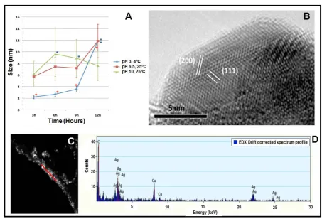
As mentioned above, nanoparticle formation was achieved under all conditions applied, but since our interest was to establish a protocol to obtain small uniform size of NPs, only promising results (NPs synthesized at 4˚C and pH 3, 25˚C and pH 6.5, and at 25˚C and pH 10) were further analyzed via HRTEM and EDS. Successful biosynthesis of AgNPs was already indicated by the color change of the solution from clear yellow to darker yellow upon exposure to the AgNO3 precursor solution (insets of Figures 1(A)-(C). Synthesized NPs were mainly quasi-spherical in shape in all cases (Figures 1 (A)-(C)).Figures 1(D)-(F)show TEM images of higher resolution to confirm the quasi-spherical character of the synthesized silver NPs. At least 100 particles were measured and the corresponding histograms are shown inFigures 1(G)-(I). Significant differences according to Tukey’s HSD post hoc test are marked with an asterisk inFigure 2(A). Applying a temperature of 4˚C and pH 3 resulted in the formation of very small AgNPs in a main size range of 1 to 6 nm (Figure 1(A) and Figure 1(G)) being significantly smaller than the other samples (red asterisks,Figure 2(A)); however, after more than 9 hours, AgNPs showed significantly bigger sizes (blue asterisk,Figure 2(A)). At 25˚C and unmodified pH (6.5), AgNPs of mainly 1 to 13 nm were synthesized (Figure 1(B)and Figure 1(H)), also with the appearance of significantly bigger NPs after 12 hours (blue asterisk, Figure 2(A)), while at pH 10 and 25˚C resulted in the formation of AgNPs of mainly 1 to 10 nm (Figure 1(C)and Figure 1(I)). Here, significantly bigger AgNPs were formed af-ter 6 and 9 hours compared to 3 hours (blue asaf-terisks,Figure 2(A)) while after 12 hours, the significant differ-ence in particle size could not be observed. Determination of lattice fringe spacing of AgNPs (Figure 2(B)) con-firms the planes (111) and (200) of face-centered cubic (fcc) metal silver. EDS analysis was carried out in a group of particles (Figure 2(C)) and the elemental character of synthesized AgNPs was confirmed (Figure 2(D)
Table 1. Silver NPs obtained at different incubation time and conditions at the ratio 1:3 (fungal extract: metal precursor; v/v).
Temperature (˚C) pH Value 3 h 6 h 9 h 12 h
4
3 1.9 - 5.9 nm 1.7 - 7.2 nm 2.1 - 9.2 nm 3.7 - 28.3 nm
Unmodified (6.5) 2.0 - 8.2 nm 3.4 - 10.3 nm 3.3 - 40.2 nm 7.5 - 110.3 nm
10 3.5 - 10.4 nm 2.1 - 6.8 nm 5.9 - 15.8 nm 10.0 - 41.3 nm
25
3 2.1 - 20.1 nm 2.5 - 24.0 nm 2.5 - 25.0 nm 2.1 - 19.9 nm
Unmodified (6.5) 1.7 - 22.5 nm 2.5 - 17.4 nm 1.9 - 21.0 nm 1.9 - 19.5 nm
10 1.0 - 22.5 nm 1.7 - 21.5 nm 2.1 - 21.7 nm 1.4 - 21.0 nm
37
3 7.1 - 43.1 nm 6.5 - 36.5 nm 3.3 - 34.3 nm 4.3 - 62.8 nm
Unmodified (6.5) 3.3 - 59.9 nm 3.2 - 21.9 nm 2.3 - 32.0 nm 2.9 - 26.3 nm
10 7.5 - 96.5 nm 4.4 - 148.0 nm 2.5 - 44.3 nm 2.9 - 44.9 nm
60
3 11.6 - 330.8 nm 1.8 - 308.7 nm 2.1 - 12.3 nm 1.4 - 88.7 nm
Unmodified (6.5) 3.6 - 44.7 nm 4.1 - 46.1 nm 2.7 - 36.2 nm 4.3 - 124.9 nm
10 4.2 - 47.9 nm 1.4 - 93.6 nm 1.4 - 73.8 nm 1.8 - 8.1 nm
80
3 11.6 - 330.8 nm 1.8 - 308.7 nm 2.1 - 12.3 nm 1.4 - 88.7 nm
Unmodified (6.5) 2.4 - 20.5 nm 4.3 - 101.0 nm 2.7 - 23.9 nm 2.7 - 20.5 nm
Figure 1. TEM images of silver nanoparticles synthesized by N. crassa extract at 4˚C and 25˚C and different pH values. 4˚C and pH 3 (A and D), 25˚C and pH 5.5 (B and E), and 25˚C pH 10 (C and F). Insets: color change of fungal extract-metal precursor solution from before (clear yellow; left image) and after (darker yellow; right image) incubation. (G-I) Corres-ponding size distribution histograms of produced nanoparticles.
The copper signal in the spectrum corresponds to the Cu-grid used to put the sample.
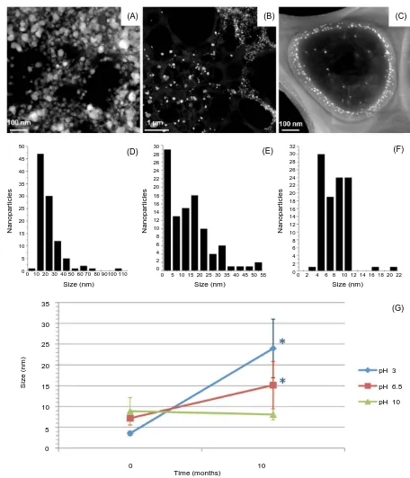
The size of AgNPs was analyzed over time; HAADF-TEM images show that NPs synthesized at 4˚C and pH 3 incremented in size over time (Figure 3(A) andFigure 3(D)), revealing significantly bigger sizes after 10 months storage (Figure 3(G)). A similar effect was observed for AgNPs synthesized at 25˚C and pH 6.5 (Figure 3(B)and Figure 3(E)), although the difference is not as big as in the sample synthesized at 4˚C and pH 3 (Figure 3(G)). However, AgNPs synthesized at 25˚C and pH 10 (Figure 3(C)and Figure 3(F)) kept their small size range without aggregation or agglomeration (Figure 3(G)).
Size (nm) N anopar ti c les
Size (nm) Size (nm)
N anopar ti c les N anopar ti c les 32 30 28 26 24 22 20 18 16 14 12 10 8 6 4 2 0 34 32 30 28 26 24 22 20 18 16 14 12 10 8 6 4 2 0 34 32 30 28 26 24 22 20 18 16 14 12 10 8 6 4 2 0
Figure 2. Analysis of size and elemental character of produced AgNPs. (A) Analysis of variance (ANOVA) of average sizes of AgNPs at different incubation times with respect to different pH values. Significant differences are marked with an aste-risk. Confidence intervals were set to 0.95; error bars represent the standard deviation. (B) HRTEM images of a quasi- spherical silver nanoparticle with plane information. (C) HAADF image of silver nanoparticles. (D) EDS spectrum showing the presence of silver in the sample shown in (C). Red line in (A) indicates where the analysis was carried out.
4. Discussion
Neurospora crassa extract was used to biosynthesize AgNPs under different environmental conditions in order to produce small particles with narrow size dispersion. The obtained results in this study indicate that the forma-tion of different sizes can be tuned by combining a range of temperatures with varying pH value. It was already shown that by changing the pH value and/or temperature, the resulting shapes and/or sizes of metal NPs synthe-sized by fungi vary [1] [7] [11] [12]; however, each fungal strain produces different metabolites and results dif-fer among the species used. For example, optimal pH values for nanoparticle synthesis were shown to be alka-line for the fungus Isaria fumosorosea [13], pH 6.0 for Penicillium fellutanum [14] and acidic for Fusarium acuminatum [15].
In the case of N. crassa extract, it was clear that using temperatures higher than 25˚C led to the production of bigger AgNPs, contrary to the results found by Fayaz et al. [11]. Also, at lower temperatures they found the formation of silver nanoplates while in our study, under all experimental conditions mainly quasi-spherical par-ticles were obtained.
Figure 3. Analysis of AgNP size over time. HAADF-TEM micrographs and size distribution histograms of silver NPs syn-thesized at 4˚C and pH 3 (A and D), 25˚C and pH 5.5 (B and E), and 25˚C and pH 10 (C and F) after 10 months of storage. (G) ANOVA of average sizes of silver nanoparticles after 10 months of storage compared to average sizes at time point zero (freshly synthesized). Significant differences are marked with an asterisk. Confidence intervals were set to 0.95. Error bars represent the standard deviation.
donate electrons quite easily while releasing a proton. Nitrogen is regarded as a less soft, or borderline base and Nath et al. [18] realized studies about the grade of softness of gold and silver by determining the binding affinity of these metals to nitrogen and sulfur; the latter being a softer Pearson-base. The authors showed that silver is a (C) (B)
(A)
Size (nm) Size (nm) Size (nm)
(D) (E) (F)
(G) 0 10 20 30 40 50 60 70 80 90100 110 0 5 10 15 20 25 30 35 40 45 50 55 0 2 4 6 8 10 12 14 16 18 20 22
N anopar ti c les N anopar ti c les N anopar ti c les S iz e ( nm ) 30 28 26 24 22 20 18 16 14 12 10 8 6 4 2 0 50 45 40 35 30 25 20 15 10 5 0 32 30 28 26 24 22 20 18 16 14 12 10 8 6 4 2 0
pH 3
pH 6.5
pH 10
less soft Pearson-acid than gold, possessing much stronger affinity to the less soft nitrogen atom, while the softer gold bound stronger to the softer sulfur atom. Therefore, due to its higher binding affinity, the bioreduction process between amino groups and silver ions might be faster due to a quicker immobilization of the silver ions by the amino groups of the proteins, which thereby results in isotropic growth of AgNPs even at low pH values, based on the statement of Li and Kaner [19], declaring that in a faster reaction, it is more likely that the embryo-nic nuclei evolve to create homogenous nuclei before they can diffuse to heterogeneous nucleation sites to nuc-leate. As stated above, carboxyl groups are less effective stabilizers at lower pH values, which might be the rea-son of variations in AgNPs sizes between the samples.
The exact mechanisms are still not fully understood and much more investigation needs to be done to gain a better understanding and the ability to further manipulate size and shape of biosynthesized metal NPs. Mean-while, the results in this study indicate that under certain temperatures and pH values small size range of par-ticles with very good stability can be produced.
5. Conclusion
Silver nanoparticles of small size have been successfully produced using a simple green methodology. Further-more, we have demonstrated that biosynthesis of nanoparticles using fungal extract can be optimized and that under specific conditions of pH and temperature, the size of particles can be maintained for at least 10 months without aggregation or agglomeration.
Acknowledgements
We thank CONACyT for a scholarship for KQ. We also thank LINAN for providing access to electron micro-scopy facilities, Dr. Nicolás Cayetano Castro and Dr. Héctor Gabriel Silva Pereyra (LINAN-IPICyT) for helping in TEM characterization. Part of this work was funded by a SEP-CONACyT grant (CB2011/169154).
References
[1] Xie, J., Lee, J.Y., Wang, D.I. and Ting, Y.P. (2007) High-Yield Synthesis of Complex Gold Nanostructures in a Fungal System. The Journal of Physical Chemistry C, 111, 16858-16865. http://dx.doi.org/10.1021/jp0752668
[2] Ahmad, A., Mukherjee, P., Mandal, D., Senapati, S., Khan, M.I., Kumar, R. and Sastry, M. (2002) Enzyme Mediated Extracellular Synthesis of CdS Nanoparticles by the Fungus, Fusarium oxysporum. Journal of the American Chemical Society, 124, 12108-12109. http://dx.doi.org/10.1021/ja027296o
[3] Mukherjee, P., Roy, M., Mandal, B.P., Dey, G.K., Mukherjee, P.K., Ghatak, J., Tyagi, A.K. and Kale, S.P. (2008) Green Synthesis of Highly Stabilized Nanocrystalline Silver Particles by a Non-Pathogenic and Agriculturally Impor-tant Fungus T. asperellum. Nanotechnology, 19, 075103. http://dx.doi.org/10.1088/0957-4484/19/7/075103
[4] Vigneshwaran, N., Kathe, A.A., Varadarajan, P.V., Nachane, R.P. and Balasubramanya, R.H. (2007) Silver-Protein (Core-Shell) Nanoparticle Production Using Spent Mushroom Substrate. Langmuir, 23, 7113-7117.
http://dx.doi.org/10.1021/la063627p
[5] Gericke, M. and Pinches, A. (2006) Biological Synthesis of Metal Nanoparticles. Hydrometallurgy, 83, 132-140.
http://dx.doi.org/10.1016/j.hydromet.2006.03.019
[6] Das, S.K., Das, A.R. and Guha, A.K. (2010) Microbial Synthesis of Multishaped Gold Nanostructures. Small, 6, 1012- 1021. http://dx.doi.org/10.1002/smll.200902011
[7] Sanghi, R. and Verma, P. (2010) pH Dependant Fungal Proteins in the “Green” Synthesis of Gold Nanoparticles. Ad-vanced Materials Letters, 1, 193-199. http://dx.doi.org/10.5185/amlett.2010.5124
[8] Qian, Y., Yu, H., He, D., Yang, H., Wang, W., Wan, X. and Wang, L. (2013) Biosynthesis of Silver Nanoparticles by the Endophytic Fungus Epicoccum nigrum and Their Activity against Pathogenic Fungi. Bioprocess and Biosystems Engineering, 36, 1613-1619. http://dx.doi.org/10.1007/s00449-013-0937-z
[9] Quester, K., Avalos-Borja, M., Vilchis-Nestor, A.R., Camacho-López, M.A. and Castro-Longoria, E. (2013) SERS Properties of Different Sized and Shaped Gold Nanoparticles Biosynthesized under Different Environmental Condi-tions by Neurospora crassa Extract. PLoS ONE, 8, e77486. http://dx.doi.org/10.1371/journal.pone.0077486
[10] Bradford, M.M. (1976) A Rapid and Sensitive Method for the Quantitation of Microgram Quantities of Protein Utiliz-ing the Principle of Protein-Dye BindUtiliz-ing. Analytical Biochemistry, 72, 248-254.
http://dx.doi.org/10.1016/0003-2697(76)90527-3
Nanopar-ticles—An Effect of Temperature on the Size of Particles. Colloids and Surfaces B: Biointerfaces, 74, 123-126.
http://dx.doi.org/10.1016/j.colsurfb.2009.07.002
[12] Philip, D. (2009) Biosynthesis of Au, Ag and Au-Ag Nanoparticles Using Edible Mushroom Extract. Spectrochimica Acta, Part A: Molecular and Biomolecular Spectroscopy, 73, 374-381. http://dx.doi.org/10.1016/j.saa.2009.02.037
[13] Banu, A.N. and Balasubramanian, C. (2014) Optimization and Synthesis of Silver Nanoparticles Using Isaria fumoso-rosea against Human Vector Mosquitoes. Parasitology Research, 113, 3843-3851.
http://dx.doi.org/10.1007/s00436-014-4052-0
[14] Kathiresan, K., Manivannan, S., Nabeel, M.A. and Dhivya, B. (2009) Studies on Silver Nanoparticles Synthesized by a Marine Fungus, Penicillium fellutanum Isolated from Coastal Mangrove Sediment. Colloids and Surfaces B: Biointer-faces, 71, 133-137. http://dx.doi.org/10.1016/j.colsurfb.2009.01.016
[15] Tidke, P.R., Gupta, I., Gade, A.K. and Rai, M. (2014) Fungus-Mediated Synthesis of Gold Nanoparticles and Standar-dization of Parameters for its Biosynthesis. IEEE Transactions on NanoBioscience, 13, 397-402.
[16] Pearson, R.G. (1993) The Principle of Maximum Hardness. Accounts of Chemical Research, 26, 250-255.
http://dx.doi.org/10.1021/ar00029a004
[17] Mukherjee, P., Senapati, S., Mandal, D., Ahmad, A., Khan, M.I., Kumar, R. and Sastry, M. (2002) Extracellular Syn-thesis of Gold Nanoparticles by the Fungus Fusarium oxysporum.ChemBioChem, 3, 461-463.
http://dx.doi.org/10.1002/1439-7633(20020503)3:5<461::AID-CBIC461>3.0.CO;2-X
[18] Nath, S., Ghosh, S.K., Kundu, S., Praharaj, S., Panigrahi, S. and Pal, T. (2006) Is Gold Really Softer than Silver? HSAB principle revisited. Journal of Nanoparticle Research, 8, 111-116. http://dx.doi.org/10.1007/s11051-005-8025-1