http://dx.doi.org/10.4236/ojo.2016.68033
Postoperative Mechanical Axis Alignment
and Components Position after Conventional
and Patient-Specific Total Knee Arthroplasty
Mohamed Mosa Mohamed
1, Maher A. El Assal
2, Ahmed M. Abdel Aal
2, Yaser E. Khalifa
2,
Mahmoud A. Hafez
31Orthopaedic Department, Alazhar University (Assiut Branch), Assiut, Egypt 2Orthopaedic Department, Assiut University, Assiut, Egypt
3Orthopaedic Department, October 6 University, Cairo, Egypt
Received 28 June 2016; accepted 9 August 2016; published 12 August 2016
Copyright © 2016 by authors and Scientific Research Publishing Inc.
This work is licensed under the Creative Commons Attribution International License (CC BY). http://creativecommons.org/licenses/by/4.0/
Abstract
Introduction: Accurate postoperative alignment and implant positioning are determinant factors for successful total knee arthroplasty (TKA). Patient-specific template (PST) is a technique that uses computer technology for the planning, deigning and production of cutting guides. This study aims to compare PST to conventional technique in terms of mechanical axis alignment and com-ponent positioning. Patients and method: 109 TKA were performed for 78 patients in 2 groups. Group A included 69 conventional TKA in 55 patients and Group B included 40 patient-specific TKA in 23 patient. Postoperative long-film X-rays were done for all patients to observe the me-chanical axis, anatomical axis, lateral distal femoral meme-chanical angle and medial proximal tibial angle. Results: No statistically significant difference was found between the two groups regarding alignment or component positioning. Conclusion: Both techniques have shown similar results in restoring the mechanical axis and alignment after TKA. However, PST had the advantages of re-duced blood loss and shorter operative time.
Keywords
Total Knee Arthroplasty, Patient-Specific Templates, Conventional TKA, Mechanical Axis, Limb Alignment, Component Position
1. Introduction
re-lieve the pain and restore the function of the joint. The procedure has a high success rate and patient satisfaction (82%) as reported by the Swedish Knee Arthroplasty Registry [1].
Accurate postoperative alignment and implant positioning are key elements to achieve optimal results from the surgery. Conventional TKA is the standard technique in registry data. However, computer technologies, such as patient specific templates (PST) and navigation, have been introduced in joint-replacement surgeries to offer more precision and easier setup [2].
PST is an evolving technology that aims at simplifying TKA by decreasing surgical steps making the proce-dure less invasive. The technique has been practiced with reportedly minimized bone injury and infection rate. Thus, the technique was found to be more useful than the conventional for patients with severe comorbidities as the later requires intra- and/or extra-medullary rods with more pain and higher chance for infection [3].
PST relies on preoperative CT or MRI imaging and 3D printing technology to produce patient-tailored tem-plates (tibial/femoral) which act as cutting guides (for direct bone cutting through the temtem-plates) or as pin loca-tor (pin-placement guides) [4].
Controversy exists about the efficacy of PST to restore the mechanical axis and alignment of the knee in comparison to the conventional instrumentation. Some mixed results have been reported regarding the number of outliers (<3˚ varus/valgus) and the overall postoperative alignment [5].
The aim of this work was to compare the postoperative mechanical axis alignment and components position-ing after TKA.
2. Patients and Methods
This is a prospective comparative study on 109 knees operated with TKA (2014) in 78 patients whom were di-vided into 2 groups. Group A included 69 TKA in 55 patients done by conventional instruments under supervi-sion of 3 senior orthopaedic surgeons whom are experienced in TKA using conventional method; while Group B included 40 TKA in 23 patients done by another senior surgeon (head of arthroplasty unit) who is experienced in TKA by conventional, navigation and PST techniques. Patients were divided into the two groups according to their presentation time to the centers. Ethical Committee approval was achieved prior to TKA procedures.
The mean age was 54.8 years and 58.9 years for Groups A and B, respectively. Female percentage was 74.6% (41 patients) and 91.3% (20 patients) in Groups A and B, respectively.
All cases in both groups were similar in diagnosis and inclusion criteria, and they were all indicated for pri-mary TKA due to pripri-mary OA, secondary OA or inflammatory arthritis; while revision cases were excluded.
Standing long-leg X-rays were obtained for all patients postoperatively. The X-rays were assesses through a digital software (screen scale). Pre- and postoperative mechanical axis deviation (MAD) and anatomical axis deviation (AAD) were measured.
Accordingly, the number of outliers was also obtained, that is, knees with mechanical axis deviation more than 30 varus or valgus. Lateral distal femoral angle (LDFA) and medial proximal tibial angle (MPTA) were measured in both groups by the same software, and t and chi-square tests were done for comparison. Statistical analysis of the data was performed using SPSS_16 software package. Statistical analysis was done for preopera-tive demographic data, preoperapreopera-tive diagnosis, type of deformity, anesthesia used and number of deformed knees.
3. Results
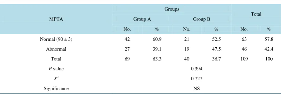
No statistically significant difference between both groups was found in the mean postoperative mechanical axis and number of outliers. The number of outliers decreased in both groups postoperatively from 88.4% and 92.5% to 42% and 45% in Groups A and B, respectively with P value 0.762 (see Table 1 and Figure 1). The mean MAD was found to be 3.19 for Group A and 3.91 for Group B with P value of 0.202 (see Table 2).
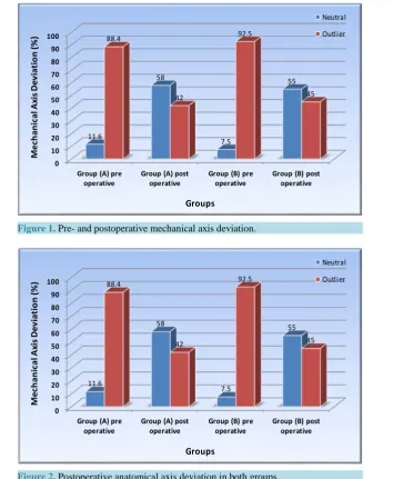
No statistically significant difference was found in the postoperative anatomical axis (see Figure 2).
Also, LDFA and MPTA were not statically different between both groups with P value of 0.122 and 0.107, respectively, and the number of knees with abnormal LDFA and MPTA in both groups had P value of 0.215 and 0.394, respectively (see Table 3 and Table 4).
4. Discussion
Table 1. Pre- and postoperative mechanical axis deviation.
MAD
Groups
Preoperative Postoperative
Group A Group B Group A Group B
Neutral 8 (11.6%) 3 (7.5%) 40 (58.0%) 22 (55.0)
Outliers 61 (88.4%) 37 (92.5%) 29 (42.0%) 18 (45.0%)
P value 0.495 0.762
Χ2 0.46 0.21
Significance NS NS
Data are presented in the form of frequency and percent. Chi square was used to test the significance between groups. NS: Not significant.
Table 2. Postoperative mechanical axis deviation in terms of mean values and standard deviation.
Angles
Groups
P value Significance
Group A Group B
Mean SD Mean SD
MAD 3.19 2.52 3.91 3.01 0.202 NS
[image:3.595.87.539.559.710.2]T test was used to test the significance between groups. NS: Not significant.
Table 3. Postoperative lateral distal femoral angle.
LDFA
Groups
Total
Group A Group B
No. % No. % No. %
Normal (90˚ ± 3˚) 54 78.3 27 67.5 81 74.3
Abnormal 15 21.7 13 32.5 28 25.7
Total 69 63.3 40 36.7 109 100
P value 0.215
Χ2 1.536
Significance NS
Data are presented in the form of frequency and percent. Chi was used to test square the significance between groups. NS: Not significant.
Table 4. Postoperative medial proximal tibial angle.
MPTA
Groups
Total
Group A Group B
No. % No. % No. %
Normal (90 ± 3) 42 60.9 21 52.5 63 57.8
Abnormal 27 39.1 19 47.5 46 42.4
Total 69 63.3 40 36.7 109 100
P value 0.394
Χ2
0.727
Significance NS
0 10 20 30 40 50 60 70 80 90 100
Group (A) pre
operative Group (A) post operative Group (B) pre operative Group (B) post operative
[image:4.595.141.496.83.515.2]11.6 58 7.5 55 88.4 42 92.5 45 M ec ha nic al A xis D ev ia tio n (% ) Groups Neutral Outlier
Figure 1. Pre- and postoperative mechanical axis deviation.
0 10 20 30 40 50 60 70 80 90 100
Group (A) pre
operative Group (A) post operative Group (B) pre operative Group (B) post operative
[image:4.595.157.469.297.512.2]11.6 58 7.5 55 88.4 42 92.5 45 M ec ha nic al A xis D ev ia tio n (% ) Groups Neutral Outlier
Figure 2. Postoperative anatomical axis deviation in both groups.
decades with reliable postoperative outcomes. However, PST has been introduced as an alternative for conven-tional instrumentation to eliminate some drawbacks of the old technique such as the required perforation of fe-moral and tibial medullary canals which potentially leads to more bleeding, fat embolism, DVT, infection and inaccuracy or malalignment [6]. PST was also introduced to decrease the number of instruments routinely used by the conventional technique, reduce the operative time and increase learning curve [7].
However, conventional TKA seems more favorable for many surgeons especially in developing countries due to financial limitations of computer-assisted settings. Several studies have compared both techniques; however, this paper has a main focus on the mechanical axis and postoperative alignment.
In this series, no statistically significant difference was found between PST and conventional TKA regarding the mean of postoperative mechanical axis deviation, percentage of postoperative outliers and components posi-tion.
Postoperatively, there is a marked reduction in the number of outliers in both groups; thus, both techniques seem to have nearly the same ability to correct deformity and restore knee alignment and accuracy of compo-nents position.
A comparative study between three groups, group 1 conventional, group 2 PST restoring mechanical axis (as group B in our study) and group 3 PST restoring kinematic axis has been conducted earlier and found no statis-tical significant difference between groups 1 and 2 in the postoperative alignment while group 3 gave a worse result [6].
Chen et al. conducted a comparative study between PST and conventional TKA and concluded that PST gave more postoperative outliers than conventional; accordingly, PST seemed less advisable in the authors’ view [7]. On the contrary, a randomized control trial on 222 TKA (108 conventional and 114 PST) by Anderl et al. stated that PST had given more accurate results regarding postoperative alignment and components’ position al-though no significant clinical difference was found between both techniques [8].
A meta-analysis on 7 studies (Level I and Level II evidence, 559 patients) compared PST and conventional TKA and found no statistical significant difference between both groups regarding the number of outliers and mean postsoperative mechanical axis [9] [10].
In light of our results, it is controversial that although PST uses a computerized preoperative tool in planning and manufacturing the cutting guides for each patient, its ability to restore mechanical axis and components po-sition has yet not proven superior than conventional technique. PST is further thought to have the disadvantage of lacking intraoperative guidance compared to navigation technique, thus it lacks the property of immediate correction of any inaccurate in bone cuts [11]-[13].
As secondary endpoints of this study, there was no statistically significant difference regarding the patients’ demographic parameters (age and sex). Primary osteoarthritis represented the highest percentage of preoperative diagnosis in both groups: 89.9% (49 patients) and 80% (18 patients) in Groups A and B, respectively; followed by rheumatoid arthritis: 10.1% (5 patients) and 17.5% (4 patients) in Groups A and B, respectively; while sec-ondary osteoarthritis was only 2.5% (1 patient) in Group B only. Chi square test was done for these diagnostic data and showed no significant difference with P value of 0.266.
The percentage of the number of deformed knees preoperatively was 88.4% (61 knees) and 92.5% (37 knees) in groups A and B, respectively, with no statistically significant difference.
On the other hand, there was significant difference between both groups regarding type of anesthesia and number of bilateral TKAs. In Group A, all patients had received spinal anesthesia; while in Group B, 40% re-ceived epidural, 50% rere-ceived epidural and general, 5% rere-ceived spinal and 5% rere-ceived GA. Also, it was no-ticed that the percentage of bilateral TKAs was markedly higher in Group B (82.5%; 19 patients) than in Group A (17.5%; 10 patients).
Limitations of this study include the low number of patients. Surgeries were done by different surgeons and thus the setup for each surgery was not standardized.
5. Conclusion
PST has the same results as conventional TKA regarding postoperative alignment and component position. However, PST is more applicable in severely deformed knees, revision TKA and bilateral simultaneous proce-dures.
References
[1] Bäthis, H., Perlick, L., Tingart, M., Lüring, C., Zurakowski, D. and Grifka, J. (2004) Alignment in Total Knee Arth-roplasty. A Comparison of Computer-Assisted Surgery with the Conventional Technique. The Journal of Bone & Joint
Surgery (Br), 86, 682-687. http://dx.doi.org/10.1302/0301-620X.86B5.14927
[2] Pitto, R.P., Graydon, A.J., Bradley, L., Malak, S.F., Walker, C.G. and Anderson, I.A. (2006) Accuracy of a Comput-er-Assisted Navigation System for Total Knee Replacement. The Journal of Bone & Joint Surgery (Br), 88, 601-605. http://dx.doi.org/10.1302/0301-620X.88B5.17431
[3] Cheng, T., Zhao, S. and Peng, X.C. (2012) Does Computer-Assisted Surgery Improve Postoperative Leg Alignment and Implant Positioning Following Total Knee Arthroplasty? A Meta-Analysis of Randomized Controlled Trials? Knee
Surgery, Sports Traumatology, Arthroscopy, 20, 1307-1322. http://dx.doi.org/10.1007/s00167-011-1588-8
[4] Conteduca, F., Iorio, R. and Mazza, D. (2014) Patient-Specific Instruments in Total Knee Arthroplasty. International
Orthopaedics (SICOT), 38, 259-265. http://dx.doi.org/10.1007/s00264-013-2230-9
[6] Nunley, R.M., Ellison, B.S., Zhu, J., Ruh, E.L., Howell, S.M. and Barrack, R. (2012) Do Patient-Specific Guides Im-prove Coronal Alignment in Total Knee Arthroplasty? Clinical Orthopaedics and Related Research, 470, 895-902. http://dx.doi.org/10.1007/s11999-011-2222-2
[7] Chen, J.Y., Yeo, S.J., Yew, A.K., Tay, D.K., Chia, S.L., Lo, N.N. and Chin, P.L. (2014) The Radiological Outcomes of Patient-Specific Instrumentation versus Conventional Total Knee Arthroplasty. Knee Surgery, Sports Traumatology,
Arthroscopy, 22, 630-635. http://dx.doi.org/10.1007/s00167-013-2638-1
[8] Anderl, W., Pauzenberger, L., Kölblinger, R., Kiesselbach, G., Brandl, G., Laky, B., Kriegleder, B., Heuberer, P. and Schwameis, E. (2014) Patient-Specific Instrumentation Improved Mechanical Alignment, While Early Clinical Out-come Was Comparable to Conventional Instrumentation in TKA. Knee Surgery, Sports Traumatology, Arthroscopy.
[9] Russell, R., Brown, T., Huo, M. and Jones, R. (2014) Patient-Specific Instrumentation Does Not Improve Alignment in Total Knee Arthroplasty. Journal of Knee Surgery, 27, 501-504. http://dx.doi.org/10.1055/s-0034-1368143
[10] Hafez, M.A. (2012) Patient Specific Instruments: Past, Present and Future. In: Thienpont, E., Ed, Improving Accuracy
in Knee Arthroplasty, Taypee Brothers Medical Publishers, New Delhi, 150-168.
[11] Isaacson, M., Bunn, K. and Incavo, S. (2015) Patient-Specific Instrumentation and Custom Total Knee Implants in To-tal Knee Arthroplasty. Current Orthopaedic Practice, 26, 224-227. http://dx.doi.org/10.1097/BCO.0000000000000228
[12] Gray, C.F. and Bozic, K.J. (2015) Epidemiology, Cost, and Health Policy Related to Total Knee Arthroplasty. Current
Orthopaedic Practice, 26, 212-216. http://dx.doi.org/10.1097/BCO.0000000000000229
[13] Amanatullah, D.F., et al. (2015) Perioperative Management in Total Knee Arthroplasty: Patient Selection, Pain Man-agement, Thromboprophylaxis, and Rehabilitation. Current Orthopaedic Practice, 26, 217-223.
http://dx.doi.org/10.1097/BCO.0000000000000230
Submit or recommend next manuscript to SCIRP and we will provide best service for you:
Accepting pre-submission inquiries through Email, Facebook, LinkedIn, Twitter, etc. A wide selection of journals (inclusive of 9 subjects, more than 200 journals) Providing 24-hour high-quality service
User-friendly online submission system Fair and swift peer-review system
Efficient typesetting and proofreading procedure
Display of the result of downloads and visits, as well as the number of cited articles Maximum dissemination of your research work