http://dx.doi.org/10.4236/wjcs.2016.65012
How to cite this paper: Bellouize, S., et al. (2016) Single-Stage Repair of a Critical Aortic Coarctation, a Bicuspid Aortic Ste-nosis and an Ascending Aortic Aneurysm. World Journal of Cardiovascular Surgery, 6, 73-78.
http://dx.doi.org/10.4236/wjcs.2016.65012
Single-Stage Repair of a Critical Aortic
Coarctation, a Bicuspid Aortic Stenosis
and an Ascending Aortic Aneurysm
Siham Bellouize
1,2, Younes Moutakiallah
1,2*, Mahdi Ait Houssa
1,2, Aniss Seghrouchni
1,2,
Noureddine Atmani
1,2, Mohamed Drissi
2,3, Abdedaïm Hatim
2,3, Ilyass Asfalou
2,4,
Jamal El Fenni
2,5, Abdelatif Boulahya
1,21Department of Cardiovascular Surgery, Mohammed V Teaching Military Hospital, Rabat, Morocco 2Faculty of Medicine and Pharmacy of Rabat, Mohammed V University, Rabat, Morocco
3Intensive Care of Cardiovascular Surgery, Mohammed V Teaching Military Hospital, Rabat, Morocco 4Department of Cardiology, Mohammed V Teaching Military Hospital, Rabat, Morocco
5Department of Radiology, Mohammed V Teaching Military Hospital, Rabat, Morocco
Received 22 February 2016; accepted 19 May 2016; published 24 May 2016
Copyright © 2016 by authors and Scientific Research Publishing Inc.
This work is licensed under the Creative Commons Attribution International License (CC BY). http://creativecommons.org/licenses/by/4.0/
Abstract
We report a 26-year-old man with critical aortic coarctation, severe bicuspid aortic valve stenosis, infective endocarditis and ascending aortic aneurysm. He underwent simultaneously in single- stage a Bentall’s procedure and an extra-anatomic ascending-descending aortic bypass grafting by 14-mm Dacron tube, through median sternotomy. The immediate postoperative outcome was fa-vourable. The CT scan control for 7 years after surgery showed a good patency of the extra-ana- tomic bypass.
Keywords
Aortic Coarctation, Bicuspid Aortic Stenosis, Ascending Aortic Aneurysm, Single Stage, Extra-Anatomic Bypass
1. Introduction
The surgical management of uncorrected Aortic Coarctation (AC), seen in the adulthood, is difficult because of
*
the collateral arteries and the fragility of vascular tissues. Moreover, this management become more challenging in case of associated cardiovascular abnormalities such as septal defect and aortic root pathology, which is commonly known and well described. The best surgical approach is still uncertain since there are no guidelines about this topic [1]. However, it seems that the single-stage approach for simultaneous repair of cardiac lesions and ascending-to-descending aortic extra-anatomic bypass through median sternotomy is a safe technique, which avoids the complications of the anatomic repair and the double-stage surgery [2] [3]. The aim of this ob-servation is to underline the excellent result of the single-stage surgery with a long term patency of the extra- anatomic graft.
2. Case Report
We report a 26-year-old man with a recently discovered hypertension, admitted in our hospital for invalidating shortness of breath, congestive heart failure and fever. On admission, the blood pressure was 190/110 mmHg in the left arm and 150/85 mmHg in lower limbs, with very faint femoral pulses and no palpable distal pulses in the lower extremities. The cardiac auscultation revealed a pansystolic murmur of aortic stenosis and a diastolic murmur of Aortic Regurgitation (AR). The resting electrocardiography showed normal sinus rhythm with left ventricular hypertrophy. The X-ray chest showed a significant cardiomegaly (cardio-thoracic index was 72%). The transthoracic echocardiography revealed an aortic coarctation with a 60 mmHg gradient, and showed an Ascending Aortic Aneurysm (AAA). The aortic root and the valsalva sinus measured respectively 60 and 45mm in diameter. Besides, it revealed a Bicuspid Aortic Valve (BAV) with a severe Aortic Stenosis (AS) and a 3+ Aortic Regurgitation (AR) with an abscess of aortic annulus, vegetations and valvular lesions indicating an in-fective endocarditis. In addition, the left ventricular end-diastolic diameter measured 72 mm with concentric left ventricular hypertrophy and the ejection fraction was 50%. The CT scan showed a critical coarctation of the aor-tic isthmus, distal to the left subclavian artery simulating a quasi-interruption of the aorta with an internal di-ameter less than 1mm and very well developed collateral circulation. The AAA measured 63mm in maximal diameter and involved the ascending aorta until 2 cm before the innominate artery origin.
At the admission, the patient had no fever and the lab tests showed normal leukocytes (9875/mm3) and the C reactive protein was at 7 mg/L. No germ had been isolated on blood culture test. The patient had received anti-biotic therapy by combination of Ampicillin (12 g daily) for 4 weeks and Gentamicin (240mg daily) for 2 weeks.
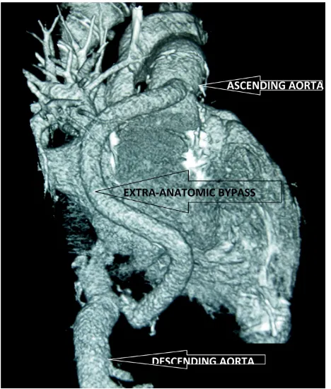
Even if the patient was hemodynamically stable, we decided to operate precocily because of the aortic abscess and the worseness of the infectious lesions of the aortic valve which became more regurgitant. We proceeded with single stage surgery through median sternotomy to repair simultaneously the AC, the ascending aortic an-eurysm and the aortic valve by modified Bentall procedure and ascending-descending aortic extra-anatomic by-pass. After a routine monitoring and anaesthetic procedure, a classical median sternotomy was made. The aneu-rysm involved all the ascending aortic including the sinus of valsalva. The Cardio-pulmonary bypass (CPB) was established by arterial cannulation of the ascending aorta and the left femoral artery to enable whole-body perfu-sion during CPB. The venous cannulation was made by dual-stage right atrial cannulae; and the left ventricle was vented by a left superior pulmonary venting cannulae. The CPB was conducted under total hemodilution and systemic cooling to 30˚C. The ascending aorta was cross-clamped and transversally sectioned; an antegrade cold crystalloid cardioplegia was infused directly into the coronary ostia with topical ice hypothermia. The aor-tic valve was bicuspid and seriously damaged with a residual abscess cavity, and subsequently not suitable for a valve-sparing root replacement. Therefore, the ascending aortic aneurysm and the aortic valve were excised, and a 23-mm ATS valved conduit was inserted and followed by coronary re-implantation. After de-airing, we car-ried out by aortic clamp removal and cardiac defibrillation. Under CPB, the heart was retracted and the scending aorta was approached via a longitudinal incision of the posterior pericardium behind the heart. The de-scending aorta was controlled and a lateral aortic clamp applied. A 14-mm Dacron graft was anastomosed end- to-side to the aorta by continuous 4-0 polypropylene suture, the graft was tunnelled around the right heart border, over the inferior vena cava, brought laterally from the right atrium and anastomosed to the neo-ascending aortic graft (Figure 1). The patient was weaned from CPB uneventfully under low dose of Dobutamin and electro- systolic training by an external pace maker. Total aortic cross-clamp and CPB time was respectively 120 and 210 minutes. There was no residual pressure gradient between radial and femoral arterial line pressure.
postoperative course was marked by a complete atrioventricular block that required a pacemaker implantation with good outcome. The patient was discharged 22 days after surgery in good conditions. After 7 years, the pa-tient regained physical activity and social life quite normal under antihypertensive and oral anticoagulant ther-apy. The clinical examination showed a normal femoral pulse and a normal ankle brachial systolic pressure in-dex at 1.15. The blood pressure in the left arm was 129/84 mmHg and 148/89 mmHg in the left leg. The control CT scan showed the patency of the extra-anatomic bypass. However, there was no change in collateral arteries (Figure 2).
Figure 1. Operative view of the extra-anatomic aortic bypass. The tube is tunnelled around the right heart border.
[image:3.595.198.432.428.703.2]3. Discussion
Aortic Coarctation is a well known congenital cardiovascular malformation, which can remain asymptomatic and unknown until adulthood and result in cardiovascular complications and premature death [4]. It can be asso-ciated in more than 50% of patients to major cardiac pathologies such as bicuspid aortic valve diseases and ven-tricular septal defect, which makes the surgery more challenging [4]. In our case, the AC was well tolerated until the 3rd decade of life, when the patient begun to have shortness of breath probably because of the aortic valve disease.
Three therapeutic strategies have been proposed for the repair of the association of AC and intra-cardiac anomalies. As first choice, we have catheter-based intervention by percutaneous dilatation-stenting of the AC, followed by surgical repair of the aortic valve and the aortic root by median sternotomy. The second alternative is a double-stage procedure using sternotomy and thoracotomy. The third option is a single-stage simultaneous repair of all pathologies by a single incision: median sternotomy [2] [5]-[7].
Certainly, the AC repair can be achieved by transcatheter dilation and stenting [8]. However, this procedure is not practical or suitable in all patients, and long-term outcome data are not available [1] [9]. In our case, the percutaneous dilatation was impossible because of the severity of the AC.
The second therapeutic option is a double-stage surgery, which consists to operate firstly the AC by left tho-racotomy, then the aortic root repair by median sternotomy; or operate the aortic valve then the AC. Mulay re-ported 3 patients who underwent staged procedures by repair of cardiac abnormalities followed after 6 weeks by the AC repair. He suggested that the staged repair allowed myocardial recovery and coronary blood flow redi-stribution during the interval between the two operations; while simultaneous repair may cause a sudden de-crease in systemic vascular resistance. He has also pointed out that the lower-body perfusion was not of great concern during CPB because of the good collateral circulation in adult patients [5] [7].
Finally, the third option is a single-stage surgery for simultaneous repair of the aortic root followed by an ex-tra-anatomic aortic bypass grafting between the ascending aorta and the descending aorta post-coarctation by a median sternotomy.
Certainly, the AC can be treated by the classical anatomic repair through a left thoracotomy. However, it re-quires an extensive dissection to control the aorta and the collateral blood vessels with the risk of important blood loss, pulmonary injuries, recurrent laryngeal or phrenic nerves damage and chylothorax; but the most feared complications still the spinal cord ischemia and paraplegia [1] [10]. The probability of these complica-tions increases with prolonged aortic clamping time and older age [1] [10]. Various techniques have been pro-posed to decrease those potential complications ([5]-[7] such as the use of deep hypothermic circulatory arrest [5] or the extra-anatomic ascending-to-descending aortic bypass [1]-[4] [8] [11]. Ananiadou reported excellent re-sults with no surgical mortality or graft-related complications [3]. Thanks to lateral partial aortic clamp that al-lows continuation of blood flow to the posterior wall of the aorta, paraplegia is rarely described with the ex-tra-anatomic bypass [2] [4] [8]. Connolly has reported no serious complications with this technique and espe-cially no cases of spinal cord ischemia [1].
po-tential complications of graft kinking and narrowing. Additional popo-tential late complications include infection, false aneurysms, and anastomotic dehiscence with pseudoaneurysm formation in patients who have considerable somatic growth after repair [1] [14].
Arterial cannulation is a crucial step for this kind of procedure because of the risk of perfusional problems especially of upper extremities and brain in case of femoral cannulation or hypo-perfusion of the mesenteric and renal arteries in case of cannulation of ascending aorta or the right axillary artery. The double arterial cannula-tion of the ascending aorta and the femoral artery can provide an effective whole-body perfusion and can avoid adverse perfusional sequelae [5] [15] as elucidated our case, we had no complications related to the CPB.
Certainly, the valve-sparing aortic root replacement is an attractive option for treating aortic root pathology with best long-term results, especially in young patients. But in our case, this option was not possible because of the severity of aortic disease with extensive destruction of the leaflets by the infective endocarditis.
The choice of the diameter of the tube to be used for extra-anatomic bypass does not obey to well-known and elaborated rules, it remains rather dependent on the surgeon's preference but generally it ranges between 14 and 20mm with a trend for large calibres. In our case, we chose a tube of 14 mm because the heart was frankly di-lated and occupied the entire pericardial cavity, and also the risk of incongruence between a large tube, and an underdeveloped descending aorta which may cause dehiscence and anastomotic pseudo aneurysm. On the other hand, a small tube exposes the patient to the risk of thrombosis of the graft. The CT control showed a good patency of the extra-anatomic bypass and the arterial doppler showed no residual blood pressure gradient be-tween upper and lower extremities with a normal index of systolic.
4. Conclusion
The complex pathology of AC with aortic root aneurysm, presenting in adulthood, poses a tactical challenge [3]. The single-stage procedure, with combination of an extra-anatomic aortic bypass and concomitant cardiovascu-lar disorders repair via median sternotomy, might be a useful and a safe alternative, which can be performed ef-ficiently with low morbidity and mortality [1] [3].
5. Disclosures
All authors declare that there is no conflict of interest in this article.
The authors declare that the patient has given his approval and consent to the publication of this article.
References
[1] Connolly, H.M., Schaff, H.V., Izhar, U., Dearani, J.A., Warnes, C.A. and Orszulak, T.A. (2001) Posterior Pericardial Ascending-to-Descending Aortic Bypass. An Alternative Surgical Approach for Complex Coarctation of the Aorta.
Circulation, 104, I133-I137. http://dx.doi.org/10.1161/hc37t1.094897
[2] Morris, R.J., Samuels, L.E. and Brockman, S.K. (1998) Total Simultaneous Repair of Coarctation and Intracardiac Pa-thology in Adult Patients. The Annals of Thoracic Surgery, 65, 1698-1702.
http://dx.doi.org/10.1016/S0003-4975(98)00291-4
[3] Ananiadou, O.G., Koutsogiannidis, C., Ampatzidou, F. and Drossos, G.E. (2012) Aortic Root Aneurysm in an Adult Patient with Aortic Coarctation: A Single-Stage Approach. Interactive CardioVasc Thoracic Surgery, 15, 534-536.
http://dx.doi.org/10.1093/icvts/ivs235
[4] McKellar, S.H., Schaff, H.V., Dearani, J.A., Daly, R.C., Mullany, C.J., Orszulak, T.A., et al. (2007) Intermediate-Term Results of Ascending-Descending Posterior Pericardial Bypass of Complex Aortic Coarctation. The Journal of
Tho-racic and Cardiovascular Surgery, 133, 1504-1509. http://dx.doi.org/10.1016/j.jtcvs.2006.11.011
[5] Narin, C., Ege, E., Orhan, A. and Yeniterzi, M. (2008) Repair of Coarctation-Related Aortic Arch Aneurysm and Ven-tricular Septal Defect in an Adolescent. Texas Heart Institute Journal, 35, 466-469.
[6] Yilmaz, M., Polat, B. and Saba, D. (2006) Single-Stage Repair of Adult Aortic Coarctation and Concomitant Cardi-ovascular Pathologies: A New Alternative Surgical Approach. Journal of Cardiothoracic Surgery, 1, 18.
http://dx.doi.org/10.1186/1749-8090-1-18
[7] Mulay, A.V., Ashraf, S. and Watterson, K.G. (1997) Two-Stage Repair of Adult Coarctation of the Aorta with Conge-nital Valvular Lesions. The Annals of Thoracic Surgery, 64, 1309-1311.
http://dx.doi.org/10.1016/S0003-4975(97)00814-X
Bypass with Valve-Sparing Root Replacement for Coarctation with Aortic Root Aneurysm. The Journal of Thoracic
and Cardiovascular Surgery, 143, 514-515. http://dx.doi.org/10.1016/j.jtcvs.2011.07.047
[9] Weber, H. and Cyran, S. (1999) Initial Results and Clinical Follow-Up after Balloon Angioplasty for Native Coarcta-tion. American Journal of Cardiology, 84, 113-116. http://dx.doi.org/10.1016/S0002-9149(99)00207-6
[10] Grinda, J.M., Macé, L., Dervanian, P., Folliguet, T.A. and Neveux, J.Y. (1995) Bypass Graft for Complex Forms of Isthmic Aortic Coarctation in Adults. The Annals of Thoracic Surgery, 60, 1299-1302.
http://dx.doi.org/10.1016/0003-4975(95)00557-2
[11] Wang, R., Sun, L.Z., Hu, X.P., Ma, W.G., Chang, Q., Zhu, J.M., et al. (2010) Treatment of Complex Coarctation and Coarctation with Cardiac Lesions Using Extra-Anatomic Aortic Bypass. Journal of Vascular Surgery, 51, 1203-1208.
http://dx.doi.org/10.1016/j.jvs.2009.12.027
[12] Vijayanagar, R., Natarajan, P., Eckstein, P.F., Bognolo, D.A. and Toole, J.C. (1980) Aortic Valvular Insufficiency and Postductal Aortic Coarctation in the Adult: Combined Surgical Management through Median Sternotomy: A New Sur-gical Approach. The Journal of Thoracic and Cardiovascular Surgery, 79, 266-268.
[13] Powell, W., Adam, P. and Cooley, D. (1983) Repair of Coarctation of the Aorta with Intra-Cardiac Repair. Texas Heart
Institute Journal, 10, 409-413.
[14] Pethig, K., Wahlers, T., Tager, S. and Borst, H.G. (1996) Perioperative Complications in Combined Aortic Valve Re-placement and Extra-Anatomic Ascending-Descending Bypass. The Annals of Thoracic Surgery, 61, 1724-1726.
http://dx.doi.org/10.1016/0003-4975(96)00140-3
[15] Heper, G., Yorukoglu, Y. and Korkmaz, M.E. (2005) Clinical and Hemodynamic Follow-Up of a Patient after Opera-tion for DissecOpera-tion of an Ascending Aortic Aneurysm Secondary to CoarctaOpera-tion of the Aorta. International Heart
