J
OURNAL OFC
LINICALM
ICROBIOLOGY,
0095-1137/97/$04.00
1
0
Sept. 1997, p. 2337–2341
Vol. 35, No. 9
Copyright © 1997, American Society for Microbiology
Identification of Clinically Relevant Viridans Group
Streptococci to the Species Level by PCR
FABIEN GARNIER, GUY GERBAUD, PATRICE COURVALIN,
ANDMARC GALIMAND*
Unite´ des Agents Antibacte´riens and National Reference Center for Antibiotics,
Institut Pasteur, 75724 Paris Cedex 15, France
Received 13 September 1996/Returned for modification 9 December 1996/Accepted 25 June 1997
A PCR assay that allows identification of clinically relevant viridans group streptococci (
Streptococcus
gordonii
,
S. mitis
,
S. mutans
,
S. oralis
,
S. salivarius
, and
S. sanguis
) to the species level and identification of milleri
group streptococci (
S. anginosus
,
S. constellatus
, and
S. intermedius
) to the group level was developed. This assay
was based on specific amplification of internal fragments of genes encoding
D-alanine:
D-alanine ligases which
are species specific and ubiquitous in prokaryotes possessing peptidoglycan. The specificity of this assay was
tested on 9 reference strains and 91 characterized clinical isolates. This assay offers a specific and rapid
alternative to phenotypic or DNA-DNA hybridization methods for identification of clinically relevant viridans
group streptococci.
Viridans group streptococci form an important part of the
normal flora of the human oral cavity. They are responsible for
several infections including purulent infections (14),
endocar-ditis (7), septicemia (4), and meningitis (3). Viridans
strepto-cocci do not possess a specific group antigen and show variable
reactions with Lancefield antisera (11). Their identification is
based on different physiological and biochemical
characteris-tics, but conventional phenotypic identification methods are
sometimes unable to differentiate established species. First,
not all strains in a species may be positive for a common trait
(2, 19), and second, the same strain may give different results
with repeated tests in the absence of changes in the
corre-sponding genes (15, 25). Thus, rapid systems using standard
phenotypic tests, such as API-20STREP or rapid ID 32 Strep,
that are used in clinical laboratories are not totally satisfactory
for accurate identification at the species level (16, 18). Species
identification of viridans streptococci is useful in cases of
in-fective endocarditis when the patient has relapsed and in cases
of positive blood cultures and in assessing the involvement of
a given strain in an infection.
PCR has been extensively applied to species identification of
infectious agents (8, 13, 21). PCR allows amplification of a
pre-selected DNA region and is a highly specific and sensitive
tech-nique (20). In many instances, the target genes are involved in
pathogenicity (13). In other cases, the target is a random cloned
fragment from a genomic library selected by differential
hybrid-ization to the pathogen and its close relatives (23).
In this study, we have selected the gene encoding a
D-ala-nine:
D-alanine (
D-Ala:
D-Ala) ligase which is species specific
and ubiquitous in prokaryotes possessing peptidoglycan. The
D
-Ala:
D-Ala ligase catalyzes synthesis of the terminal dipeptide
D-alanine–
D-alanine of peptidoglycan precursors (26). We
have developed a PCR assay which allows identification of six
species (
Streptococcus gordonii
,
S. mitis
,
S. mutans
,
S. oralis
,
S.
salivarius
, and
S. sanguis
) to the species level and identification
of three species (
S. anginosus
,
S. constellatus
, and
S.
interme-dius
) to the group level.
MATERIALS AND METHODS
Bacterial strains, plasmids, and culture conditions.The reference strains of
Streptococcusspp. used in this study were as follows:S. anginosusATCC 33397,
[image:1.612.61.558.539.674.2]S. bovisNCTC 8177,S. constellatusATCC 27823,S. gordoniiATCC 10558,S.
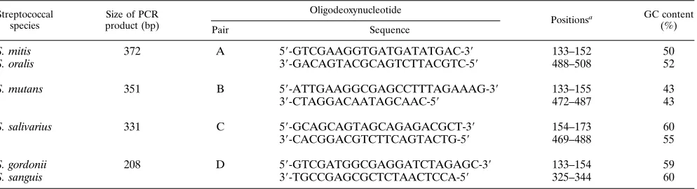
TABLE 1. Oligodeoxynucleotide primers for the first PCR
Streptococcal species
Size of PCR product (bp)
Oligodeoxynucleotide
Positionsa GC content
(%)
Pair Sequence
S. mitis
372
A
5
9
-GTCGAAGGTGATGATATGAC-3
9
133–152
50
S. oralis
3
9
-GACAGTACGCAGTCTTACGTC-5
9
488–508
52
S. mutans
351
B
5
9
-ATTGAAGGCGAGCCTTTAGAAAG-3
9
133–155
43
3
9
-CTAGGACAATAGCAAC-5
9
472–487
43
S. salivarius
331
C
5
9
-GCAGCAGTAGCAGAGACGCT-3
9
154–173
60
3
9
-CACGGACGTCTTCAGTACTG-5
9
469–488
55
S. gordonii
208
D
5
9
-GTCGATGGCGAGGATCTAGAGC-3
9
133–154
59
S. sanguis
3
9
-TGCCGAGCGCTCTAACTCCA-5
9
325–344
60
aPositions were derived from the alignment in Fig. 1.
* Corresponding author. Mailing address: Unite
´ des Agents
Anti-bacte
´riens, Institut Pasteur, 28, rue du Docteur Roux, 75724 Paris
Cedex 15, France. Phone: (33) 01 45 68 83 18. Fax: (33) 01 45 68 83 19.
E-mail: galimand@pasteur.fr.
2337
on May 15, 2020 by guest
http://jcm.asm.org/
intermediusATCC 27335,S. mitisNCTC 12261,S. mutansNCTC 10449,S. oralis
NCTC 7864,S. salivariusATCC 9758, andS. sanguisNCTC 7863. A total of 91
Streptococcus strains from the bioMe´rieux collection (La Balme-les-Grottes,
France) were also tested. The species and number of the 91 strains tested were as follows:S. anginosus, 9;S. constellatus, 10;S. gordonii, 10;S. intermedius, 6;S.
mitis, 10;S. mutans, 10;S. oralis, 10;S. salivarius, 10;S. sanguis, 10; andS.
vestibularis, 6. Plasmid pCRII (Invitrogen, San Diego, Calif.) was used for the
cloning of the PCR products.Escherichia coliINVaF9One Shot (Invitrogen) was the host strain for recombinant plasmids. The recombinant plasmids were pAT437 (ddlgene fromS. bovis[ddlS. bovis]), pAT438 (ddlS. gordonii), pAT439
(ddlS. mitis), pAT440 (ddlS. mutans), pAT441 (ddlS. oralis), pAT442 (ddlS. salivarius),
pAT443 (ddlS. sanguis), pAT444 (ddlS. anginosus), pAT445 (ddlS. constellatus), and
pAT446 (ddlS. intermedius). All strains were grown at 37°C in brain heart infusion
broth and on agar (Difco Laboratories, Detroit, Mich.) not supplemented or supplemented with horse blood (5% [vol/vol]). For milleri group streptococci, the incubation was under anaerobic conditions.
DNA manipulation. Total DNA from streptococci was prepared by the cetyldimethylethyl-ammonium bromide method (1). Degenerate oligode-oxynucleotides V1 [GGIGA(A/G)GA(T/C)GGI(T/A)(C/G)I(T/C/A)TICA(A/ G)GG] and V2 [TT(A/G)TGI(T/A/G)AIGGICCIAA(A/G)TG] (10), where I stands for inosine, are complementary to sequences encoding conserved amino acid motifs inD-Ala:D-Ala ligases ofE. coli(22, 28) and the related glycopeptide resistance enzyme VanA (9). Amplification of DNA fragments by PCR using ca. 50 ng of template DNA and primers V1 and V2 at the concentration of 0.1mM each in a total volume of 100ml was performed with a DNA thermal cycler (model 2400; Perkin-Elmer Cetus, Emeryville, Calif.) as described previously (10). TheTaqDNA polymerase was purchased from Amersham Life Science (Cleveland, Ohio). The PCR conditions were as follows: 2 min at 94°C for the first step; 30 cycles, with 1 cycle consisting of 1 min at 94°C, 1 min at 54°C, and 1 min at 72°C; and 10 min at 72°C for the last step. The PCR products were purified by agarose gel electrophoresis followed by extraction from the cut-out low-melting-point agarose block with the Sephaglas Kit (Pharmacia, Uppsala, Sweden). Recombinant plasmids were prepared by the Wizard Miniprep proce-FIG. 1. Comparison of the sequences of fragments within genes coding for
D-Ala:D-Ala ligases fromS. anginosus(ATCC 33397),S. constellatus(ATCC
27823),S. gordonii(ATCC 10558),S. intermedius(ATCC 27335),S. mitis(NCTC 12261),S. mutans(NCTC 10449),S. oralis(NCTC 7864),S. salivarius(ATCC 9758), andS. sanguis(NCTC 7863). Asterisks indicate nucleotides that were identical in all sequences. Dots indicate gaps introduced to optimize alignment.
on May 15, 2020 by guest
http://jcm.asm.org/
dure (Promega, Madison, Wis.). The DNA sequences of PCR products were determined by the dideoxynucleotide chain terminator technique (24) by using universal or specific oligodeoxynucleotides (Unite´ de Chimie Organique, Institut Pasteur, Paris, France) as the primers, [a-35S]dATP (Amersham Radiochemical
Centre, Amersham, England), and T7 DNA polymerase (T7 Sequencing Kit; Pharmacia) according to the manufacturer’s recommendations. For Southern hybridization, DNA was transferred by vacuum onto Nytran membranes (Schlei-cher and Schuell, Dassel, Germany). Prehybridization and hybridization were performed under stringent conditions at 68°C in 0.1% sodium dodecyl sulfate– 0.05% nonfat dry milk–63SSC (13SSC is 0.15 M NaCl plus 0.015 M sodium citrate) for 3 and 18 h, respectively. Probes were labeled with [a-32P]dCTP
(Amersham Radiochemical Centre) by the nick translation procedure (Nick Translation Kit; Amersham International, Little Chalfont, England).
Sequence analysis.Nucleotide sequences were analyzed with Genetics Com-puter Group software (6), and the phylogenetic analysis was performed with the PHYLIP program package (12).
Nucleotide sequence accession numbers.The sequences were submitted to GenBank and were assigned the following accession numbers: U69162 (ddlS. bovis), U69163 (ddlS. gordonii), U69164 (ddlS. mitis), U69165 (ddlS. mutans), U69166
(ddlS. oralis), U69167 (ddlS. salivarius), U69168 (ddlS. sanguis), U91912 (ddlS. anginosus),
U91913 (ddlS. intermedius), and U91914 (ddlS. constellatus).
RESULTS AND DISCUSSION
Design of oligodeoxynucleotides.
Internal portions (ca. 600
bp) of the genes coding for
D-Ala:
D-Ala ligases in nine species
of streptococci (
S. anginosus
,
S. constellatus
,
S. gordonii
,
S.
intermedius
,
S. mitis
,
S. mutans
,
S. oralis
,
S. salivarius
, and
S.
sanguis
) were amplified by PCR with oligodeoxynucleotides V1
and V2 and cloned into the pCRII vector. Southern
hybridiza-tion with total DNA of each strain was carried out to confirm
the origins of the PCR products (data not shown) that were
subsequently sequenced on both strands. Sequence
compari-son indicated that the inserts corresponded to internal portions
of
ddl
genes.
[image:3.612.56.557.82.242.2]The partial sequences of the nine
ddl
genes were aligned
(Fig. 1) and showed high degrees of identity. Pairs of
oligode-oxynucleotides, each intended to prime amplification of a
frag-ment within a
ddl
gene, were selected in nonconserved regions.
FIG. 2. First-PCR analysis of total DNA from reference and wild strains of viridans group streptococci. Lanes: 1,S. mitisNCTC 12261; 2,S. oralisNCTC 7864; 3,
S. mutansNCTC 10449; 4,S. salivariusATCC 9758; 5,S. gordoniiATCC 10558; 6,S. sanguisNCTC 7863; 7 and 8,S. mitis; 9 and 10,S. oralis; 11 and 12,S. mutans;
[image:3.612.106.509.492.680.2]13 and 14,S. salivarius; 15 and 16,S. gordonii; 17 and 18,S. sanguis; M, bacteriophagelDNA (Pharmacia) digested withPstI used as size standards. PCR products were resolved by electrophoresis on a 2% agarose–Tris–borate–EDTA gel containing 0.5mg of ethidium bromide per ml. The sizes of the PCR products (in base pairs) are indicated to the sides of the gel.
TABLE 2. Oligodeoxynucleotide primers for the second PCR
Streptococcal group and species
Size of PCR product (bp)
Oligodeoxynucleotide
Positionsa GC content
(%)
Pair Sequence
Group A or milleri group
217
E
5
9
-TGCAGAAGTAGAGGCAAATC-3
9
162–181
45
3
9
-TTCCTCGGTTTTCGTCAACCG-5
9
362–382
52
Group B
S. mitis
259
F
5
9
-TGAAATCGAGGTTGGCCTAC-3
9
333–352
50
3
9
-TTCCC(G/T)CTCTAAAAGGATTTGC-5
9
571–592
45
S. oralis
563
G
5
9
-CTTATGTCGGCTGCAATATCC-3
9
23–43
47
3
9
-TTCCC(G/T)CTCTAAAAGGATTTGC-5
9
571–592
45
Group C
S. gordonii
260
H
5
9
-GTCGATGGCGAGGATCTAGAGC-3
9
133–154
59
3
9
-CAGAAGGTCCCCTTCAACAA-5
9
377–396
50
S. sanguis
374
I
5
9
-GTCGATGGCGAGGATCTAGAGC-3
9
133–154
59
3
9
-GACTACGCAGTTTTACGTCTC-5
9
490–510
47
aPositions were derived from the alignment in Fig. 1.
V
OL. 35, 1997
IDENTIFICATION OF VIRIDANS GROUP STREPTOCOCCI BY PCR
2339
on May 15, 2020 by guest
http://jcm.asm.org/
Primers of similar sizes and with a GC content ranging from 43
to 60% were designed to avoid variations in annealing
temper-ature and to allow their simultaneous use in a single reaction
mixture. However, due to the small sizes of the internal
frag-ments sequenced (600 bp), the high degrees of identity for the
nine sequences, and the fact that each amplification product
should be assigned to a species on the basis of its size, a
two-stage PCR appeared necessary. In the first PCR with
oli-godeoxynucleotide pairs A to D, the amplification products
obtained with pairs B and C could be assigned to a single
species (Table 1). Pair A amplified
S. oralis
and
S. mitis
, and
pair D amplified both
S. gordonii
and
S. sanguis
. A second PCR
using primers F and G or H and I (Table 2) allowed the
differentiation of
S. oralis
from
S. mitis
or
S. gordonii
from
S.
sanguis
, respectively, whereas pair E (Table 2), used alone,
allowed identification of the species from the milleri group.
PCR experiments.
PCRs were performed with DNA from
every reference strain as a template. Occurrence of nonspecific
bands led us to modify the PCR conditions as follows: (i) for
the first PCR and for the second PCR with oligonucleotide
pairs F and G or H and I, 2 min at 94°C for the first step; 20
cycles, with 1 cycle consisting of 1 min at 94°C, 1 min at 56°C,
and 1 min at 72°C; and 10 min at 72°C for the last step; and (ii)
for the second PCR with oligonucleotide pair E, 2 min at 94°C
for the first step; 30 cycles, with 1 cycle consisting of 1 min at
94°C, 1 min at 50°C, and 1 min at 72°C; and 10 min at 72°C for
the last step.
The sizes of the amplification products obtained under these
conditions differed sufficiently to allow identification of the
reference strains (data not shown).
[image:4.612.352.516.68.296.2]We finally investigated our strategy by testing 91
character-ized strains of viridans group streptococci. The PCR results
confirmed the identification of 60 strains (10
S. gordonii
, 10
S.
mitis
, 10
S. mutans
, 10
S. oralis
, 10
S. salivarius
, and 10
S.
sanguis
strains) to the species level (Fig. 2 and 3A and B and
data not shown) and the assignment of 25 strains (10
S.
angi-nosus
, 9
S. constellatus
, and 6
S. intermedius
strains) to the
milleri group (Fig. 3C and data not shown), whereas the 6
strains of
S. vestibularis
were identified as
S. salivarius
(data not
shown). A relatively high degree of relatedness has been
ob-served by DNA-DNA hybridization between strains of
S.
ves-tibularis
and
S. salivarius
(5, 27). These data are consistent with
FIG. 3. Second-PCR analysis of total DNA from wild and reference strainsof viridans group streptococci. (A) PCR with oligonucleotide pairs F and G. Lanes: 1,S. mitisNCTC 12261; 2 to 4,S. mitis; 5,S. oralisNCTC 7864; 6 to 8,S.
oralis. (B) PCR with oligonucleotide pairs H and I. Lanes: 1,S. gordoniiATCC
10558; 2 to 4,S. gordonii; 5,S. sanguisNCTC 7863; 6 to 8,S. sanguis. (C) PCR with oligonucleotide pair E. Lanes: 1,S. anginosusATCC 33397; 2,S. constellatus
ATCC 27823; 3,S. intermediusATCC 27335; 4 and 5,S. anginosus; 6 and 7,S.
constellatus; 8 and 9,S. intermedius. In all panels, lanes M contained
[image:4.612.56.297.77.569.2]bacterio-phage lDNA (Pharmacia) digested withPstI used as size standards. PCR products were resolved by electrophoresis on a 2% agarose–Tris–borate–EDTA gel containing 0.5mg of ethidium bromide per ml. The sizes of the PCR products (in base pairs) are indicated to the sides of the gels.
FIG. 4. Phylogenetic relationship among streptococci. The tree was con-structed by the neighbor-joining method and slightly modified taking into ac-count the results of maximum-parsimony and bootstrapping analysis.
on May 15, 2020 by guest
http://jcm.asm.org/
the observation that the oligonucleotides designed for
S.
sali-varius
also amplified a fragment of total DNA from
S.
vestibu-laris
. However, to the best of our knowledge,
S. vestibularis
has
not been reported to be responsible for purulent infections,
endocarditis, septicemia, or meningitis and is thus unlikely to
be isolated from foci of infection.
Phylogenetic analysis.
The amino acid sequence deduced
from the DNA region between oligonucleotides V1 and V2 of
10 species (
S. anginosus
,
S. bovis
,
S. constellatus
,
S. gordonii
,
S.
intermedius
,
S. mitis
,
S. mutans
,
S. oralis
,
S. salivarius
, and
S.
sanguis
) was used for phylogenetic analysis (Fig. 4). The
phy-logeny obtained was compared with that derived from 16S
rRNA sequences (17). The topologies of the two trees
ob-tained with the neighbor-joining method (12) were
superim-posable except for
S. gordonii
and
S. constellatus.
The
differ-ence concerns the position of nodes of these two species.
Identification of viridans group streptococci to the species
level is required for certain infections. DNA-DNA
hybridiza-tion with the type strain is the “gold standard” technique for
identification to the species level. However, this method
re-quires radioisotopes and involves complex procedures, and its
application is thus limited to research or reference
laborato-ries. Our PCR assay provides a specific and rapid alternative to
phenotypic or DNA-DNA hybridization methods for
identifi-cation of clinically relevant viridans group streptococci to the
species or group level within 48 h from the time of isolation of
the microorganism.
ACKNOWLEDGMENT
We thank bioMe
´rieux (La Balme-les-Grottes, France) for the gift of
Streptococcus
strains.
REFERENCES
1.Ausubel, F. M., R. Brent, R. E. Kingston, D. D. Moore, J. G. Seidman, J. A. Smith, and K. Struhl.1992. Current protocols in molecular biology, vol 1, p. 2–33. Wiley, New York, N.Y.
2.Beighton, D., J. M. Hardie, and R. A. Whiley.1991. A scheme for the identification of viridans streptococci. J. Med. Microbiol.35:367–372. 3.Beighton, D., A. D. Carr, and B. A. Oppenheim.1994. Identification of
viridans streptococci associated with bacteraemia in neutropenic cancer pa-tients. J. Med. Microbiol.40:202–204.
4.Bochud, P.-Y., P. Eggiman, T. Calandra, G. Van Melle, L. Saghafi, and P. Francioli.1994. Bacteremia due to viridans streptococcus in neutropenic patients with cancer: clinical spectrum and risk factors. Clin. Infect. Dis. 18:25–31.
5.Coykendall, A. L.1989. Classification and identification of the viridans strep-tococci. Clin. Microbiol. Rev.2:315–328.
6.Devereux, J., P. Haerberli, and O. Smithies.1984. A comprehensive set of sequence analysis programs for the VAX. Nucleic Acids Res.12:387–395. 7.Douglas, C. W. I., J. Heath, K. K. Hampton, and F. E. Preston.1993. Identity
of viridans streptococci isolated from cases of infective endocarditis. J. Med. Microbiol.39:179–182.
8.Dutka-Malen, S., S. Evers, and P. Courvalin.1995. Detection of glycopep-tide resistance genotypes and identification to the species level of clinically relevant enterococci by PCR. J. Clin. Microbiol.33:24–27.
9.Dutka-Malen, S., C. Molinas, M. Arthur, and P. Courvalin.1990. The VanA glycopeptide resistance protein is related toD-alanine:D-alanine ligase cell-wall biosynthesis enzymes. Mol. Gen. Genet.224:364–372.
10. Dutka-Malen, S., C. Molinas, M. Arthur, and P. Courvalin.1992. Sequence of thevanCgene ofEnterococcus gallinarumBM4174 encoding aD-alanine:
D-alanine ligase-related protein necessary for vancomycin resistance. Gene 112:53–58.
11. Facklam, R. R.1977. Physiological differentiation of viridans streptococci. J. Clin. Microbiol.5:184–201.
12. Felsenstein, J.1993. PHYLIP version 3.5c. University of Washington, Seat-tle.
13. Frankel, G., J. A. Giron, J. Valmassoi, and G. K. Schoolnik.1989. Multigene amplification: simultaneous detection of three virulence genes in diarrhoeal stool. Mol. Microbiol.3:1729–1734.
14. Gossling, J.1988. Occurrence and pathogenicity of theStreptococcus milleri
group. Rev. Infect. Dis.10:257–285.
15. Hillman, J. D., S. W. Andrews, S. Painter, and P. Stashenko.1989. Adap-tative changes in a strain ofStreptococcus mutansduring colonization of the human oral cavity. Microb. Ecol. Health Dis.2:231–239.
16. Hinnebusch, C. J., D. M. Nikolai, and D. A. Bruckner.1991. Comparison of API Rapid STREP, Baxter Microscan Rapid Pos ID panel, BBL Minitek Differential Identification system, IDS RapID STR system, and Vitek GPI to conventional biochemical tests for identification of viridans streptococci. Am. J. Clin. Pathol.96:459–463.
17. Kawamura, Y., X. G. Hou, F. Sultana, H. Miura, and T. Ezaki.1995. De-termination of 16S rRNA sequences ofStreptococcus mitisandStreptococcus
gordoniiand phylogenetic relationships among members of the genus
Strep-tococcus. Int. J. Syst. Bacteriol.45:406–408.
18. Kikuchi, K., T. Enari, K. I. Totsuka, and K. Shimizu.1995. Comparison of phenotypic characteristics, DNA-DNA hybridization results, and results with a commercial rapid biochemical and enzymatic reaction system for identifi-cation of viridans group streptococci. J. Clin. Microbiol.33:1215–1222. 19. Kilian, M., L. Mikkelsen, and J. Henrichsen.1989. Taxonomic study of
viridans streptococci: description of Streptococcus gordonii sp. nov. and emended descriptions ofStreptococcus sanguis (White and Niven 1946),
Streptococcus oralis(Bridge and Sneath 1982), andStreptococcus mitis
(An-drewes and Horder 1906). Int. J. Syst. Bacteriol.39:471–484.
20. Mullis, K. B., and F. A. Faloona.1987. Specific synthesis of DNA in vitro via a polymerase chain reaction. Methods Enzymol.155:335–350.
21. Oyofo, B. A., S. A. Thornton, D. H. Burr, T. J. Trust, O. R. Pavlovskis, and P. Guerry.1992. Specific detection of Campylobacter jejuniand
Campy-lobacter coliby using polymerase chain reaction. J. Clin. Microbiol.30:2613–
2619.
22. Robinson, A. C., D. J. Kenan, J. Sweeney, and W. D. Donachie.1986. Further evidence for overlapping transcriptional units in anEscherichia coli cell envelope-cell division gene cluster: DNA sequence and transcriptional or-ganization of theddl ftsQregion. J. Bacteriol.167:809–817.
23. Rosa, P. A., and T. G. Schwan.1989. A specific and sensitive assay for the Lyme disease spirocheteBorrelia burgdorferiusing the polymerase chain reaction. J. Infect. Dis.160:1018–1029.
24. Sanger, F., S. Nicklen, and A. R. Coulson.1977. DNA sequencing with chain-terminating inhibitors. Proc. Natl. Acad. Sci. USA74:5463–5467. 25. Tardif, G., M. C. Sulavik, G. W. Jones, and D. B. Clewell.1989. Spontaneous
switching of the sucrose-promoted colony phenotype inStreptococcus san-guis. Infect. Immun.57:3945–3948.
26. Walsh, C. T.1989. Enzymes in theD-alanine branch of bacterial cell wall
peptidoglycan assembly. J. Biol. Chem.264:2393–2396.
27. Whiley, R. A., and J. M. Hardie.1988.Streptococcus vestibularissp. nov. from the human oral cavity. Int. J. Syst. Bacteriol.38:335–339.
28. Zawadzke, L. E., T. D. H. Bugg, and C. T. Walsh.1991. Existence of two
D-alanine:D-alanine ligases inEscherichia coli: cloning and sequencing of the
ddlAgene, and purification and characterization of the DdlA and DdlB enzymes. Biochemistry30:1673–1682.