Role of AMP-activated protein kinase in
mechanism of metformin action
Gaochao Zhou, … , Laurie J. Goodyear, David E. Moller
J Clin Invest.
2001;
108(8)
:1167-1174.
https://doi.org/10.1172/JCI13505
.
Metformin is a widely used drug for treatment of type 2 diabetes with no defined cellular
mechanism of action. Its glucose-lowering effect results from decreased hepatic glucose
production and increased glucose utilization. Metformin’s beneficial effects on circulating
lipids have been linked to reduced fatty liver. AMP-activated protein kinase (AMPK) is a
major cellular regulator of lipid and glucose metabolism. Here we report that metformin
activates AMPK in hepatocytes; as a result, acetyl-CoA carboxylase (ACC) activity is
reduced, fatty acid oxidation is induced, and expression of lipogenic enzymes is
suppressed. Activation of AMPK by metformin or an adenosine analogue suppresses
expression of SREBP-1, a key lipogenic transcription factor. In metformin-treated rats,
hepatic expression of SREBP-1 (and other lipogenic) mRNAs and protein is reduced;
activity of the AMPK target, ACC, is also reduced. Using a novel AMPK inhibitor, we find
that AMPK activation is required for metformin’s inhibitory effect on glucose production by
hepatocytes. In isolated rat skeletal muscles, metformin stimulates glucose uptake
coincident with AMPK activation. Activation of AMPK provides a unified explanation for the
pleiotropic beneficial effects of this drug; these results also suggest that alternative means of
modulating AMPK should be useful for the treatment of metabolic disorders.
Article
Find the latest version:
Introduction
Metformin is widely used for the ther-apy of type 2 diabetes mellitus (DM2) (1). Metformin ameliorates hyper-glycemia without stimulating insulin secretion, promoting weight gain, or causing hypoglycemia (2, 3). In addi-tion, metformin has beneficial effects on circulating lipids linked to increased cardiovascular risk (2–4).
Although used as a drug since 1957, the mechanism(s) by which metformin lowers glucose and lipids remains an enigma. Two effects, decreased hepatic glucose production (2, 5, 6) and increased skeletal myocyte glucose uptake (7, 8), have been implicated as major contributors to glucose-lowering
efficacy. Metformin also decreases hepatic lipids in obese mice (9). Met-formin is, however, a low-potency com-pound that is used at high doses, result-ing in only modest net efficacy; in addition, significant side effects can occur (2). Thus, an understanding of the molecular basis for metformin’s effects on glucose and lipid homeostasis is a critical focus of research directed toward improved therapeutic approaches to DM2 and related disorders.
AMP-activated protein kinase (AMPK) provides a candidate target capable of mediating the beneficial metabolic effects of metformin. AMPK is a multisubunit enzyme that is rec-ognized as a major regulator of lipid
biosynthetic pathways due to its role in the phosphorylation and inactiva-tion of key enzymes such as acetyl-CoA carboxylase (ACC) (10). More recent data strongly suggest that AMPK has a wider role in metabolic regulation (10, 11): this includes fatty acid oxidation, muscle glucose uptake (12–14), expres-sion of cAMP-stimulated gluco-neogenic genes such as PEPCK and G6Pase (15), and glucose-stimulated genes associated with hepatic lipogen-esis, including fatty acid synthase (FAS), Spot-14 (S14), and L-type pyru-vate kinase (16). Chronic activation of AMPK may also induce the expression of muscle hexokinase and glucose transporters (Glut4), mimicking the effects of extensive exercise training (17). Thus, it has been predicted that AMPK activation would be a good approach to treat DM2 (11). In this report we tested the hypothesis that activation of AMPK mediates the ben-eficial metabolic effects of metformin.
Methods
Measurements of AMPK, ACC, and fatty acid oxidation in primary hepatocytes. Hepatocytes were isolated from male Sprague Dawley (SD) rats by collage-nase digestion (18). For the AMPK assay, cells were seeded in six-well plates at 1.5 ×10⁶cells/well in DMEM
contain-ing 100 U/ml penicillin, 100 µg/ml streptomycin, 10% FBS, 100 nM insulin,
100 nM dexamethasone, and 5 µg/ml
transferrin for 4 hours. Cells were then cultured in serum-free DMEM for 16 hours followed by treatment for 1 hour or 7 hours with control medium, 5-amino-imidazole carboxamide
ribo-Role of AMP-activated protein kinase
in mechanism of metformin action
Gaochao Zhou,
1Robert Myers,
1Ying Li,
1Yuli Chen,
1Xiaolan Shen,
1Judy Fenyk-Melody,
1Margaret Wu,
1John Ventre,
1Thomas Doebber,
1Nobuharu Fujii,
2Nicolas Musi,
2Michael F. Hirshman,
2Laurie J. Goodyear,
2and David E. Moller
11Departments of Molecular Endocrinology, Metabolic Disorders, and Comparative Medicine, Merck Research Laboratories, Rahway, New Jersey, USA
2Joslin Diabetes Center and Harvard Medical School, Boston, Massachusetts, USA
Address correspondence to: Gaochao Zhou, Merck Research Laboratories, Rahway, New Jersey 07065, USA. Phone: (732) 594-4782; Fax: (732) 594-5700; E-mail: gaochao_zhou@merck.com.
Received for publication June 13, 2001, and accepted in revised form August 28, 2001.
Metformin is a widely used drug for treatment of type 2 diabetes with no defined cellular mechanism of action. Its glucose-lowering effect results from decreased hepatic glucose production and increased glucose utilization. Met-formin’s beneficial effects on circulating lipids have been linked to reduced fatty liver. AMP-activated protein kinase (AMPK) is a major cellular regulator of lipid and glucose metabolism. Here we report that metformin activates AMPK in hepatocytes; as a result, acetyl-CoA carboxylase (ACC) activity is reduced, fatty acid oxidation is induced, and expression of lipogenic enzymes is suppressed. Activation of AMPK by metformin or an adenosine analogue suppresses expres-sion of SREBP-1, a key lipogenic transcription factor. In metformin-treated rats, hepatic expression of SREBP-1 (and other lipogenic) mRNAs and protein is reduced; activity of the AMPK target, ACC, is also reduced. Using a novel AMPK inhibitor, we find that AMPK activation is required for metformin’s inhibitory effect on glucose production by hepatocytes. In isolated rat skeletal muscles, metformin stimulates glucose uptake coincident with AMPK activation. Acti-vation of AMPK provides a unified explanation for the pleiotropic beneficial effects of this drug; these results also suggest that alternative means of modu-lating AMPK should be useful for the treatment of metabolic disorders.
This article was published online in advance of the print edition. The date of publication is available from the JCI website, http://www.jci.org.
J. Clin. Invest.108:1167–1174 (2001). DOI:10.1172/JCI200113505.
Rapid Publication
side (AICAR), or metformin at concen-trations indicated. For a 39-hour treat-ment, cells for both control and met-formin (10 or 20 µM) groups were cultured in DMEM plus 5% FBS and 100 nM insulin, and the fresh control and metformin-containing medium were replaced every 12 hours (last medi-um change was 3 hours before harvest). After treatment, the cells were directly lysed in digitonin-containing and phos-phatase inhibitor–containing buffer A (19), followed by precipitation with ammonium sulfate at 35% saturation. AMPK activity was determined by meas-urement of phosphorylation of a syn-thetic peptide substrate, SAMS
(HMR-SAMSGLHLVKRR) (20). For ACC assay, the 35% ammonium sulfate precipitate from digitonin-lysed hepatocytes (4 µg each) was used for determination of ACC activity via 14CO
2fixation in the
presence of 20 mM citrate as done pre-viously (19). For fatty acid oxidation, the oxidation of 14C-oleate to acid-soluble
products was performed as done previ-ously (21), but in medium M199 in the absence of albumin.
AMPK partial purification and in vitro kinase assay. Liver AMPK was partially purified from male SD rats (22) to the blue-Sepharose step. The 100-µl reac-tion mixture contained 100 µM AMP, 100 µM ATP (0.5 µCi 33P-ATP per
reac-tion), and 50 µM SAMS in a buffer (40 mM HEPES, pH 7.0, 80 mM NaCl, 0.8
mM EDTA, 5 mM MgCl2, 0.025% BSA,
and 0.8 mM DTT). The reaction was initiated with addition of the enzyme. After 30-minute incubation at 30°C, the reaction was stopped by addition of 80 µl 1% H3PO4. Aliquots (100 µl) were
transferred to 96-well MultiScreen plates (MAPHNOB50; Millipore Corp., Bedford, Massachusetts, USA). The plate was washed three times with 1% H3PO4followed by detection in a
Top-count. The in vitro AMPK inhibition data obtained with compound C — (6- [4-(2-Piperidin-1-yl-ethoxy)-phenyl)]-3-pyridin-4-yl-pyyrazolo[1,5-a]
pyrimi-Figure 1
[image:3.576.61.512.262.716.2]dine — were fit to the following equa-tion for competitive inhibiequa-tion by non-linear regression using a least-squares Marquardt algorithm in a computer program written by N. Thornberry of Merck Research Laboratories: Vi/Vo=
(Km+ S)/[S+ Km×(1 + I/Ki)], where Viis
the inhibited velocity, Vois the initial
velocity, Sis the substrate (ATP) con-centration, Km is the Michaelis
con-stant for ATP, Iis the inhibitor (com-pound C) concentration, and Kiis the
dissociation constant for compound C.
Quantitation of mRNA. Rat hepato-cytes were seeded and starved in M199 medium as described (16). Cells were then treated for 6 hours with the same serum-free and hormone-containing medium containing 25 mM glucose in the presence or absence of AICAR or increasing concentrations of met-formin as indicated. Total RNAs were extracted from cultured hepatocytes using the guanidine thiocyanate method (TRIZOL; Life Technologies Inc., Gaithersburg, Maryland, USA). The mRNA was quantitated using a TaqMan One Step Gold RT-PCR kit (Applied Biosystems, Branchburg, New Jersey, USA). Primers were as follows:
for S14, S14p (6FAM-
TGGTGATGATC-CCCAGCCTTCTGAG), S14F ( TGTGGT-GCGGAACATGGA), and S14R ( CTCCG-GACCCACTCAGCTC); for FAS, FASp
(6FAM-TCCGCC
AGAGCCCTTTGTTAA-TTGG), FASF (
AACTGAACGGCATTACT-CGGTC), and FASR (GTGTCCCATGTTG-GATTTGGT); for sterol regulatory element-binding protein 1 (SREBP1),
SREBP1p (6FAM-
TCCACCATCGGCAC-CCACTGCT), SREBP1F ( AGGACCCAAG-GTGACACCTG), SREBP1R ( GCCG-GACGGG-TACATCTTT).
Hepatocyte glucose production. Hepato-cytes from 24-hour starved rats were incubated at 2 ×10⁶cell/ml in
bicar-bonate-buffered saline medium con-taining 10 mM L-lactate, 1 mM
pyru-vate, 0.3 µM glucagon, with a gas
phase of O2/CO2(19:1). Glucose was
measured using glucose oxidase kit from Sigma Chemical Co. (St. Louis, Missouri, USA).
Measurements of muscle AMPK activity and glucose uptake. Isolated rat epitrochlearis muscles were incubated for 3 hours with metformin (2 mM) or control medium followed by measure-ment of AMPKα1 or AMPKα2 activities as described (23). For glucose uptake, insulin (300 nM) was present where indicated for the last 30 minutes of the 3-hour incubation. Then, 3-0 -methyl-glucose uptake was measured using a 10-minute incubation in the absence or presence of metformin and/or insulin as described previously (23).
Animal experiments. Oral gavage was used to administer 1 ml of metformin (100 mg/ml) or water alone to male SD rats (300–350 g, n = 7–8). Rats were treated once (see Table 1and Figure 5b) or twice (see Figure 6) a day for 5 days. Rats were starved for 20 hours and then re-fed for 2 hours before the final dose; 4 hours after final dose, the ani-mals were anesthetized and livers rap-idly removed by freeze clamping
fol-lowed by blood withdrawal. RNA was prepared from the freeze-clamped liver by Ultraspec RNA isolation reagent (Biotecx Laboratories Inc., Houston, Texas, USA). Nuclear extracts were pre-pared from a pool of seven rat livers (24). Glucose levels were determined using the standard glucose oxidase assay kit; β-hydroxybutyrate concen-trations were assayed by measuring the reduction of NAD to NADH with a standard assay kit (Sigma Chemical Co.). FFA levels were measured with the assay kit from Boehringer Mannheim GmbH (Mannheim, Germany). Triglyceride levels were assayed with a kit from Roche Diagnostics (Indi-anapolis, Indiana, USA). Insulin con-centrations were measured with the enzyme-immunoassay kit from ALPCO Ltd. (Windham, New Hamp-shire, USA). All in vivo experiments were approved by the Institutional Ani-mal Care and Use Committee.
Immunoblot analysis. For phospho-AMPK (Thr172) detection, the 35% ammonium sulfate precipitate from treated rat hepatocytes was used for Western blot analysis using polyclonal
Ab’s to phospho AMPKα (Thr172;
Cell Signaling Technology, Beverly, Massachusetts, USA). For SREBP-1,
100 µg nuclear extracts from
[image:4.576.103.494.51.209.2]met-formin-treated rat livers were similarly analyzed, but using monoclonal anti–SREBP-1 Ab (prepared from hybridoma CRL-2121; American Type Culture Collection [ATCC], Rockville, Maryland, USA).
Figure 2
Immunoprecipitation-AMPK assay. Ten micrograms of 35% ammonium sul-fate precipitate (containing AMPK) from AICAR- or metformin-treated rat hepatocytes was immunoprecipitated using polyclonal Ab’s raised against
AMPKα1 (NH2
-DFYLATSPPDSFLD-DHHLTR-OH) or AMPKα2 (NH2
-MDDSAMHIPPGLKPH-OH), fol-lowed by AMPK assay.
Results
Metformin promotes AMPK activation in primary rat hepatocytes. Although steady-state plasma levels of metformin in humans are reported to be around 10 µM (25, 26) to as high as 40 µM (27), studies in rats showed that levels in liver are severalfold higher than in plas-ma (28). Thus, liver levels of greater than 180 µM can be achieved in rats after a 50 mg/kg dose (29); this dose is lower than doses typically required for
efficacy in diabetic rats (30, 31). In addition to the fact that high concen-trations may be achieved in target tis-sues, membrane permeability of met-formin is a time-dependent and slow process (32). Therefore, we anticipated that high concentrations and/or longer-term experiments might be required in order to see effects of met-formin on AMPK in cells or tissues. In primary cultured hepatocytes from both rats and humans, metformin acti-vated AMPK in a concentration- and time-dependent manner (Figure 1a and data not shown). AICAR was used as positive control. AICAR is a cell-per-meable adenosine analogue that can be phosphorylated to ZMP, an AMP ana-logue and known AMPK activator (10). After a 1-hour treatment, 500 µM met-formin was required to significantly activate AMPK, whereas after a 7-hour treatment, 50 µM was sufficient to
sig-nificantly activate the enzyme. Maxi-mal AMPK stimulation, comparable to
the effect of 500 µM AICAR, was
achieved with metformin concentra-tions of 2,000 µM (1 hour) or 500 µM (7 hours). To ensure that lower con-centrations could mediate a similar effect, rat hepatocytes were incubated with 10 µM or 20 µM metformin for 39
hours. Both 10 µM and 20 µM
met-formin produced significant AMPK activation (1.3-fold, P = 0.0062, and 1.6-fold, P = 0.0007, respectively; Figure 1a). Given the time-dependent effects of metformin, we relied on relatively high concentrations in several shorter-term experiments described below in order to augment the ability to see rel-evant effects. In Figure 1b, we used an immunoprecipitation-AMPK assay to show that metformin as well as AICAR
activated AMPK in both AMPKα1 and
AMPKα2 complexes efficiently. AMP activates AMPK by promoting its phosphorylation at Thr 172 and by direct activation via an allosteric AMP site. Since enzyme activity shown in Figure 1, a and b, was measured after precipitation from hepatocyte lysates and in the presence of AMP, it is like-ly that metformin promotes AMPK phosphorylation. In keeping with this notion, we observed that treatment of hepatocytes with either metformin or AICAR resulted in slower elec-trophoretic mobility of AMPKα1 and
AMPKα2, which is consistent with
[image:5.576.63.374.48.288.2]increased Ser/Thr phosphorylation (data not shown). More specifically, both metformin and AICAR treat-ment induced AMPK Thr172 phos-phorylation demonstrated by using anti–phospho-AMPK Ab (Figure 1c). Recent observations suggest that metformin can impair oxidative phosphorylation by inhibiting mito-chondrial phosphorylation complex 1 (32, 33). Although some have reported that very high metformin concentrations (10 mM) can suppress total cellular ATP levels (25, 33), Owen et al. (32) pointed out that more subtle changes in the free ATP/ADP ratio might occur with concentrations of metformin that do not suppress total ATP, but do inhib-it gluconeogenesis. We also found that measured total ATP concentra-tions from metformin-treated Figure 3
(500–2,000 µM) rat hepatocytes were not affected (data not shown). Thus, it is still plausible that metformin could produce a subtle decline in the free ATP/ADP and ATP/AMP ratios or a change in local concentrations of nucleotides, serving as a stimulus for AMPK phosphorylation and activa-tion. Alternatively, metformin could directly activate AMPK kinase (AMPKK) or promote AMPK phos-phorylation by binding to AMPK and making it a better substrate for AMPKK or worse substrate for phos-phatase PP2C (10). The possibility that metformin could be a direct allosteric AMPK activator was exclud-ed by showing that, when incubatexclud-ed with partially purified rat liver AMPK, metformin (from 4 µM to 12 mM), unlike AMP, did not activate the enzyme (Figure 1d).
Metformin induces ACC inactivation in primary hepatocytes. As a result of AMPK activation, hepatocyte ACC activity was markedly suppressed by metformin treatment, paralleling concentrations and time points asso-ciated with AMPK activation (Figure 1e). ACC catalyzes the biosynthesis of malonyl-CoA from acetyl-CoA. Mal-onyl-CoA is an initial substrate for de novo fatty acid biosynthesis, and it serves as a potent inhibitor of carni-tine palmitoyl transferase I (CPT-I), a rate-limiting step for fatty acid oxida-tion (34). Thus, decreased malonyl-CoA concentrations, as a result of ACC inactivation, could lead to
decreased lipid synthesis and an increased rate of fatty acid oxidation. Indeed, like AICAR, metformin stim-ulated fatty acid oxidation in isolated rat hepatocytes (Figure 1f).
Metformin and AICAR suppress lipogenic gene expression. AMPK is also involved in gene regulation (10). As noted above, activation of AMPK can sup-press the exsup-pression of several glucose-activated lipogenesis-associated genes. Similar to the effect of AICAR, pro-gressive inhibition of glucose-induced FAS and S14 expression was measured in hepatocytes in response to increas-ing concentrations of metformin (Fig-ure 2). As a control, expression of HMG-CoA reductase (HMGR), a gene not affected by AICAR, was not affect-ed by metformin.
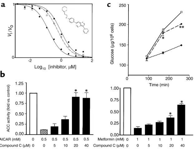
Identification and characterization of AMPK inhibitor. To firmly establish the role of AMPK in mediating met-formin’s effects, a high-throughput in vitro assay was used to identify small-molecule AMPK inhibitors. Screening of a compound library containing more than 10,000 mole-cules lead to the discovery and char-acterization of compound C (inset in Figure 3a). Experiments conducted using variable ATP concentrations revealed that compound C is a potent reversible inhibitor that is competi-tive with ATP, with Ki= 109 ± 16 nM
in the absence of AMP (Figure 3a). In in vitro assays, compound C did not exhibit significant inhibition of sev-eral structurally related kinases
including ZAPK, SYK, PKCθ, PKA,
and JAK3. Thus, to our knowledge, this is the first description of a potent and selective small-molecule AMPK inhibitor that may be useful as a means to further assess the phys-iologic effects of this pathway. Incu-bation of cultured hepatocytes with compound C inhibited ACC inactiva-tion by either AICAR or metformin (Figure 3b). Compound C also atten-uated AICAR and metformin’s effect to increase fatty acid oxidation or suppress lipogenic genes in hepato-cytes (not shown).
Metformin-mediated hepatocyte glucose production requires AMPK activation. It is believed that metformin-mediated inhibition of hepatic glucose produc-tion (HGP) plays a major role in its glucose-lowering efficacy (2, 3, 6). Here, we determined that both met-formin and AICAR were able to inhib-it cumulative glucose production in primary cultured rat hepatocytes stimulated with glucagon (Figure 3c and data not shown). Importantly, coincubation of hepatocytes with compound C was able to attenuate the effects of metformin to decrease glu-cose production in these cells (Figure 3c), demonstrating that AMPK activa-tion was required for metformin’s inhibitory effect on hepatocyte glu-cose production.
[image:6.576.111.485.519.700.2]Metformin activates muscle AMPK and promotes glucose uptake. Since AMPK activation is implicated as a mecha-nism for stimulation of glucose
Figure 4
uptake in skeletal muscle (12–14), we assessed the effect of metformin on glucose uptake and AMPK activity in intact rat epitrochlearis muscles. Incubation of isolated muscles with metformin resulted in an increase in the activity of both catalytic subunits of AMPK (Figure 4a). This was coinci-dent with a significant increase in glucose uptake that was also observed to be additive with the effect of insulin stimulation (Figure 4b). In
previous studies, AMPKα2 was
stim-ulated to a greater extent than
AMPKα1 by AICAR and other stress
(23). Further studies will be required to assess this difference.
AMPK activation suppresses SREBP-1. SREBP-1c is an important insulin-stimulated transcription factor that is implicated in the pathogenesis of insulin resistance, dyslipidemia, and DM2 (35, 36). Target genes that are induced by SREBP-1 include those that encode lipogenic enzymes, such as FAS and S14. Under conditions where SREBP-1 was induced by
insulin, incubation of rat hepatocytes with metformin or AICAR strongly suppressed SREBP-1 mRNA expres-sion (Figure 5a). Thus, a new pathway by which AMPK can mediate effects on lipogenic gene expression was also identified via these experiments.
Assessment of metformin’s in vivo effects. To assess whether selected effects of metformin described above also occurred in vivo, SD rats were studied (Table 1). Rats were orally dosed with metformin or vehicle
(H2O) for 5 days. Rats were starved
for 20 hours and then re-fed for 2 hours before the final dose. Four hours after the final dose, tissue and blood samples were obtained for analysis (see Methods). During star-vation, there should be very little lipid synthesis. Upon refeeding, hepatic lipid synthesis should be dra-matically induced. Metformin’s effects were examined under re-fed conditions. Along with modest decreases in plasma insulin and triglycerides, a small, but significant
increase in β-hydroxybutyrate was
present, suggesting that hepatic fatty acid oxidation was induced in met-formin-treated rats. Furthermore, metformin treatment produced sig-nificant decreases in hepatic expres-sion of mRNAs for SREBP-1, FAS, and S14 that were consistent with effects documented in cells (Table 1). The mature SREBP-1 protein in rat liver nuclear extracts was examined using an anti-SREBP1 Ab (Figure 5b). As anticipated (35), SREBP-1 mature-form protein was not detected in hepatic nuclear extracts from starved animals. In re-fed animals, mature SREBP-1 protein had accumulated consistent with an increase in lipid synthesis under this condition. Treat-Figure 5
[image:7.576.113.318.46.296.2]Metformin and AICAR downregulate hepatic SREBP-1. (a) Metformin (500 µM) and AICAR (500 µM) have similar effects to suppress SREBP-1 mRNA expression in rat hepatocytes. Mean ± SEM (n = 3 replicate assays) are shown. Insulin significantly increased SREBP-1 mRNA (P = 0.03 vs. control medium by Student’s ttest). Both AICAR and metformin signif-icantly decreased SREBP-1 mRNA with Pvalues of 0.0046 and 0.01, respectively, versus insulin. (b) Metformin prevents re-fed stimulated accumulation of mature SREBP-1 in nuclear extracts from treated rats. Western blot analysis using a monoclonal anti–SREBP-1 Ab (pre-pared from hybridoma CRL-2121) was performed. Similar results were obtained from a sep-arate in vivo experiment.
Table 1
In vivo effects of metformin on selected parameters known to be downstream of AMPK activation
Serum Vehicle Metformin
Glucose (mg/dl) 134.24 ± 4.82 126.33 ± 7.19 TG (mg/dl) 40.97 ± 2.81 30.2 ± 3.49A
FFA (mM) 0.17 ± 0.04 0.2 ± 0.04 Insulin (ng/ml) 1.48 ± 0.19 0.49 ± 0.09A
β-hydroxybutyrate (mg/dl) 0.94 ± 0.06 1.5 ± 0.15A
RNA Fold Fold
SREBP-1 1 ± 0.06 0.5 ± 0.03A
FAS 1 ± 0.06 0.35 ± 0.02A
S14 1 ± 0.06 0.43 ± 0.03A
Normal SD rats were treated as described in Methods. In rats studied in the fed state, serum samples were analyzed for glucose, triglyceride (TG), FFA, insulin, and β-hydroxybutyrate. Hepatic mRNA levels were quantitated. Results are expressed as mean values from seven rats in each group. AP < 0.05 vs.
[image:7.576.226.536.563.695.2]ment with metformin prevented this accumulation. Additional results obtained using hepatic nuclear extracts from re-fed rats after treat-ment with AICAR (500 mg/kg/day) also showed that the presence of SREBP-1 mature-form protein was ablated (data not shown).
Measurement of AMPK activation in liver ex vivo is difficult because brief hypoxia is known to produce marked activation of the enzyme (ref. 37 and data not shown). Thus we used liver tissue derived from metformin-treat-ed rats to determine that ACC activity was decreased significantly at several tested citrate concentrations (Figure 6). The greatest ACC activity reduc-tion was at a citrate concentrareduc-tion of 1 mM (from 54.6 ± 11.8 to 35.6 ± 7.7 nmol/mg/min; P < 0.01). These results are consistent with metformin having produced in vivo AMPK activation and ACC inactivation.
Discussion
To summarize, results presented here are consistent with a model (Figure 7) in which increased phosphorylation and activation of AMPK by met-formin leads to the effects on glucose and lipid metabolism as follows. Phosphorylation and inactivation of ACC, as a result of AMPK activation, serves to inhibit the proximal and rate-limiting step of lipogenesis. Reduced synthesis of the ACC prod-uct, malonyl-CoA, is also predicted to relieve inhibition of CPT-1, resulting in increased fatty acid oxidation. These effects are likely to contribute to metformin’s in vivo ability to lower triglycerides and VLDL.
AMPK mediates a decrease in SREBP-1 mRNA and protein expres-sion. Known target genes for SREBP-1, which include FAS and S14, are also downregulated in liver, further con-tributing to metformin’s effects to modulate circulating lipids and to reduce hepatic lipid synthesis and fatty liver. It should be noted that increased SREBP-1 is postulated as a central mediator of insulin resistance in DM2 and related metabolic disor-ders (35, 36) and that increased liver lipid content is implicated in hepatic insulin resistance (38).
Metformin-mediated effects on hepatic glucose production con-tribute to its glucose-lowering effica-cy. We demonstrated (using an AMPK inhibitor, Figure 3) that AMPK acti-vation is required for inhibition of
hepatocyte glucose production by metformin under the test conditions we employed. Additional studies will be required to further elucidate pre-cise mechanism(s) by which met-formin-stimulated AMPK activation could result in inhibition of hepatic glucose production.
AMPK activation is implicated as a mechanism for the induction of skele-tal muscle glucose uptake; this effect is also additive with insulin (12). Thus, the observed association of increased glucose uptake and AMPK activation in isolated skeletal muscles suggests that metformin’s effect to augment muscle insulin action in vivo may be attributed to AMPK as well.
A major mechanism (AMPK activa-tion) by which metformin produces beneficial metabolic effects has been characterized, along with discovery of the mechanism (SREBP-1 suppres-sion), by which AMPK inhibits the expression of lipogenic genes. Attempts to generate novel therapies for metabolic disease via AMPK acti-vation may be worthy of pursuit.
Acknowledgments
[image:8.576.64.258.55.222.2]We thank Shiying Chen for character-izing anti-AMPK Ab’s, Marcie Donnel-ly for technical support related to ani-mal studies, Mark Fraley for compound synthesis, and Georgianna Harris and Denis McGarry for helpful discussions and intellectual support. Figure 6
Metformin inhibits hepatic ACC activity in vivo. ACC activity was analyzed in the presence or absence of the indicated citrate concentrations using liver sam-ples from fed rats treated for 5 days with control vehicle (filled squares, n = 7) or treated with 200 mg/kg (twice daily oral gav-age) metformin (open squares,
n = 8). **P < 0.01.
Figure 7
[image:8.576.252.526.498.697.2]1. 2000. IMS HEALTH’s National Prescription Audit PlusTM. http://www.ims-global.com.
2. Stumvoll, M., Nurjhan, N., Perriello, G., Dailey, G. and Gerich, J.E. 1995. Metabolic effects of met-formin in non-insulin-dependent diabetes melli-tus. N. Engl. J. Med.333:550–554.
3. Wiernsperger, N.F., and Bailey, C.J. 1999. The antihyperglycaemic effect of metformin: thera-peutic and cellular mechanisms. Drugs. 58:31–39. 4. Wu, M.S., et al. 1990. Effect of metformin on car-bohydrate and lipoprotein metabolism in NIDDM patients. Diabetes Care.13:1–8. 5. Schafer, G. 1983. Biguanides. A review of history,
pharmacodynamics and therapy. Diabete Metab.
9:148–163.
6. Hundal, R.S., et al. 2000. Mechanism by which metformin reduces glucose production in type 2 diabetes.Diabetes.49:2063–2069.
7. Hundal, H.S., Ramlal, T., Reyes, R., Leiter, L.A., and Klip, A. 1992. Cellular mechanism of met-formin action involves glucose transporter translocation from an intracellular pool to the plasma membrane in L6 muscle cells. Endocrinol-ogy.131:1165–1173.
8. Galuska, D., Nolte, L.A., Zierath, J.R., and Wall-berg-Henriksson, H. 1994. Effect of metformin on insulin-stimulated glucose transport in iso-lated skeletal muscle obtained from patients with NIDDM. Diabetologia.37:826–832.
9. Lin, H.Z., et al. 2000. Metformin reverses fatty liver disease in obese, leptin-deficient mice. Nat. Med.6:998–1003.
10. Hardie, D.G., and Carling, D. 1997. The AMP-activated protein kinase: fuel gauge of the mam-malian cell? Eur. J. Biochem.246:259–273. 11. Winder, W.W., and Hardie, D.G. 1999.
AMP-acti-vated protein kinase, a metabolic master switch: possible roles in type 2 diabetes. Am. J. Physiol.
277:E1–E10.
12. Hayashi, T., Hirshman, M.F., Kurth, E.J., Winder, W.W., and Goodyear, L.J. 1998. Evidence for 5′ AMP-activated protein kinase mediation of the effect of muscle contraction on glucose trans-port. Diabetes.47:1369–1373.
13. Merrill, G.F., Kurth, E.J., Hardie, D.G., and Winder, W.W. 1997. AICA riboside increases AMP-activated protein kinase, fatty acid oxida-tion, and glucose uptake in rat muscle. Am. J. Physiol. 273:E1107–E1112.
14. Goodyear, L.J. 2000. AMP-activated protein kinase: a critical signaling intermediary for
exer-cise-stimulated glucose transport?Exerc. Sport Sci. Rev. 28:113–116.
15. Lochhead, P.A., Salt, I.P., Walker, K.S., Hardie, D.G., and Sutherland, C. 2000. 5-aminoimida-zole-4-carboxamide riboside mimics the effects of insulin on the expression of the 2 key gluco-neogenic genes PEPCK and glucose-6-phos-phatase. Diabetes.49:896–903.
16. Foretz, M., Carling, D., Guichard, C., Ferre, P., and Foufelle, F. 1998. AMP-activated protein kinase inhibits the glucose-activated expression of fatty acid synthase gene in rat hepatocytes. J. Biol. Chem. 273:14767–14771.
17. Holmes, B.F., Kurth-Kraczek, E.J., and Winder, W.W. 1999. Chronic activation of 5′ -AMP-acti-vated protein kinase increases GLUT-4, hexoki-nase, and glycogen in muscle.J. Appl. Physiol.
87:1990–1995.
18. Pollard, J.W., and Walker, J.M., editors. 1992. Basic cell culture protocols.Methods in Molecular Biolo-gy series, Volume 75. Humana Press Inc. Totowa, New Jersey, USA. 145–150.
19. Witters, L.A., and Kemp, B.E. 1992. Insulin acti-vation of acetyl-CoA carboxylase accompanied by inhibition of the 5′-AMP-activated protein kinase. J. Biol. Chem. 267:2864–2867.
20. Davies, S.P., Carling, D., and Hardie, D.G. 1989. Tissue distribution of the AMP-activated protein kinase, and lack of activation by cyclic-AMP-dependent protein kinase, studied using a specif-ic and sensitive peptide assay. Eur. J. Biochem.
186:123–128.
21. Mannaerts, G.P., Debeer, L.J., Thomas, J., and DeSchepper, P.J. 1979. Mitochondrial and perox-isomal fatty acid oxidation in liver homogenates and isolated hepatocytes from control and clofi-brate-treated rats. J. Biol. Chem.254:4585–4595. 22. Carling, D., Clarke, P.R., Zammit,V.A., and Hardie, D.G. 1989. Purification and characteri-zation of the AMP-activated protein kinase. Cop-urification of acetyl-CoA carboxylase kinase and 3-hydroxy-3-methylglutaryl-CoA reductase kinase activities. Eur. J. Biochem. 186:129–136. 23. Hayashi, T., et al. 2000. Metabolic stress and
altered glucose transport: activation of AMP-acti-vated protein kinase as a unifying coupling mech-anism. Diabetes.49:527–531.
24. Sheng, Z., Otani H., Brown, M.S., and Goldstein, J.L. 1995. Independent regulation of sterol regu-latory element-binding proteins 1 and 2 in ham-ster liver. Proc. Natl. Acad. Sci. USA. 92:935–938.
25. Cusi, K., and DeFronzo, R.A. 1998. Metformin: a review of its metabolic effects. Diabetes Reviews.
6:89–131.
26. Wiernsperger, N.F. 1999. Membrane physiology as a basis for the cellular effects of metformin in insulin resistance and diabetes. Diabetes. Metab.
25:110–127.
27. Sum, C.F., et al. 1992. The effect of intravenous metformin on glucose metabolism during hyper-glycaemia in type 2 diabetes. Diabet. Med.9:61–65. 28. Wilcock, C., Wyre, N.D., and Bailey, C.J. 1991. Subcellular distribution of metformin in rat liver. J. Pharm. Pharmacol.43:442–444.
29. Wilcock, C., and Bailey, C.J. 1994. Accumulation of metformin by tissues of the normal and dia-betic mouse. Xenobiotica.24:49–57.
30. Hermann, L.S. 1979. Metformin: a review of its pharmacological properties and therapeutic use. Diabete Metab.5:233–245.
31. Sterne, J., Duval, D., and Junien, J.L. 1979. Met-formin therapy. Research Clinical Forums1:13. 32. Owen, M.R., Doran, E., and Halestrap, A.P. 2000.
Evidence that metformin exerts its anti-diabetic effects through inhibition of complex 1 of the mitochondrial respiratory chain. Biochem. J.
348:607–614.
33. El-Mir, M.Y., et al. 2000. Dimethylbiguanide inhibits cell respiration via an indirect effect tar-geted on the respiratory chain complex I. J. Biol. Chem. 275:223–228.
34. McGarry, J.D., and Brown, N.F. 1997. The mito-chondrial carnitine palmitoyltransferase system. From concept to molecular analysis. Eur. J. Biochem. 244:1–14.
35. Shimomura, I., et al. 2000. Decreased IRS-2 and increased SREBP-1c lead to mixed insulin resist-ance and sensitivity in livers of lipodystrophic and ob/ob mice. Mol. Cell.6:77–86.
36. Kakuma, T., et al.2000. Leptin, troglitazone, and the expression of sterol regulatory element bind-ing proteins in liver and pancreatic islets. Proc. Natl. Acad. Sci.97:8536–8541.
37. Davies, S.P., Carling, D., Munday, M.R., and Hardie, D.G. 1992. Diurnal rhythm of phospho-rylation of rat liver acetyl-CoA carboxylase by the AMP-activated protein kinase, demonstrated using freeze-clamping. Effects of high fat diets. Eur. J. Biochem.203:615–623.
38. McGarry, J.D. 1992. What if Minkowski had been ageusic? An alternative angle on diabetes. Science.