B cells from patients with systemic lupus
erythematosus display abnormal antigen
receptor-mediated early signal transduction
events.
S N Liossis, … , G M Kammer, G C Tsokos
J Clin Invest.
1996;
98(11)
:2549-2557.
https://doi.org/10.1172/JCI119073
.
To understand the molecular mechanisms that are responsible for the B cell overactivity that
is observed in patients with SLE, we have conducted experiments in which the surface
immunoglobulin (sIg)-mediated early cell signaling events were studied. The
anti-sIgM-mediated free intracytoplasmic calcium ([Ca2+]i) responses were significantly higher in SLE
B cells compared with responses of normal individuals and to those of patients with other
systemic autoimmune rheumatic diseases. The anti-IgD mAb induced [Ca2+]i responses
were also higher in lupus B cells than in controls. The magnitude of anti-sIgM-mediated
Ca2+ release from intracellular stores was also increased in B cells from SLE patients
compared with normal controls. The amount of inositol phosphate metabolites produced
upon crosslinking of sIgM was slightly higher in patients with lupus than in normal controls,
although the difference was not statistically significant. In contrast, the degree of
anti-sIgM-induced protein tyrosine phosphorylation was obviously increased in lupus patients. Our
study demonstrates clearly for the first time that SLE B cells exhibit aberrant early signal
transduction events, including augmented calcium responses after crosslinking of the B cell
receptor and increased antigen-receptor-mediated phosphorylation of protein tyrosine
residues. Because the above abnormalities did not correlate with disease activity or
treatment status, we propose that they may have pathogenic significance.
Research Article
Find the latest version:
J. Clin. Invest.
© The American Society for Clinical Investigation, Inc. 0021-9738/96/12/2549/09 $2.00
Volume 98, Number 11, December 1996, 2549–2557
B Cells from Patients with Systemic Lupus Erythematosus Display Abnormal
Antigen Receptor–mediated Early Signal Transduction Events
Stamatis-Nick C. Liossis,* Birgit Kovacs,* Greg Dennis,‡ Gary M. Kammer,§ and George C. Tsokos*‡
*Department of Clinical Physiology, Walter Reed Army Institute of Research, Washington, DC 20307-5100; ‡Departments of Clinical
Investigation and Medicine, Walter Reed Army Medical Center, Washington, DC 20307-5001; and §Department of Medicine, Bowman
Gray School of Medicine, Wake Forest University, Winston-Salem, North Carolina 27157-1058
Abstract
To understand the molecular mechanisms that are responsi-ble for the B cell overactivity that is observed in patients with SLE, we have conducted experiments in which the sur-face immunoglobulin (sIg)-mediated early cell signaling events were studied. The anti–sIgM-mediated free intracy-toplasmic calcium ([Ca21]
i) responses were significantly
higher in SLE B cells compared with responses of normal individuals and to those of patients with other systemic au-toimmune rheumatic diseases. The anti-IgD mAb induced [Ca21]
i responses were also higher in lupus B cells than in
controls. The magnitude of anti–sIgM-mediated Ca21 re-lease from intracellular stores was also increased in B cells from SLE patients compared with normal controls. The amount of inositol phosphate metabolites produced upon crosslinking of sIgM was slightly higher in patients with lu-pus than in normal controls, although the difference was not statistically significant. In contrast, the degree of anti– sIgM-induced protein tyrosine phosphorylation was obvi-ously increased in lupus patients. Our study demonstrates clearly for the first time that SLE B cells exhibit aberrant early signal transduction events, including augmented cal-cium responses after crosslinking of the B cell receptor and increased antigen–receptor-mediated phosphorylation of protein tyrosine residues. Because the above abnormalities did not correlate with disease activity or treatment status, we propose that they may have pathogenic significance. (J. Clin. Invest. 1996. 98:2549–2557.) Key words: lymphocyte
signaling • calcium responses • tyrosine phosphorylation •
inositol phosphate metabolites • human autoimmunity
Introduction
SLE is considered to be the archetypal human autoimmune disease, and it is characterized by a variety of abnormalities of the immune system (1, 2). Both in human (3) and in murine models of the disease (4), B cell overactivity is considered to be responsible for the hypergammaglobulinemia and the
pro-duction of a large variety of autoantibodies, some of which are convincingly involved in the pathogenesis of certain types of immune complex–mediated histological damage (5, 6). Differ-ent factors contribute to lupus B cell overactivity. Among these are defective suppressor/inducer T cell subset (7),
exces-sive help provided by certain T cell subsets (8, 9), defective Fcg
receptor-mediated suppression (10), and overreactivity to cy-tokines delivered to B cells in an endocrine, paracrine, or auto-crine fashion (11, 12).
B cell function itself has so far been considered to be intact, but the conclusion that B lymphocytes are not intrinsically de-fective has been inferred indirectly (13–15). Recent evidence produced from the study of murine models suggests that B cells may be intrinsically defective (16–20). To further investigate possible inherent lupus B cell abnormalities, we examined a central aspect of their function, namely the early events of the
B cell receptor (BCR)1-mediated signal transduction.
Stimulation of B cells with antigens (Ags) or mAbs results in a well-regulated cascade of intracellular events, which lead to cell activation and proliferation. The ligation of the BCR complex initially causes the activation of protein tyrosine ki-nases (PTK) (21, 22) which, in turn, induces among other events the phosphorylation and activation of phospholipase C
(PLC) isozymes PLCg1 and PLCg2. The latter PLC isozyme
predominates in B cells (23). Activated PLC acts on
mem-brane phosphatidylinositol 4,5-biphosphate (PIP2) that leads
to the production of diacylglycerol (DAG) and inositol
1,4,5-trisphosphate (InsP3) (24–26). DAG activates protein kinase C
(PKC) (27), whereas InsP3 binds to its receptor on the
endo-plasmic reticulum and causes the release of free Ca21 from
in-tracellular stores (28). The increase of free intracytoplasmic Ca21 concentration ([Ca21]
i) and activated PKC act on a
vari-ety of genes, resulting in their transcription, cellular activation,
and proliferation (29, 30). Ca21 is an important intracellular
messenger which plays a role in a spectrum of cellular events that range from egg fertilization to apoptosis, via the regula-tion of signal transducregula-tion (31–35).
The BCR-complex-mediated early signal transduction events in lupus have not been investigated so far. In this com-munication, we present studies in which we examined the
BCR-initiated [Ca21]
i responses, tyrosine phosphorylation and
InsP3 production in fresh, unmanipulated B cells from SLE
pa-tients, disease and normal controls. Our results show that only Address correspondence to George C. Tsokos, Department of
Clini-cal Physiology, Bldg. 40, Rm. 3078, Walter Reed Army Institute of Research, Washington, DC 20307-5100. Phone: 202-782-9146; FAX: 202-782-3160; E-mail: gtsokos@mem.po.com
Received for publication 21 August 1996 and accepted in revised
form 2 October 1996. 1. Abbreviations used in this paper: BCR, B cell receptor; [Ca21] i, free
intracytoplasmic concentration of Ca21; HRPO, horseradish
peroxi-dase; InsP1, inositol phosphate-1; InsP2, Ins phosphate-2; InsP3, Ins
1,4,5-triphosphate; InsP3R, InsP3 receptor; ITAM, Ig(superfamily)
lupus B cells display aberrant BCR-induced early cell signaling events, which may contribute to the abnormal function of SLE B cells.
Methods
Patients and controls. 21 SLE patients (18 women, 3 men) were stud-ied. All subjects fulfilled at least 4 of the 11 revised criteria of the American Rheumatism Association for the Classification of SLE (36). The age of the patients ranged from 20 to 76 yr (mean6SD 5 44614.2). Patients who were receiving glucocorticoids were studied at least 24 h after their last dose. Disease activity was measured using the SLEDAI score (37) (mean6SD 5 6.465.7). 15 patients were on prednisone, 13 on hydroxychloroquine, and 4 were on cytotoxic med-ications (1 on cyclophosphamide, 1 on methotrexate, and 2 were re-ceiving azathioprine). An additional sample of 12 patients with other systemic autoimmune rheumatic diseases (disease-control group) was also studied. Of those patients, nine had rheumatoid arthritis, one had juvenile chronic arthritis, one had systemic sclerosis, and one had an undifferentiated connective tissue disease. Of these, 10 were women and 2 were men. The age range was similar to that of the SLE patient group (mean6SD 5 43614.5). The protocol was approved by the Health Use Committee at Walter Reed Army Medical Center or the Bowman Gray School of Medicine, and informed consent was ob-tained from all patients. 17 healthy volunteers were included in the control group.
Cells. Heparinized peripheral venous blood was obtained from study subjects. Leukopheresis samples were obtained from several healthy control individuals as well as certain SLE patients. PBMC were obtained by standard Ficoll-Hypaque gradient centrifugation. In most instances, this cell population was enriched in B cells by ro-setting once or twice with sheep red blood cells, as described (38). Monocytes were depleted by adherence to plastic (39). The cell popu-lation was rested overnight in a concentration of 1 3 106 cells/ml in
RPMI-1640 containing 10% FCS.
Reagents and mAbs. The F(ab9)2 fragment of an affinity-purified
goat anti-human m heavy chain Ab (Jackson Immunoresearch Labo-ratories Inc., Avondale, PA), and two murine anti–human IgD mono-clonal antibodies, ( CBDA-4E5-4C7 and dIAG.2, both were generous gifts of Dr. Fred Finkelman) were used for the stimulation of lympho-cytes. The fluorochrome-conjugated mAbs used were: anti-CD3 (IgG1)-FITC (Coulter, Hialeah, FL), anti-CD16 (Leu-11c)-phyco-erythrin (PE) (a murine IgG1 mAb) and anti-CD14 (Leu-M3)-PE, (murine IgG2b mAb) (Becton-Dickinson Immunocytometry Sys-tems, San Jose, CA). The antiphosphotyrosine horseradish peroxi-dase (HRPO)-conjugated mAb (4G10) was purchased from Upstate Biotechnology Inc. (Lake Placid, NY). The dye Indo-1 acetoxy meth-ylester (Molecular Probes, Eugene, OR) was dissolved in DMSO and stored at 2708C until use. Ficoll (Lymphoprep; Nycomed Pharma AS, Oslo, Norway), BSA, EGTA, DMSO, lithium chloride, calcium chloride, sodium formate, formic acid, disodium tetraborate, and am-monium formate were purchased from Sigma Chemical Co. (St. Louis, MO). Chloroform was purchased from J.T. Baker Chemical Company (Phillipsburg, NJ), methanol was purchased from Mallin-krodt (Paris, KY), and RPMI-1640, FCS, glutamine, streptomycin, penicillin, HBSS and Hepes were purchased from Life Technologies (Grand Island, NY).
[Ca21]
i measurement using flow cytometry. Cells were suspended at 5 3 106 cells/ml in RPMI-1640 supplemented with 2 mM glutamine,
100 mg/ml streptomycin, 100 U/ml penicillin, 25 mM Hepes, pH 7.4, and 0.1% FCS. The fluorescent dye Indo-1 acetoxy methylester (Indo-1 AM) was used to measure the [Ca21]
i, as previously described
(40, 41). At the beginning of each analysis, gating was performed on the lymphocyte population in order to select negatively the B cell subpopulation. T cells (stained with CD3-FITC mAb), monocytes (stained with CD 14-PE mAb) and natural killer cells (stained with
CD 16-PE mAb) were gated out as previously described (42). For the first 60 s the cells were left to run unstimulated to record their base fluorescence ratio, which represents the resting [Ca21]
i levels. Cells
were then stimulated with an anti-sIg mAb and left to run for a total of 10 min. Data were analyzed using the Multitime analysis program (Phoenix Flow Systems, San Diego, CA). In all experiments, the mean fluorescence ratio for the negatively selected subpopulation was plotted over time and the mean peak/base ratios were recorded.
Dose–response curves. To define the dose of the stimulus that causes the optimal Ca21 mobilization response, we used increasing
doses of all three Abs to stimulate normal PBMC, recorded the peak fluorescence ratio of Ca21 obtained for each dose as described above,
and constructed a curve. The doses used were: 2 mg/ml, 10 mg/ml, 20 mg/ml, 40 mg/ml, and 100 mg/ml for each used Ab.
Inositol phosphate metabolite measurement. The inositol phos-phate metabolites were measured as previously described (43) with minor modifications. Briefly, PBMC were washed twice with HBSS supplemented with 25 mM Hepes, antibiotics, and 1 mg/ml BSA. They were incubated overnight at a concentration of 10 3 106 cells/ml
in RPMI-1640 supplemented with 10% FCS, 50 mCi/ml
myo-[1,2-3H]inositol (sp act 18.3 Ci/mmol; DuPont NEN, Boston, MA),
antibi-otics, 25 mM Hepes, and 2 mM glutamine in a humidified, 5% CO2
atmosphere incubator. Next, cells were washed twice with HBSS con-taining 25 mM Hepes, antibiotics, 1 mg/ml BSA, and 10 mM LiCl, and then resuspended at 15 3 106 cells/ml (0.2 ml final volume) in
RPMI-1640, 25 mM Hepes, 1 mg/ml BSA, and 10 mM LiCl, and incu-bated at 378C for 15 min. Subsequently, cells were stimulated with the F(ab9)2 fragment of a goat anti–human m chain (20 mg) or medium
(0.02 ml) for different time periods at 378C. The reaction was termi-nated by the addition of 0.75 ml of chloroform/methanol (1:2) fol-lowed by 0.25 ml of water and 0.25 ml of chloroform, as described (43). A 0.2-ml portion of the upper-phase, which contained the water-soluble inositol phosphates (InsP) was diluted first with 2.3 ml of wa-ter and was then applied to anion-exchange columns, prepared with AG 1-X8 formate form resin (100-200 mesh; Bio-Rad Laboratories, Richmond, CA).
The columns were washed, the different InsP metabolites were eluted and the amount of radioactivity was measured as described (41). Levels of the different InsP metabolites are expressed as per-centage of increase of the specific metabolite in 3 3 106 cells
stimu-lated with the Ab compared with cells stimustimu-lated for the same time with medium alone.
Antiphosphotyrosine immunoblotting. Five million enriched B cells suspended in 0.5 ml volume of RPMI-1640, 10% FCS were incu-bated at 378C for 30 min. 20 mg of a F(ab9)2 fragment of a goat anti–
human m chain or medium (0.02 ml) was added to the cells at 378C, and the reaction was terminated after 60 s by the addition of 1 ml of ice-cold stop buffer (10 mM Tris, 50 mM NaCl, 5 mM EDTA) con-taining various protease inhibitors. The sample was centrifuged for 1 min at 11,000 g at 48C, and the pellet was lysed with sonication in 0.1 ml of stop buffer containing 1% Triton X-100. After centrifugation at 12,000 g for 15 min at 48C, insoluble material was removed and the protein content of the lysates was determined using the Bio-Rad pro-tein assay (Bio-Rad, Hercules, CA) according to the manufacturer’s instructions. Protein (30 mg) from each lysate was resolved by 10% polyacrylamide/SDS gel electrophoresis. Resolved proteins from the gels were transferred onto nitrocellulose membranes using a semi-dry transfer system. The membrane was blocked for 2 h with Tris-buf-fered saline, 2% BSA and 0.25% Tween. It was then washed and in-cubated overnight with 1:1,000 dilution of an HRPO-conjugated an-tiphosphotyrosine mAb in Tris-buffered saline, 1% BSA, 0.25% Tween. After washing, the tyrosine-phosphorylated proteins were de-tected using the enhanced chemiluminescence detection system (Am-ersham Life Sciences, Buckinghamshire, UK), according to the man-ufacturer’s instructions.
Results
SLE B cells display higher BCR-mediated [Ca21]
i responses than B cells from normal individuals and patients with other systemic autoimmune rheumatic diseases. Five million PBMC enriched in B cells were loaded with the calcium-binding dye Indo-1 AM and stained with anti–CD3-FITC, anti–CD14-PE, and anti–CD16-PE to gate out T cells, monocytes, and natural killer cells, respectively. This negative selection enabled us to
measure changes in the levels of [Ca21]
i, in fresh,
unmanipu-lated B cells which were the only unstained cells in the lym-phocyte subpopulation. Stimulation of cells was performed
with an F(ab9)2 fragment of goat-anti–human m chain Ab at 40
mg/ml, which was found to be the optimal concentration for
in-ducing maximal [Ca21]
i responses in normal B cells (Fig. 1 A).
A representative experiment is shown in Fig. 2 A. The baseline
and peak [Ca21]
i responses were recorded in samples from 21
SLE patients, 14 patients with other autoimmune rheumatic diseases, and 17 healthy individuals. There were no significant
differences when the resting [Ca21]
i recordings of the lupus
patients (7.2760.32) and the normal group (7.3060.27)
were compared (P , 0.95), or between the disease-control
(6.8560.38) and the SLE group (P , 0.43), or between the
normal group and the nonlupus patients (P, 0.35).
Although the peak [Ca21]
i recordings were similar between
the normal individuals and the disease controls (10.4160.53 vs
10.6560.59, P, 0.78), the peak [Ca21]
i responses were
signifi-cantly higher (14.6060.90) in the SLE patient group. The peak
response of lupus B cells was higher compared to the group of
healthy volunteers (P , 0.002) and was also significantly
higher in comparison with the group of patients with other
auto-immune diseases (P, 0.005) (Fig. 3).
Any sIg isotype can participate in the formation of the BCR complex and the initiation of the B-cell signaling process, but the vast majority of B-cells carry BCRs comprising sIgM
and sIgD (44). To examine whether the abnormal [Ca21]
i
re-sponse was restricted to sIgM-BCR, we used two different anti-sIgD murine mAbs to stimulate human B cells and
re-corded the changes in the levels of [Ca21]
i. The two murine
mAbs, CBDA-4E5-4C7 and dIAG.2, were used at
concentra-Figure 1. Dose–response curves of anti–sIg-induced increases in [Ca21]
i. Three anti-sIg Abs were used in increasing concentrations to
stimulate B cell–enriched PBMC from healthy individuals. The F(ab9)2 fragment of a goat anti–human m heavy chain Ab (A), the
dIAG.2 murine anti–human IgD mAb (B), and the CBDA-4E5-4C7 murine anti–human IgD mAb (C), produced maximal peaks of [Ca21]
i at concentrations of 40 mg/ml each. [Ca21]i was measured as
[image:4.612.317.554.59.379.2]the fluorescence ratio (381/525 nm).
Figure 2. Representative anti–sIg-induced [Ca21]
i responses in lupus
patients and normal individuals. Five million PBMC enriched in B cells were loaded with the calcium-binding dye Indo-1 AM and fluo-rochrome-conjugated mAbs against non–B cell surface markers. Neg-atively selected B cells were stimulated with anti-BCR Abs (40 mg/ml each) after 60 s of resting (arrow), and the response was recorded for a total of 10 min. The [Ca21]
i is proportional to the recorded mean
[image:4.612.55.300.59.372.2]tions of 40 mg/ml each (Fig. 1, B and C). The CBDA-4E5-4C7
mAb induced significantly higher peak [Ca21]
i responses in 12
of 14 SLE patients examined (Fig. 4) compared with 10
healthy individuals (13.6461.03 vs 10.0461.00, P, 0.01). The
dIAG.2 anti-sIgD mAb induced higher peak [Ca21]
i responses
in 9 out of the 14 SLE patients examined compared with 10 normal individuals (Fig. 5) but the difference did not reach
sta-tistical significance (P , 0.07). Representative experiments
with the use of these two anti-IgD mAbs are shown in Figs. 2,
B and C. The representative curves of Fig. 2 also show that the
[Ca21]
i responses were also prolonged in lupus B cells. This
was true for all those SLE patients which displayed higher
Ca21 fluxes than their respective controls regardless of the
stimulatory Ab used.
These experiments demonstrate clearly that fresh B cells from patients with SLE display increased BCR-mediated [Ca21]
i responses that are not restricted to only one sIg isotype.
Of importance is the fact that increased [Ca21]
i responses were
observed only in lupus patients. sIgM-initiated [Ca21]
i changes due to release from intracel-lular stores are higher in B cells from SLE patients. Changes
in the concentration of [Ca21]
i induced upon BCR stimulation
reflect release of Ca21 from intracellular stores and influx of
Ca21 from the extracellular space (28). The InsP
3 that is
pro-duced after ligation of the BCR complex induces the release of
Ca21 from the intracellular stores. To examine this specific
component of the calcium response, we used EGTA to chelate
the extracellular Ca21. The BCR-mediated changes in [Ca21]
i
would thus reflect the component of the response that is due to
the intracellularly stored Ca21 only. Levels of the
intracellu-larly stored Ca21 can be affected by changes in the
extracellu-lar concentration of the ion. Therefore, we used 5 mM EGTA
immediately before the beginning of the Ca21 measurement.
The anti–sIgM-induced peak [Ca21]
i response in the presence
of 5 mM EGTA is significantly higher in B cells from SLE pa-tients compared to the response of the normal individuals as shown in Table I. These experiments indicate that increased
release of Ca21 from its intracellular stores contributes
signifi-cantly to the observed augmentation of BCR-mediated [Ca21]
i
response in lupus B cells.
Anti–sIgM-induced InsP production. The release of
intra-cellularly stored Ca21 from the endoplasmic reticulum to the
cytoplasm occurs upon binding of the second messenger InsP3
[image:5.612.311.548.58.248.2]to its receptor (InsP3R). To investigate whether the increased
Figure 3. Anti-sIgM induced [Ca21]
i responses in control, disease
control, and SLE B cells. PBMC enriched in B cells were stimulated with 40 mg/ml of the F(ab9)2 fragment of a goat-anti–human m Ab.
The base line and the peak responses of intracellular free Ca21 were
recorded. While the base line values obtained were similar between the three groups, the peak [Ca21]
i responses were significantly higher
in the lupus patients group compared to the control (P , 0.002), as well as to the disease control group (P , 0.005). Bars represent the mean6SEM.
Figure 4. Anti-sIgD initiated [Ca21]
i responses using the
CBDA-4E5-4C7 murine anti–human mAb. Base line values and peak [Ca21] i
re-sponses were recorded for B cells obtained from normal individuals and from lupus patients. Stimulation was performed with 40 mg/ml of the Ab. Significantly higher peak free intracellular Ca21 responses
[image:5.612.57.285.59.247.2]were observed for the SLE patients group (P , 0.01). Bars represent the mean6SEM.
Figure 5. Anti-sIgD initiated [Ca21]
i responses using the dIAG.2
[image:5.612.57.292.475.662.2][Ca21]
i response in lupus B cells after crosslinking of the sIgM
is due to increased InsP3 production, we incubated PBMC
en-riched in B cells with tritiated myoinositol, stimulated them
with 20 mg of the F(ab9)2 fragment of a goat-anti–human m Ab,
and measured the inositol phosphate metabolites using anion-exchange chromatography at three time-points (0, 5, and 10
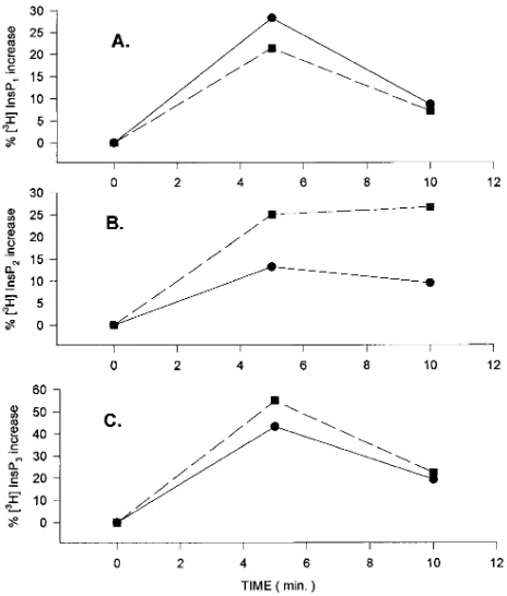
min after the addition of anti-sIgM Ab). Fig. 6 shows one rep-resentative experiment of four. The anti–sIgM-induced levels of inositol phosphate metabolites were always slightly higher than the levels that were observed in normal cells, but the dif-ference did not reach statistical significance. These experiments
show that the observed increased BCR-mediated [Ca21]
i
re-sponses in lupus B cells do not appear to be due to increased
BCR-induced InsP3 production.
The anti–sIgM-induced phosphorylation of protein tyrosine residues is increased in lupus B cells. Perhaps the earliest event after the BCR ligation is the activation of a cascade of protein tyrosine kinases, which phosphorylate various proteins on ty-rosine residues. Several quite important cellular functions are executed and regulated by the action of these kinases, the cal-cium response being one of them. As has been shown in sev-eral cell types including B cells, modulations in the action of
the “pre-Ca21” kinases may lead to altered calcium responses.
Over or underexpression of members of this pathway in im-mune cells results in increased or decreased activity of the
ki-nases and may modify the amount of Ca21 mobilization (45–
48). Since we observed increased BCR-initiated [Ca21]
i values
in lupus B cells we conducted experiments to determine whether this phenomenon was correlated with altered
BCR-mediated overall “pre-Ca21” protein tyrosine kinase (PTK)
ac-tivity.
An equal number of PBMC enriched in B cells was
stimu-lated with 20 mg of the F(ab9)2 fragment of a goat anti–human
m Ab for 60 s, a time that has been known to be required for
[image:6.612.57.296.93.166.2]maximal anti–sIgM-induced PTK activity (49). After lysing the cells, an equal amount of protein was electrophoretically sepa-rated on gels, transferred on membranes and blotted with an anti–phosphotyrosine-HRPO-conjugated mAb. Results show (Fig. 7), that in two out of three SLE patients examined, the overall degree of tyrosine phosphorylation was obviously in-creased. Although we did not notice the appearance of new Table I. Contribution of Intracellularly Stored Calcium to the
[Ca21]
i Response as Measured by EGTA Treatment of Normal and Lupus B Cells*
Peak/baseline [Ca21]
i ratio
EGTA treatment SLE (n) Control (n) P value‡
No 1.9760.11 (5) 1.2760.05 (3) 0.004
Yes 1.5960.12 (5) 1.1360.04 (3) 0.031
*Values obtained before and after the addition of 5 mM EGTA refer to the same SLE patients or normal individuals. The ratio of the peak over
the base fluorescence values for [Ca21]
i was estimated, and the
mean6SEM are represented here for each group. ‡Values of P , 0.05
were considered as statistically significant.
Figure 6. Anti-sIgM initiated production of inositol phosphate me-tabolites in B cells from a lupus patient ( j ) and a control
( d ) individual. (One representative experiment out of four with
similar results). Three million PBMC greatly enriched in B lympho-cytes were incubated with tritiated myoinositol, stimulated with 20 mg of the F(ab9)2 fragment of a goat-anti–human m Ab for 0, 5, and 10
min and lysed. The tritiated inositol phosphate metabolites [3H]InsP 1
(A), [3H]InsP
2 (B), and the [3H]InsP3 (C), were resolved by anion
[image:6.612.57.289.329.602.2]ex-change chromatography. Results are expressed as the percentage of increase in the levels of each metabolite over the levels obtained in cells stimulated with medium alone for the same time period. Lupus samples produced slightly (but not significantly) higher amounts of InsP2 and more importantly InsP3.
[image:6.612.317.555.466.624.2]bands the intensity of at least four of them, located between the 64- and the 36-kD mol wt standards, was obviously en-hanced. Moreover, the intensity of these bands (determined densitometrically) was directly proportional to the magnitude
of the Ca21 response for each individual SLE patient studied.
The activity/availability therefore of the PTKs assessed by the overall degree of tyrosine phosphorylations was positively
cor-related with the volume of the [Ca21]
i response in lupus B
cells. [Ca21]
i responses in SLE B cells do not correlate with dis-ease activity and treatment status. At the time of the study, an SLEDAI score was determined for each patient and treat-ment, if any, consisting of prednisone, hydroxychloroquine, and cytotoxic drugs (azathioprine, cyclophosphamide, meth-otrexate) was also recorded. Patients were classified as having active disease when the SLEDAI score was four or higher. Ta-ble II shows that there were no significant differences among patients with active or inactive disease at the time of the study. Moreover, treatment status did not affect the recorded re-sponses. The lupus group had significantly higher
peak/base-line ratios when compared with the control group (P5 0.001)
and to the disease–control group (P5 0.01). The lack of any
correlation between Ca21 responses and the activity of the
dis-ease suggests that the cell signaling abnormalities reported herein may be of pathogenetic importance.
Discussion
We conducted the present study to investigate the possible ab-errations in the early events of the signal transduction pathway in lupus B cells. Our purpose was to test the hypothesis that B cells in SLE are not innocent bystanders of an altered T cell ac-tivity but exhibit inherent biochemical defects. We used anti-sIgM and anti-sIgD Abs to stimulate fresh, unmanipulated B cells from lupus, disease controls, and normal individuals, and
evaluated the [Ca21]
i response, the tyrosine phosphorylation
pattern and the inositol phosphate metabolite production. We found that lupus, but not disease control, B cells exhibit signif-icantly higher responses regardless of the crosslinking anti-sIg Ab that did not correlate with disease activity or treatment status.
Our findings clearly demonstrated that the
anti–sIgM-induced peak [Ca21]
i responses were significantly higher in lupus
B cells compared with responses of B cells from the normal
group, although their resting [Ca21]
i were similar. Moreover,
the peak [Ca21]
i values recorded for lupus B cells were also
sig-nificantly higher than those observed for B cells obtained from patients with other systemic autoimmune rheumatic diseases. This latter group of patients had baseline as well as peak [Ca21]
i responses similar to the normal group. Our results
sug-gest the existence of an SLE-specific abnormality in the signal-ing events in lupus B cells.
The vast majority of B cells are sIgM and sIgD positive. No differences in the expression of these two surface immunoglob-ulins were found between lupus and normal B cells (data not shown). To test whether or not our results were sIgM specific,
we evaluated the anti–sIgD-mediated Ca21 responses using
two different murine anti-IgD mAbs. The anti–sIgD-induced responses were again significantly enhanced for one of the two mAbs used, while the other produced higher but not
statisti-cally significant elevations. The anti-sIgD recorded Ca21 fluxes
were shorter in duration than the responses to anti-sIgM, in ac-cordance with previously described data (50). These findings indicate that SLE B cells exhibit a disease-specific abnormality
in the BCR-mediated [Ca21]
i responses.
The Ca21 response to ligation of the BCR results from the
release of stored calcium from the internal stores to the cyto-plasm initially followed by influx of the ion from the extracel-lular space. By adding the chelating agent EDTA to the me-dium immediately before the recording, we were able to study the release of the ion from the endoplasmic reticulum. These
experiments yielded an augmented [Ca21]
i response in lupus B
cells, suggesting that increased release of stored Ca21
contrib-utes to the initially recorded higher peak [Ca21]i responses in
B cells from patients with SLE.
The BCR complex is a hetero-oligomer, consisting of the
surface immunoglobulin molecule and the Ig-a/b heterodimer(s)
(51, 52). The latter two molecules are responsible for the sig-nal-transducing properties of the antigen receptor (43). The signaling motif ITAM (Ig [superfamily] tyrosine-based
activa-tion motif) is present in the cytoplasmic tails of both Ig-a and
Ig-b (54). ITAM has been shown to be sufficient and necessary
to carry out signaling functions, including PTK activation,
pro-tein tyrosine phosphorylation, and Ca21 mobilization (22).
PTKs involved include the src-family members p55blk, p59fyn,
and p53/56lyn and the cytoplasmic PTK p72syk (55–57). After
sIg ligation p72syk becomes autophosphorylated first (58, 59),
which leads to binding of the src-family kinases through their
SH-2 domains, derepressing tyrosyl kinase activation (60).
This, in turn, leads to tyrosyl phosphorylation of Ig-a and Ig-b
ITAMs (61), with resultant activation, reorientation and
re-cruitment of additional src-family kinase molecules (62). The
activated src-family kinases regulate several cascades including
the activation of PLCg2, of mitogen-activated protein kinase
(MAPK), of GTPase-activating protein (GAP), of the p21ras
pathway, and of phosphoinositide 3-kinase (PI 3-K) (63–65). Ligation of either sIgM or sIgD results in the activation of the same PTKs, which in turn phosphorylate the same
sub-strates (66–68) and consequently lead to the Ca21 mobilization
[image:7.612.56.298.84.192.2]response (69). Both “precalcium” segments are qualitatively, but not quantitatively, identical, since their regulatory mecha-nisms might differ (49). The “postcalcium” events might be quite dissimilar as well (70, 71).
Table II. Correlation between Disease Activity, Treatment Regimes, and [Ca21]
i Responses in Lupus B Cells*
Peak/baseline [Ca21]
i ratio
Parameter Present (n) Absent (n) P value
SLEDAI score $ 4 2.0560.06 (15) 2.0460.22 (6) 0.97
Prednisone 2.0460.16 (15) 2.0660.21 (6) 0.94
Hydroxychloroquine 1.9360.15 (13) 2.2260.22 (8) 0.29
Cytotoxics 2.5160.46 (4) 1.9360.11 (17) 0.08
SLE 2.0460.13 (21)‡ 1.4360.06 (17)§ 0.001i
*Disease activity and form of treatment were determined at the time of the study for each patient. The ratio of the peak over the base
fluores-cence values for [Ca21]
i was estimated, and the mean6SEM are
repre-sented here for each subgroup. ‡value for the whole SLE patient group;
§value for the whole group of healthy individuals; istatistically
Therefore, BCR ligation leads to immediate activation of several PTKs and phosphorylation of substrates on tyrosine residues. To investigate the relative amount of early tyrosine phosphorylation in B cells from SLE patients, we immunoblot-ted lysates from SLE and control B cells activaimmunoblot-ted with anti-sIgM, with an antiphosphotyrosine mAb. There were no dif-ferences in the amount of tyrosine phosphorylated substrates between lupus and normal B cells in the resting state. After 60
s of stimulation with the same F(ab9)2 anti-m fragment we
ob-served markedly enhanced tyrosine phosphorylation of several protein bands from lupus samples, indicating increased PTK activation in lupus B cells compared with normal controls. Even though the cell populations used did not consist of pure
B cells (. 90% B cells determined flow cytometrically), the
stimulus that we used is B cell specific because only B cells bear sIgM. Thus, any resultant increase in tyrosine phosphory-lation of substrates, cannot be attributed to any other cell sub-population.
Interestingly, the amount of inositol phosphate metabolites
InsP1, InsP2, and, more importantly, of InsP3 produced after
sIgM stimulation, were only slightly, but not statistically signif-icantly, higher in lupus than in normal cells. One explanation
for this discrepancy between increased Ca21 responses and
normal InsP3 production may be an increased density of
InsP3R on the Ca stores in the lupus cells, as has been
de-scribed in some cell types (72, 73). Alternatively, increased
InsP3R binding resulting in enhanced release of calcium may
be present. The sensitivity of the InsP3R is regulated by InsP3
itself, by Ca21 and by PTK-mediated phosphorylation (74).
In-terestingly, antigen receptor–mediated and Fyn-dependent
phosphorylation of the InsP3R caused higher responses in T
cells and also increased the inhibitory threshold of rising [Ca21]
i on the receptor. Increased availability or activity,
therefore, of certain PTKs influences the sensitivity of the cal-cium channels and promotes increased responses upon antigen receptor stimulation. To date, similar studies have not been
performed on normal or lupus B cells. Finally, an InsP3
-inde-pendent Ca21 mobilizing mechanism may be operative in SLE
B cells. Evidence that such a mechanism might exist is pro-vided from experiments with B cells and other cell types (45– 47). This might implicate abnormal expression of certain pro-tein tyrosine kinases in SLE B cells. Such evidence has been
provided by the recent finding of overexpression of the p59fyn
in T cells from the lpr/lpr murine model of SLE and from
transfected cells overexpressing p59fyn (47, 48). At present, no
data exist for B cells.
Work from this laboratory has recently shown that a
simi-lar abnormality in the antigen receptor–mediated Ca21
re-sponse is associated with normal InsP3 production in lupus T
cells as well (41). The tyrosine phosphorylation pattern was not addressed. It may be, therefore, appropriate to assume that lupus T and B cells display similar cell signaling-associated biochemical abnormalities. Why do lupus B and T cells re-spond in an altered manner to stimuli that closely mimic physi-ologic stimuli? The recent description by Kammer et al. (42) that lupus T cells have abnormally low levels of the cAMP-dependent-PKA isozyme I activity, may offer some explana-tion. The intracellular levels of cAMP and the activity of this
enzyme can influence the Ca21 response after antigen receptor
ligation in T cells (75–77). Since increased cAMP and PKA ac-tivity can down regulate the calcium response, our observation
of heightened [Ca21]
i values might be explained by a
dimin-ished function of the cAMP/PKA system. Recently, a
cAMP-sensitive K1 channel was also described on the plasma
membrane operating in activated cells only. An increase in in-tracellular cAMP acting on this channel influences adversely
the magnitude of the free Ca21 response (78). Increased
[Ca21]
i responses as the ones we describe could be then
attrib-uted to decreased intracellular cAMP.
Provided that the same PKA enzymatic defect exists in SLE B cells, we hypothesize that the defective inhibitory cAMP/PKA-I pathway might contribute to the augmented [Ca21]
i responses of the lupus immune cells. Evidence that
fur-ther supports this hypothesis was provided from the study of a single patient, (included in our SLE patients group), who had unusually normal cAMP-dependent PKA-I activity in her T cells. As could be predicted from the above hypothesis, her
BCR-induced [Ca21]
i response was normal. More work is
nec-essary though, (a) to establish that this enzymatic defect also
characterizes lupus B cells, (b) to examine whether other
pa-tients as the one mentioned above respond in the same way
and (c) to determine the availability and/or activity of each one
of the early protein kinase systems.
The function of intracellular Ca21 underlines the
physio-logic importance of our findings. Numerous intracellular en-zymes and a spectrum of diverse and even quite opposite
cellu-lar functions are Ca21 regulated (22, 27, 79–81). Indeed,
abnormalities of some of these functions have been described in the human and the murine lupus (82–85).
In this study we have demonstrated that lupus B cells ex-hibit abnormal early signal transduction events. Because these abnormalities were not related to disease activity and because they were limited to lupus B cells, we conclude that they may be due to intrinsic defect(s) of the B cell, which may contribute to the pathogenesis of the disease.
Acknowledgments
We are indebted to Dr. Fred Finkelman for valuable comments at the beginning of the study and his generous supply of anti-IgD mAbs. The expert help of Mr. Lloyd Billups with the flow cytometer is ap-preciated.
Dr. Liossis was supported in part by a scholarship from the Hel-lenic Society of Rheumatology. This study was supported by work unit 9297 from the Department of Clinical Investigation, Walter Reed Army Medical Center. The opinions and assertions contained herein are the private views of the authors and are not to be construed as official or reflecting the views of the Department of the Army or the Department of Defense.
References
1. Tsokos, G.C. 1992. Overview of cellular immune function in systemic lu-pus erythematosus. In Systemic Lupus Erythematosus. R.G. Lahita, editor. Churchill Livingstone, New York. 15–50.
2. Theofilopoulos, A.N., G.J. Prud’Homme, and F.J. Dixon. 1985. Autoim-mune aspects of systemic lupus erythematosus. Concepts Immunopathol. 1:190– 218.
3. Ginsburg, W.W., F.D. Finkelman, and P.E. Lipsky. 1979. Circulating and pokeweed-mitogen-induced immunoglobulin-secreting cells in systemic lupus erythematosus. Clin. Exp. Immunol. 35:76–88.
4. Theofilopoulos, A.N. 1992. Murine models of lupus. In Systemic Lupus Erythematosus. R.G.Lahita, editor. Churchill Livingstone, New York. 121–194. 5. Kofler, D., V. Agnello, R. Thoburn, and H.G. Kunkel. 1971. Systemic lu-pus erythematosus: prototype of immune complex nephritis in man. J. Exp. Med. 134:169S.
7. Morimoto, C., A. Steinberg, N. Letvin, M. Hagan, T. Takeuchi, J. Daley, H. Levine, and S. Schlossman. 1987. A defect of immunoregulatory T cell sub-sets in systemic lupus erythematosus patients identified with anti-2H4 antibody.
J. Clin. Invest. 79:762–768.
8. Inghirami, G., J. Simon, J.E. Balow, and G.C. Tsokos. 1988. Activated T lymphocytes in the peripheral blood of patients with systemic lupus erythema-tosus induce B cells to produce immunoglobulin. Clin. Exp. Rheumatol. 6:269– 276.
9. Shivakumar, S., G.C. Tsokos, and S.K. Datta. 1989. T-cell receptor alpha/ beta expressing double-negative (CD42 /CD82) and CD41 T helper cells in
hu-mans augment the production of pathogenic anti-DNA autoantibodies associ-ated with lupus nephritis. J. Immunol. 143:103–112.
10. Salata, M., J. Golbus, and B.C. Richardson. 1988. Diminished response to an inhibitory signal in lymphocytes from patients with systemic lupus erythe-matosus. Clin. Exp. Immunol. 71:439–444.
11. Flescher, E., D. Fossum, A. Ballester, A. Maizel, S. Sharma, and N. Ta-lal. 1990. Characterization of B cell growth in systemic lupus erythematosus: ef-fects of recombinant 12-Kda B cell growth factor, interleukin-4 and transform-ing growth factor-b. Eur. J. Immunol. 20:2425–2430.
12. Golbus, J., M. Salata, J.Greenwood, J. Hudson, and B.C. Richardson. 1988. Increased immunoglobulin response to gamma-interferon by lympho-cytes from patients with systemic lupus erythematosus. Clin. Immunol. Immu-nopathol. 46:129–140.
13. Llorente, L., W. Zou, Y. Levy, Y. Richard-Patin, J. Wijdenes, J. Al-cover-Varela, B. Morel-Fourrier, J. Brouet, D. Alarcon-Segovia, P. Galanaud, and D. Emilie. 1995. Role of interleukin 10 in B cell overactivity and autoanti-body production of human systemic lupus erythematosus. J. Exp. Med. 181: 839–844.
14. Tsokos, G.C., and J.E. Balow. 1981. Spontaneous and pokeweed mito-gen-induced plaque-forming cells in systemic lupus erythematosus. Clin.
Immu-nol. Immunopathol. 21:172–183.
15. Tsokos, G.C., I.T. Magrath, and J.E. Balow. 1983. EpsteBarr virus in-duces normal B cell responses but defective suppressor T cell responses in pa-tients with systemic lupus erythematosus. J. Immunol. 131:1797–1801.
16. Dong, X., K.J. Hamilton, M. Satoh, J. Wang, and W.H. Reeves. 1994. Initiation of autoimmunity to the p53 tumor suppressor protein by complexes of p53 and SV40 large T antigen. J. Exp. Med. 179:1243–1252.
17. Reininger, L., T. Radaszkiewitz, M. Kosco, F. Melchers and A.G. Rolnick. 1992. Development of autoimmune disease in SCID mice populated with long-term in vitro proliferating (NZB X NZW) F1 pre-B cells. J. Exp. Med. 176:1343–1353.
18. Murakami, M., H. Yoshioka, T. Shirai, T. Tsubata, and T. Honjo. 1995. Prevention of autoimmune symptoms in autoimmune-prone mice by elimina-tion of B-1 cells. Int. Immunol. 7:877–882.
19. Wen, L., S. Roberts, F.S. Wong, C. Mallick, R.C. Findly, Q. Peng, J.E. Craft, M.J. Owen, and A.C. Hayday. 1994. Immunoglobulin synthesis and gen-eralized autoimmunity in mice congenitally deficient in alpha beta (1) T cells.
Nature (Lond.). 369:654–658.
20. Rosen, A., L. Casciola-Rosen, and J. Ahearn. 1995. Novel packages of viral and self-antigens are generated during apoptosis. J. Exp. Med. 181:1557– 1561.
21. Cambier, J.C., C.M. Pleiman, and M.R. Clark. 1994. Signal transduction by the B cell receptor and its coreceptors. Annu. Rev. Immunol. 12:457–486.
22. Weiss, A., and D.R. Littman. 1994. Signal transduction by lymphocyte antigen receptors. Cell. 76:263–274.
23. Coggeshall, K.M., J.C. McHugh, and A. Altman. 1992. Predominant ex-pression and activation-induced tyrosine phosphorylation of phospholipase C-g2 in B lymphocytes. Proc. Natl. Acad. Sci. USA. 89:5660–5664.
24. Nishizuka, Y. 1992. Intracellular signaling by hydrolysis of phospholip-ids and activation of protein kinase C. Science (Wash. DC). 258:607–614.
25. Coggeshall, K.M., and J.C. Cambier. 1984. B cell activation. VIII. Mem-brane immunoglobulins transduce signals via activation of phosphatidylinositol hydrolysis. J. Immunol. 133:3382–3386.
26. Bijsterbosch, M.K., and G.G.B. Klaus. 1985. Crosslinking of surface im-munoglobulin and Fc receptors on B lymphocytes inhibits stimulation of inosi-tol phosphate breakdown via the antigen receptors. J. Exp. Med. 162:1825– 1827.
27. Nishizuka, Y. 1992. Intracellular signaling by hydrolysis of phospholip-ids and activation of protein kinase C. Science (Wash. DC). 258:607–614.
28. Berridge, M.J. 1993. Inositol trisphosphate and calcium signaling. Na-ture (Lond.). 361:315–325.
29. Boyle, W.M., T. Smeal, L.H.K. Defize, P. Angel, J.R. Woodgett, M. Karin, and T. Hunter. 1991. Activation of protein kinase C decreases phos-phorylation of c-Jun at sites that negatively regulate its DNA-binding activity.
Cell. 64:573–584.
30. Fisher, C.L., J. Ghysdael, and J.C. Cambier. 1991. Ligation of mem-brane immunoglobulin leads to calcium mediated phosphorylation of the proto-oncogene product, Ets-1. J. Immunol. 146:1743–1749.
31. Girard, S., and D. Clapham. 1993. Acceleration of intracellular calcium waves in Xenopus oocytes by calcium influx. Science (Wash. DC). 260:229–232.
32. Gelfand, E.W., R.K. Cheung, S. Grinstein, and G.B. Mills. 1986. Char-acterization of the role for calcium influx in mitogen-induced triggering of
hu-man T cells: identification of calcium-dependent and calcium-independent sig-nals. Eur. J. Immunol. 16:907–912.
33. Lechleiter, J.D., and D. Clapham. 1992. Molecular mechanisms of intra-cellular calcium excitability in X. laervis oocytes. Cell. 69:283–294.
34. Gardner, P. 1989. Calcium and T lymphocyte activation. Cell. 59:15–20. 35. Oshimi, Y., and S. Miyazaki. 1995. Fas antigen-mediated DNA frag-mentation and apoptotic morphologic changes are regulated by elevated cyto-solic Ca11 level. J. Immunol. 154:599–609.
36. Tan, E.M., A.S. Cohen, J.F. Fries, A.T. Masi, D.J. McShane, N.F. Roth-field, J.G. Schaller, N. Talal, and R.J. Winchester. 1982. The 1982 revised crite-ria for the classification of systemic lupus erythematosus. Arthritis Rheum. 25: 1271–1277.
37. Bombardier, C., D.D. Gladman, M.B. Urowitz, D. Caron, and C.H. Chang. 1992. Derivation of the SLEDAI. A disease activity index for lupus pa-tients. Arthritis Rheum. 35:630–640.
38. Falkoff, R.M., M. Peters, and A.S. Fauci. 1982. T cell enrichment and depletion of human peripheral blood mononuclear cell preparations. Unex-pected findings in the study of the functional activities of the separated popula-tions. J. Immunol. Methods. 50:3999–4049.
39. Rabinovitch, M., and M.J. DeStefano. 1973. Macrophage spreading in vitro. Exp. Cell Res. 77:323–334.
40. Rabinovitch, P.S., C.H. June, A. Grossmann, and J.A. Ledbetter. 1986. Heterogeneity among T cells in intracellular free calcium responses after mito-gen stimulation with PHA or anti-CD3: simultaneous use of indo-1 and immuno-fluorescence with flow cytometry. J. Immunol. 137:952–961.
41. Vassilopoulos, D., B. Kovacs, and G.C. Tsokos. 1995. TCR/CD3 com-plex-mediated signal transduction pathway in T cells and T cell lines from pa-tients with systemic lupus erythematosus. J. Immunol. 155:2269–2281.
42. Kammer, G.M., I. Khan, and C. Malemud. 1994. Deficient type I protein kinase A isozyme activity in systemic lupus erythematosus T lymphocytes. J. Clin. Invest. 94:422–430.
43. Berridge, M.J., R.M. Dawson, C.P. Downes, J.P. Heslop, and R.F. Ir-vine. 1983. Changes in the levels of inositol phosphates after agonist-dependent hydrolysis of membrane phosphoinositides. Biochem. J. 212:473–482.
44. Abney, E.R., M.D. Cooper, J.F. Kearney, and A.R. Lawton. 1978. Se-quential expression of immunoglobulin in developing mouse B lymphocytes: a systematic survey that suggests a model for the generation of immunoglobulin isotype diversity. J. Immunol. 120:2041–2049.
45. Niklinska, B.B., H. Yamada, J.J. O’Shea, C.H. June, and J.D. Ashwell. 1992. Tyrosine kinase regulated and inositol phosphate independent Ca21
ele-vation and mobilization in T cells. J. Biol. Chem. 267:7154–7159.
46. Takata, M., H. Sabe, A. Hata, and T. Inazu. 1994. Tyrosine kinases Lyn and Syk regulate B cell receptor coupled Ca11 mobilization through distinct
pathways. EMBO J. 13:1341–1349.
47. Cooke, M.P., K.M. Abraham, K.A. Forbush, and R.M. Perlmutter. 1991. Regulation of T cell receptor signaling by a src family protein-tyrosine ki-nase (p59fyn). Cell. 65:281–291.
48. Katagiri, T., K.Urakawa, Y. Yamanashi, K. Semba, T. Takahashi, K.Toyoshima, T. Yamamoto, and K. Kano. 1989. Overexpression of src-family gene for tyrosine-kinase p59fyn in CD42CD82 T cells of mice with a
lymphopro-liferative disorder. Proc. Natl. Acad. Sci. USA. 86:10064–10068.
49. Kim, K.M., and M. Reth. 1995. Signaling difference between class IgM and IgD antigen receptors. Ann. NY Acad. Sci. 766:81–88.
50. Wilson, H.A., D. Greenblatt, C.W.Taylor, J.W. Putney, R.Y.Tsien, F.D. Finkelman, and T.M. Chused. 1987. The B lymphocyte calcium response to anti-Ig is diminished by membrane immunoglobulin crosslinkage to the Fcg re-ceptor. J. Immunol. 138:1712–1718.
51. Reth, M. 1992. Antigen receptors on B lymphocytes. Annu. Rev. Immu-nol. 10:97–121.
52. Reth, M., J. Hombach, J. Wienands, K.S. Campbell, N. Chien, L.B. Just-ment, and J.C. Cambier. 1991. The B cell antigen receptor complex. Immunol. Today. 12:196–201.
53. Cambier, J.C., and K.S. Campbell. 1992. Membrane immunoglobulin and its accomplices—new lessons from an old receptor. FASEB J. 6:3207–3217. 54. Reth, M. 1989. Antigen receptor tail clue. Nature (Lond.). 338:383–384. 55. Yamanashi, Y., T. Kakiuchi, J. Mizuguchi, T. Yamamoto, and K. Toyoshima. 1991. Association of B cell antigen receptor with protein tyrosine kinase Lyn. Science (Wash. DC). 251:191–194.
56. Clark, M.R., K.S.Campbell, A. Kazlauskas, S.A. Johnson, M.Hertz, T.A. Potter, C. Pleiman, and J.C. Cambier. 1992. The B-cell antigen receptor complex-association of Ig-a and Ig-b with distinct cytoplasmic effectors. Science
(Wash. DC). 258:123–126.
57. Pleiman, C.M., D. D’Ambrosio, and J.C. Cambier. 1994. The B-cell an-tigen receptor complex: structure and signal transduction. Immunol. Today. 15: 393–399.
58. Hutchcroft, J.E., M.L. Harrison, and R.L. Geahlen. 1991. B lymphocyte activation is accompanied by phosphorylation of a 72-kDa protein tyrosine ki-nase. J. Biol. Chem. 266:14846–14849.
59. Hutchcroft, J.E., M.L. Harrison, and R.L. Geahlen 1992. Association of the 72-kDa protein tyrosine kinase PTK 72 with the B cell antigen receptor. J. Biol. Chem. 267:8613–8619.
Bustelo, M. Barbacid, H. Sabe, H. Hanafusa, T. Yi et al. 1994. Specific motifs recognized by the SH2 domains of Csk, 3BP2, fps/fes, GRB-2, HCP, SHC, Syk and Vav. Mol. Cell Biol. 14:2777–2785.
61. Gold, M.R., L. Matsuchi, R.B. Kelly, and A.L. DeFranco. 1991. Ty-rosine phosphorylation of components of the B-cell antigen receptors following receptor crosslinking. Proc. Natl. Acad. Sci. USA. 88:3436–3440.
62. Burkhardt, A.L., M. Brunswick, J.B. Bolen, and J.J. Mond. 1991. Anti-immunoglobulin stimulation of B lymphocytes activates src-related protein-tyro-sine kinases. Proc. Natl. Acad. Sci. USA. 88:7410–7414.
63. Graziadei, L., K. Raibowol, and D. Bar-Sagi. 1990. Co-capping of ras proteins with surface immunoglobulins in B-lymphocytes. Nature (Lond.). 356: 396–400.
64. Padeh, S., A. Levitzki, A. Gazit, G.B. Mills, and C.M. Roifman. 1991. Activation of phospholipase C in human B cells is dependent on tyrosine phos-phorylation. J. Clin. Invest. 87:1114–1118.
65. Gold, M.R., M.T. Crowley, G.A. Martin, F. McCormick, and A.L. De-Franco. 1993. Targets of lymphocyte-B antigen receptor signal transduction in-clude the p21(ras) GTP-ase activating protein (GAP) and 2 GAP-associated proteins. J. Immunol. 150:377–386.
66. Gold, M.R., D.A. Law, and A.L. DeFranco. 1990. Stimulation of protein tyrosine phosphorylation by the B lymphocyte antigen receptor Nature (Lond.). 345:810–813.
67. Burkhardt, A.L., M. Brunswick, J.B. Bolen, and J.J. Mond. 1991. Anti-immunoglobulin stimulation of B lymphocytes activates src-related protein ty-rosine kinases. Proc. Natl. Acad. Sci. USA. 88:7410–7414.
68. Ales-Martinez, J.E., D.W. Scott, R.P. Phipps, J.E. Casnellie, G. Kroe-mer, C. Martinez, and L. Pezzi. 1992. Crosslinking of surface IgM or IgD causes differential biological effects in spite of overlap in tyrosine (de)phosphorylation profile. Eur. J. Immunol. 22:845–850.
69. Justement, L.B., J. Wienands, J. Hombach, M. Reth, and J.C. Cambier. 1990. Membrane IgM and IgD molecules fail to transduce Ca11 mobilizing
sig-nals when expressed on differentiated B lineage cells. J. Immunol. 144:3272– 3280.
70. Tisch, R., C.M. Roifman, and N. Hozumi. 1988. Functional differences between immunoglobulins M and D expressed on the surface of an immature B cell line. Proc. Natl. Acad. Sci. USA. 85:6914–6918.
71. Carsetti, R., G. Kohler, and M.C. Lamers. 1993. A role for immunoglob-ulin D: interference with tolerance induction. Eur. J. Immunol. 23:168–178.
72. Hirose, K., and M. Iino. 1995. Heterogeneity of channel density in inosi-tol-1,4,5,-trisphosphate-sensitive Ca11 stores. Nature (Lond.). 372:791–794.
73. Kasai, H., and O. Petersen. 1994. Spatial dynamics of second
messen-gers: IP3 and cAMP as long range and associative messengers. Trends. Neuro-sci. 17:95–101.
74. Thottala, J., K. Ondrias, E. Ondriasova, and A.R. Marks. 1996. Regula-tion of the inositol 1,4,5,-trisphosphate receptor by tyrosine phosphorylaRegula-tion.
Science (Wash. DC). 272:1492–1495.
75. Papadogiannakis, N., T.E. Nordstrom, L.C. Anderson, and C.H. Wolff. 1989. cAMP inhibits the OKT3-induced increase in free intracytoplasmic cal-cium in the Jurkat T cell line: the degree of inhibition correlates inversely with the amount of CD3 binding ligand used. Eur. J. Immunol. 19:1953–1956.
76. Park, D.J., H.K. Min, and S.G. Rhee. 1992. Inhibition of CD3-linked phospholipase C by phorbol ester and by cAMP is associated with decreased phosphotyrosine and increased phosphoserine contents of PLC-gamma 1. J. Biol. Chem. 267:1496–1501.
77. Alava, M.A., K.E. DeBell, A. Conti, T. Hoffman, and E. Bonvini. 1992. Increased intracellular cAMP inhibits inositol phospholipid hydrolysis induced by perturbation of the T cell receptor/CD3 complex but not by G-protein stim-ulation: association with protein kinase A-mediated phosphorylation of phos-pholipase C-gamma 1. Biochem. J. 284:189–199.
78. Oleson, D.R., L.J. DeFelice, M.F. Quinn, and R.M. Donahoe. 1996. cAMP increases the open probability of potassium channels in activated human T cells. J. Immunol. 157:1080–1086.
79. Premack, B.A., and P. Gardner. 1992. Signal transduction by T cell re-ceptors: mobilization of Ca and regulation of Ca-dependent effector molecules.
Am. J. Physol. 263:C1119–C1140.
80. Schulman, H. 1993. The multifunctional Ca11/calmodulin-dependent
protein kinases. Curr. Opin. Cell Biol. 5:247–253.
81. Trump, B.F., and I.K. Berezesky. 1992. The role of cytosolic Ca11 in cell
injury,necrosis and apoptosis. Curr. Opin. Cell Biol. 4:227–232.
82. Linker-Israeli, M. 1992. Cytokine abnormalities in human lupus. Clin.
Immunol. Immunopathol. 63:10–12.
83. Emlen, W., J. Niebur, and R. Kadera. 1994. Accelerated in vitro apopto-sis of lymphocytes from patients with systemic lupus erythematosus. J. Immu-nol. 152:3685–3692.
84. Russell, J.H., B. Rush, C. Weaver, and R.Wang. 1993. Mature T cells from autoimmune lpr/lpr mice have a defect in antigen-stimulated suicide.
Proc. Natl. Acad. Sci. USA. 90:4409–4413.
85. Bossu, P., G.G. Singer, P. Andres, R. Ettinger, A. Marshak-Rothstein, and A.K. Abbas. 1993. Mature CD41 T lymphocytes from MLR/lpr mice are
![Figure 2. Representative anti–sIg-induced [Ca2�]i responses in lupus patients and normal individuals](https://thumb-us.123doks.com/thumbv2/123dok_us/8220331.821550/4.612.317.554.59.379/figure-representative-induced-responses-lupus-patients-normal-individuals.webp)
![Figure 5. Anti-sIgD initiated [Ca2�]i responses using the �IAG.2 mu-rine anti–human mAb](https://thumb-us.123doks.com/thumbv2/123dok_us/8220331.821550/5.612.57.292.475.662/figure-anti-sigd-initiated-responses-using-iag-human.webp)
![Table II. Correlation between Disease Activity, Treatment Regimes, and [Ca2�]i Responses in Lupus B Cells*](https://thumb-us.123doks.com/thumbv2/123dok_us/8220331.821550/7.612.56.298.84.192/table-correlation-disease-activity-treatment-regimes-responses-lupus.webp)
