Interleukin 6 causes growth impairment in
transgenic mice through a decrease in
insulin-like growth factor-I. A model for stunted growth
in children with chronic inflammation.
F De Benedetti, … , G Ciliberto, E Fattori
J Clin Invest.
1997;
99(4)
:643-650.
https://doi.org/10.1172/JCI119207
.
Stunted growth is a major complication of chronic inflammation and recurrent infections in
children. Systemic juvenile rheumatoid arthritis is a chronic inflammatory disorder
characterized by markedly elevated circulating levels of IL-6 and stunted growth. In this
study we found that NSE/hIL-6 transgenic mouse lines expressing high levels of circulating
IL-6 since early after birth presented a reduced growth rate that led to mice 50-70% the size
of nontransgenic littermates. Administration of a monoclonal antibody to the murine IL-6
receptor partially reverted the growth defect. In NSE/hIL-6 transgenic mice, circulating IGF-I
levels were significantly lower than those of nontransgenic littermates; on the contrary, the
distribution of growth hormone pituitary cells, as well as circulating growth hormone levels,
were normal. Treatment of nontransgenic mice of the same strain with IL-6 resulted in a
significant decrease in IGF-I levels. Moreover, in patients with systemic juvenile rheumatoid
arthritis, circulating IL-6 levels were negatively correlated with IGF-I levels. Our findings
suggest that IL-6-mediated decrease in IGF-I production represents a major mechanism by
which chronic inflammation affects growth.
Research Article
Find the latest version:
J. Clin. Invest.
© The American Society for Clinical Investigation, Inc. 0021-9738/97/02/0643/08 $2.00
Volume 99, Number 4, February 1997, 643–650
Interleukin 6 Causes Growth Impairment in Transgenic Mice through a Decrease
in Insulin-like Growth Factor-I
A Model for Stunted Growth in Children with Chronic Inflammation
Fabrizio De Benedetti,* Tonino Alonzi,‡ Antonia Moretta,* Domenico Lazzaro,‡ Patrizia Costa,‡ Valeria Poli,‡ Alberto Martini,*
Gennaro Ciliberto,‡ and Elena Fattori‡
*Clinica Pediatrica, Università degli Studi di Pavia, IRCCS Policlinico San Matteo, 27100 Pavia, Italy; and ‡Istituto di Ricerca di Biologia
Molecolare (IRBM) P. Angeletti, 00040 Pomezia (Roma), Italy
Abstract
Stunted growth is a major complication of chronic inflam-mation and recurrent infections in children. Systemic juve-nile rheumatoid arthritis is a chronic inflammatory disorder characterized by markedly elevated circulating levels of IL-6 and stunted growth. In this study we found that NSE/hIL-6 transgenic mouse lines expressing high levels of circulating IL-6 since early after birth presented a reduced growth rate that led to mice 50–70% the size of nontransgenic litter-mates. Administration of a monoclonal antibody to the mu-rine IL-6 receptor partially reverted the growth defect. In NSE/hIL-6 transgenic mice, circulating IGF-I levels were significantly lower than those of nontransgenic littermates; on the contrary, the distribution of growth hormone pitu-itary cells, as well as circulating growth hormone levels, were normal. Treatment of nontransgenic mice of the same strain with IL-6 resulted in a significant decrease in IGF-I levels. Moreover, in patients with systemic juvenile rheumatoid ar-thritis, circulating IL-6 levels were negatively correlated with IGF-I levels. Our findings suggest that IL-6–mediated de-crease in IGF-I production represents a major mechanism by which chronic inflammation affects growth. (J. Clin. In-vest. 1997. 99:643–650.) Key words: interleukin 6 • insulin-like growth factor-I • growth disorders • juvenile
rheuma-toid arthritis
Introduction
Stunted growth is one of the major complications of chronic inflammation and infection in children. Although several causes, including poor nutrition, immobilization, and malab-sorption, have been hypothesized, the mechanism responsible for growth impairment in these conditions is unknown (1).
Systemic juvenile rheumatoid arthritis (s-JRA)1 is a chronic
inflammatory disease associated with stunted growth (2, 3). Growth impairment in s-JRA occurs during periods of disease activity with subsequent normalization of growth rate during remission (4, 5). Growth hormone (GH) production is normal in patients with s-JRA (6, 7), while levels of insulin-like growth factor-I (IGF-I) are decreased (7–9). IGF-I is produced by the liver in response to GH, mediates GH effects in several pe-ripheral organs including muscles and bones (10), and has a pivotal role in postnatal growth as demonstrated by the obser-vation that mice carrying a null mutation of the IGF-I gene showed markedly decreased postnatal growth (11). In s-JRA, laboratory evidence of inflammation is prominent (3), and in-terleukin 6 (IL-6) appears to be very relevant to the inflamma-tory process of s-JRA; circulating and synovial levels of IL-6 are markedly elevated in patients with active s-JRA, are signif-icantly higher than in other adult or juvenile chronic arthriti-des, and are related to clinical and laboratory parameters of disease activity; IL-6 appears to explain several clinical and laboratory features of the disease (12–17).
In this study we have found that the NSE/hIL-6 transgenic murine lines with high levels of circulating IL-6 since early phases of life show a growth defective phenotype; we demon-strate that IL-6 is associated with a growth defect and causes a decrease in IGF-I levels, without affecting GH production. Our findings show that sustained production of IL-6 may rep-resent the mechanism by which chronic inflammation affects growth.
Methods
Animals and treatments. The NSE/hIL-6 construct carries the rat neurospecific enolase (NSE) promoter driving the expression of hu-man IL-6 cDNA. The generation of NSE/hIL-6 mice has been de-scribed recently (18). Overexpression of hIL-6 in neuronal tissue re-sults in reactive astrocytosis and in an increase in ramified microglial cells, but these mice do not show histological or behavioral signs of neuron damage (18). Transgenic animals were identified by PCR anal-ysis of DNA extracted from a tail segment, as described (18). Mice were maintained in standard conditions under a 12-h light–dark cycle and were provided irradiated food (4RF21, Mucedola; Settimo Mi-lanese, Milan, Italy) and chlorinated water ad libitum. Procedures in-volving animals and their care were conducted in conformity with na-tional and internana-tional laws and policies (EEC Council Directive 86/ 609, OJ L 358, 1, December 12, 1987; Italian Legislative Decree 116/ 92, Gazzetta Ufficiale della Repubblica Italiana n. 40, February 18, 1992; National Institutes of Health Guide for the Care and Use of Laboratory Animals, publication No. 85–23, 1985). To evaluate the effect of the inhibition of hIL-6 action, NSE/hIL-6 mice were injected subcutaneously at days 2, 4, 7, 11, 14, and 20 after birth (day 0) with the neutralizing rat monoclonal antibody 15A7 to the murine IL-6 re-ceptor (19) at doses of 90 mg/mouse at day 2, 150 mg/mouse at day 4, and of 300 mg/mouse at day 7 and thereafter. The antibody was pre-Address correspondence to Gennaro Ciliberto, M.D., IRBM P.
An-geletti, Via Pontina km 30.600, 00040 Pomezia (Roma), Italy. Phone: 39-6-91093204; FAX: 39-6-91093654; E-mail: [email protected]
Received for publication 19 June 1996 and accepted in revised form 26 November 1996.
cipitated from the hybridoma cell medium in saturating ammonium sulphate, dialyzed against 50 mM Tris/HCl, pH 7.5, and loaded on a protein G–Sepharose resin (cat. No. 17-0618-02; Pharmacia, Brussels, Belgium), equilibrated with the same buffer. Elution was performed with 100 mM natrium citrate/citric acid buffer, pH 3.0, and the frac-tions collected were immediately neutralized with 3 M Tris/HCl, pH 8.9. Before injection the antibody was dialyzed against sterile gen-free saline solution. Control mice were treated with sterile pyro-gen-free saline solution. To evaluate the effect of administration of IL-6 to CB6F1 (C57Bl6xBalb/C) mice, recombinant human IL-6 (rhIL-6), obtained as described (20), resuspended in sterile pyrogen-free saline solution, was administered intraperitoneally at a dose of 10
mg/mouse to 3-wk-old animals; control mice were injected intraperi-toneally with sterile pyrogen-free saline solution. To measure food in-take and body weight, food and mice were weighed every day at the same hour. Food intake per gram of body weight was calculated by dividing the amount of food consumed over a 24-h period for the mean mouse weight at the end of the same period. Hematic glucose was determined using the Accutrend/GC apparatus and Strips Accu-trend Glucose (Boehringer-Mannheim, Mannheim, Germany). The pituitaries were carefully dissected from the cranial base and fixed in 2% paraformaldehyde for 1 h. The embedding and immunostaining were performed as described (21). Antisera specific for mouse GH and for rat thyroid stimulating hormone (TSH) were kindly provided by the National Hormone and Pituitary Program at the National In-stitute of Health (Bethesda, MD); the secondary antibodies labeled
with peroxidase were purchased from Jackson ImmunoResearch Laboratories, Inc. (West Grove, PA).
Patients. 21 patients (mean age 6.5 yr, age range 2–17 yr) fulfilling the American College of Rheumatology (formerly American Rheu-matism Association) criteria for the diagnosis of s-JRA (22) were in-cluded in the study. All patients presented active disease at time of sampling as defined by the presence of synovitis on examination and were receiving nonsteroidal antiinflammatory drugs; in addition ap-proximately half of them were also treated with low-dose steroids (on alternate day regimen in the majority); two-thirds of the patients were also receiving methotrexate. Permission for drawing of extra blood during routine venipuncture was obtained from parents of all children. Since marked changes in circulating IL-6 levels occurs dur-ing the febrile peak (12, 16), peripheral blood samples were collected during the morning hours, when all patients were afebrile.
Measurement of IGF-I, IL-6, IL-1b, and TNF-a. IGF-I was mea-sured using a commercially available radioimmunoassay, which rec-ognizes both murine and human IGF-I, according to the instructions provided by the manufacturer (Nichols, San Juan Capistrano, CA), after acid-ethanol extraction of plasma samples (anticoagulated with EDTA, final concentrations 5 mg/ml). IGF-I levels in patients with s-JRA were compared with normal values for age as determined by the manufacturer in 944 healthy controls. In patients with s-JRA, se-rum IL-6 levels were measured with a hybridoma growth factor assay using B9 cells as described previously (12); the hybridoma growth fac-tor activity in sera of patients with s-JRA was abolished by the
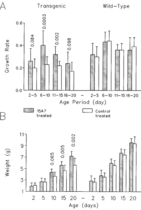
addi-Figure 1. (A) Reduced size of a 12-d-old NSE/hIL-6 mouse of line 26 (arrow) compared with an age- and sex-matched nontransgenic littermate. (B) Growth curves of transgenic (open squares) and nontransgenic (closed cir-cles) NSE/hIL-6 mice of lines 26 and 35. Numbers of each group of mice are as described in the legend to Table I. Results are shown as means6standard deviations (repre-sented by the vertical bars). *P, 0.001 and #P, 0.005
[image:3.612.56.369.337.742.2]tion of a goat antiserum to hIL-6 (not shown). In mice, IL-6 levels were determined using an immunoassay specific for biologically ac-tive human IL-6 (Boehringer-Mannheim). In patients with s-JRA, TNF-a and IL-1b were measured in plasma samples (anticoagulated with EDTA, as above) using two commercially available immunoas-says (TNF-a from Boehringer-Mannheim; IL-1b from Immunotech, Marseille, France).
Statistical analysis. Results were analyzed using the Mann-Whit-ney U test for unpaired samples and the Spearman correlation coeffi-cient, as appropriate. A P value , 0.05 was considered significant. A Z score for plasma IGF-I levels of patients with s-JRA was calculated according to the following formula: log (patient value) 2 log (mean value for the age control group)/log (standard deviations of the age control group).
Results
Growth defect in NSE/hIL-6 mice. The generation of NSE/ hIL-6 mice has been reported recently (18). Four of the seven transgenic founders generated (mice 15, 22, 26, and 35) trans-mitted the transgene to the progeny and four stable lines were established. No evident differences in body weight were re-vealed between transgenic and wild-type mice of the four lines at birth. After birth, while no differences were noted in the growth rate of transgenics of lines 15 and 22 compared with nontrans-genic littermates (not shown), transnontrans-genic mice of lines 26 and 35 presented a reduced growth rate that led to mice 50–70% the size of the age-matched littermates (Fig. 1 A). Growth rates and growth curves of lines 26 and 35 are shown in Table I and Fig. 1 B, respectively.
[image:4.612.62.557.74.222.2]Subsequent analysis showed that lines 26 and 35 had high levels of circulating hIL-6, detectable already 4 d after birth, while transgenics of lines 15 and 22 had undetectable levels (Table II). Moreover, Northern blot analysis showed that the levels of hIL-6 expression in the brain were similar in the four lines; however, while in lines 15 and 22 the expression of the transgene was confined to the central nervous system, in lines 26 and 35 hIL-6 expression was detectable in heart and lung; no hIL-6 expression was detected in liver, spleen, or kidney (18).
To verify whether the growth defect was due to the ele-vated levels of circulating IL-6, we tried to inhibit the action of the cytokine in transgenic mice of line 26 by injecting the mono-clonal antibody 15A7 neutralizing the murine IL-6 receptor,
starting at day 2 after birth. Neutralization of IL-6 activities re-sulted in a significant improvement in the growth rate, that was particularly evident from days 6 to 15 (Fig. 2 A). The nonstatis-tically significant effect on growth rate observed from days 15 to 20 may be due to some endogenous production of antibod-ies directed against the rat monoclonal antibody, and/or to the insufficiency of the dose administered relative to the increase in body weight (i.e., a constant dose of 300 mg/mice was ad-ministered at days 7, 11, 14). As shown in Fig. 2 B, the im-provement in growth rate resulted in a partial, but significant, correction of the growth defect, as indicated by the statistically significant differences in weight at days 15 (P5 0.005) and 20 (P5 0.002) of age between transgenic mice treated with the monoclonal antibody 15A7 (weight at day 20: 7.1960.84 g) and the control-treated transgenic animals (weight at day 20: 5.6361.25 g). In nontransgenic mice no significant differen-ces were found between 15A7-treated (weight at day 20: 9.5660.74 g) and control-treated animals (weight at day 20: 9.4061.21 g) (Fig. 2, A and B). These data indicate that in NSE/hIL-6 mice the growth defective phenotype is related to the elevated levels of IL-6.
Food intake and hematic glucose in NSE/hIL-6 mice. Reduc-tion of food intake and hypoglycemia are common host
re-Table I. Growth Rate of Transgenic and Nontransgenic NSE/hIL-6 Mice of Lines 26 and 35 in the Indicated Age Periods
Growth rate (mean gram increase in body weight per day)
Line 26
Age period (d) 0–10 11–20 21–30 31–40 Transgenic 0.2860.07 0.1360.10 0.3760.10 0.2060.12 Wild-type 0.5960.10 0.3660.10 0.6260.17 0.2260.13
P , 0.0001 0.0001 0.0002 0.34 Line 35
Age period (d) 0–7 8–14 15–21 22–28 29–35 Transgenic 0.7160.06 0.4160.07 0.8760.14 0.2260.08 0.0960.05 Wild-type 0.9460.08 0.5360.10 1.5260.38 0.3360.09 0.1860.05
P 0.0034 0.027 0.008 0.063 0.010
The weight was determined at the same hour of the day once every 10 d for mice of line 26 and once a week for mice of line 35 (line 26: transgenics n5 14, wild-type n5 17; line 35: transgenics n5 7, wild-type n5 6). Results are shown as means6standard deviations.
Table II. Serum hIL-6 Levels in NSE/hIL-6 Transgenic Mice at Different Ages
hIL-6 (ng/ml)
Line 15 Line 22 Line 26 Line 35
Day 4 ND ND 5.1060.84 3.9061.90 (n5 3) (n5 2) Day 11 ND ND 3.5261.75 ND
(n5 4)
Day 20 , 0.06 , 0.06 15.065.0 6.060.80 (n5 5) (n5 6)
[image:4.612.314.557.576.692.2]sponses to infection and inflammation and there is growing ev-idence that these phenomena result in part from the release of proinflammatory cytokines (23–26). Moreover, high levels of circulating IL-6 have been revealed during starvation in pa-tients with anorexia nervosa (27). Therefore, to determine whether the reduced growth rate of NSE/hIL-6 mice could be due to a behavioral disorder leading to a reduction of food take with subsequent hypoglycemia, we measured the food in-take of age-matched transgenic and wild-type mice of line 26 over a period of 7 d starting at 4 wk of age. Although the amount of food consumed daily was, as expected, lower in the small size NSE/hIL-6 transgenics than in wild-type littermates (not shown), the food intake per gram of body weight was comparable in the two groups (Fig. 3 A). Moreover, no differ-ences were found in the levels of hematic glucose (Fig. 3 B).
These results indicated that the growth defect in NSE/hIL-6 mice is not caused by a nutritional disorder.
Decreased in vivo levels of IGF-I in NSE/hIL-6 mice. The pituitary hormones GH and TSH play a major role in somatic growth; to investigate the possible effects of IL-6 overexpres-sion on pituitary functions, we evaluated the distribution of GH- and TSH-producing pituitary cells in mice of lines 26 and 35. Immunocytochemical analysis did not show differences be-tween transgenic and wild-type littermates (Fig. 4). In addi-tion, circulating levels of GH, as well as of T4, were compara-ble between transgenic and wild-type littermates (not shown).
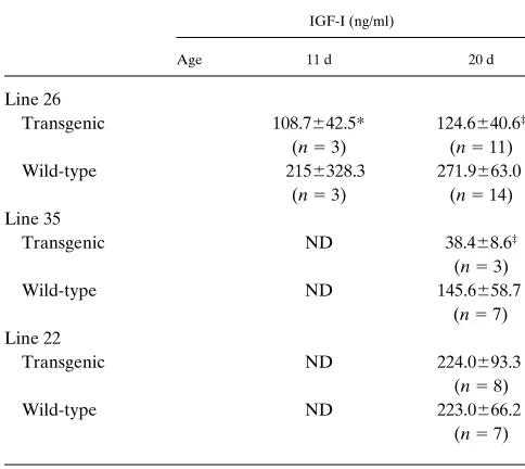
Since IGF-I mediates the great majority of the peripheral effects of GH (10) and plays a pivotal role in postnatal growth (11), and since in s-JRA the growth defect is associated with low levels of IGF-I (7–9), we measured circulating IGF-I levels in NSE/hIL-6 mice of lines 26, 35, and 22. As shown in Table III, in transgenic mice of lines 26 and 35, IGF-I levels were sig-nificantly lower than in the corresponding age-matched wild-type littermates, while no differences between transgenics and nontransgenics were found for line 22 with undetectable circu-lating IL-6 and normal growth. Further supporting the role of the decrease in IGF-I in the growth defect of line 26, a positive correlation between IGF-I levels and body weight at day 20 af-ter birth was found both in transgenic (Rs 5 0.743, P5 0.005) and nontransgenic (Rs 5 0.788, P, 0.001) mice of line 26. These results show that in both lines 26 and 35 with stunted growth, elevated circulating levels of IL-6 are associated with a decrease in IGF-I levels to values that are , 50% of those present in the corresponding nontransgenic animals.
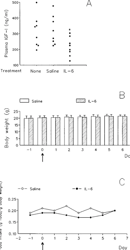
IL-6–induced decrease in in vivo IGF-I levels in CB6F1 mice. To verify the relationship between elevated levels of IL-6 and the decrease in circulating IGF-I, we injected recombinant hIL-6 in mice of the same strain of the NSE/hIL-6 transgenics. As shown in Fig. 5 A, treatment of CB6F1 mice with two intra-Figure 2.Effect of the neutralization of IL-6 activity on the growth of
NSE/hIL-6 transgenic or wild-type mice of line 26. A shows the growth rate (mean gram increase in body weight per day) in the indi-cated age periods. B shows the weight at the indicated ages. Trans-genic mice are shown on the left, and wild-type mice on the right. Sig-nificance levels (P values) of the differences of 15A7-treated animals versus corresponding control-treated animals are shown when P,
0.1. Mice were treated with the monoclonal antibody 15A7 neutraliz-ing the murine IL-6 receptor (shaded columns) or with saline (open columns), as described in Methods (control-treated transgenic n5
14; 15A7-treated transgenic n5 14; control-treated wild-type n5 20; 15A7-treated wild-type n5 19). The results are shown as means6
[image:5.612.316.552.57.238.2]standard deviations (represented by the vertical bars).
[image:5.612.60.294.58.407.2]peritoneal administrations of recombinant hIL-6 at 12-h inter-vals resulted in a significant decrease in IGF-I levels 24 h after the first treatment compared with untreated or saline-treated mice. Although a modest, albeit not significant, reduction in weight gain in the 2 d subsequent to hIL-6 administration was observed (weight gain hIL-6 treated: 0.2460.39 g; weight gain control-treated: 0.5360.38 g; P5 0.16), this did not result in a significant effect on the body weight of CB6F1 mice for the en-tire length of the period examined (Fig. 5 B). The absence of a
significant effect of this treatment on body weight suggests that prolonged overproduction of IL-6 is necessary to induce defec-tive growth. IGF-I levels are strongly reduced upon fasting both in human and rats (28). To verify if our protocol of IL-6 administration affected food intake and therefore influenced only indirectly IGF-I levels, we measured food intake in mice starting 2 d before treatment and lasting 6 d after. A reduction in food intake was observed in the IL-6–treated mice, but only starting 2 d after treatment, i.e., 24 h after the determination of IGF-I levels (Fig. 5 C). Based on these results and on the nor-mal food intake and glycemia of NSE/hIL-6 mice, we con-cluded that IL-6 affects the circulating levels of IGF-I by mechanisms that are distinct from anorexia.
Negative correlation between IL-6 and IGF-I levels in pa-tients with s-JRA. In agreement with previous studies (7–9), we found that in patients with s-JRA plasma IGF-I levels were lower than the mean normal values for age, without any rela-tion of IGF-I levels with steroid treatment (not shown). As shown in Fig. 6, IGF-I plasma levels were negatively correlated (Rs 520.667, P5 0.004) with serum IL-6 levels. A significant negative correlation of IGF-I levels was also found with C-reac-tive protein concentrations (Rs 520.499, P5 0.02). In con-trast, IGF-I levels were not correlated with IL-1b or TNF-a levels (Rs 5 0.125, P. 0.1; Rs 520.243, P. 0.1, respec-tively).
Discussion
[image:6.612.58.442.68.307.2]In this study we present the analysis of a transgenic mouse model that associates a growth defective phenotype with high circulating levels of IL-6 since birth. Several evidences demon-strate that the growth impairment present in NSE/hIL-6 mice does not result from an integration effect altering the expres-sion of genes involved in growth: (a) the growth defect oc-curred in five of the seven founder mice generated (not shown) and in two different lines of transgenic offspring; (b) NSE/hIL-6 mice of lines 15 and 22, which have expression of
Figure 4.Distribution of GH- or TSH-producing pituitary cells in NSE/hIL-6 mice. Frontal sec-tions of pituitaries from NSE/ hIL-6 (A and C) and wild-type littermates (B and D) were im-munostained with antibodies specific for GH (A and B) or TSH (C and D). n, neurohypo-physis; a, adenohypophysis. Bar 5 25 mm.
Table III. Circulating IGF-I Levels in Transgenic and Nontransgenic NSE/hIL-6 Mice of Lines 26 and 35 (Elevated Circulating hIL-6 and Growth Defect) and of Line 22 (No Circulating hIL-6 and Normal Growth), at the Indicated Ages
IGF-I (ng/ml)
Age 11 d 20 d
Line 26
Transgenic 108.7642.5* 124.6640.6‡
(n5 3) (n5 11) Wild-type 2156328.3 271.9663.0
(n5 3) (n5 14) Line 35
Transgenic ND 38.468.6‡
(n5 3) Wild-type ND 145.6658.7
(n5 7) Line 22
Transgenic ND 224.0693.3 (n5 8) Wild-type ND 223.0666.2
(n5 7)
Results are shown as means6standard deviations. *P, 0.05 and ‡P,
[image:6.612.55.297.499.715.2]the transgene confined to the CNS, grow with a normal rate, indicating that neither the presence of the transgene nor its ex-pression in the CNS is per se sufficient to cause a growth de-fect; on the contrary, in NSE/hIL-6 mice of lines 26 and 35,
which present growth impairment, hIL-6 mRNA expression is detected also in other organs (18) and this leaky expression, of unknown origin, is associated with measurable circulating IL-6 levels; and (c) the growth defect of NSE/hIL-6 mice can be partially corrected by neutralization of the murine IL-6 recep-tor, thus demonstrating that it is indeed due to IL-6 overex-pression. In addition, other IL-6 transgenic mice show defec-tive growth, including a neuron-, a keratinocyte-, and an airway epithelial cell–specific IL-6 transgenic (29–31), and al-though this aspect has never been described in detail, in some of them the defect has been reported since very early in life (29, 30). The fact that not all of the IL-6 transgenic mice de-scribed so far have growth impairment may be accounted for by different cell type and/or organ expression of IL-6, or by differential expression of the transgene with age. Indeed, in our experience, while in NSE/hIL-6 mice of lines 26 and 35 cir-culating hIL-6 in the range of nanograms per milliliter is de-tectable early after birth, MT-I/IL-6 mice, which carry the hIL-6 cDNA under the control of the mouse metallothionein-I pro-moter, do not show a growth defect and have circulating hIL-6 levels that increases markedly only after 2 mo of age (32).
The mechanism by which IL-6 causes growth defect ap-pears to involve a decrease in circulating IGF-I levels. In NSE/ hIL-6 mice of both lines 26 and 35, IGF-I levels are approxi-mately half of those of the corresponding nontransgenic litter-mates; incidentally, in both transgenic and nontransgenic mice of line 26 we found a positive correlation between IGF-I levels and weight at day 20 after birth. Moreover, treatment of CB6F1 mice with recombinant IL-6 results in a decrease in IGF-I levels. Anorexia and hypoglycemia may be observed during acute and chronic inflammation and have been related to the action of inflammatory cytokines (33, 34); in turn, ca-loric and protein restrictions have been shown to cause a de-crease in circulating IGF-I levels both in humans and rats (28). Our transgenic mice have a normal food intake, and a reduc-tion of food intake in IL-6–treated CB6F1 mice occurs only 24 h later than the decrease in IGF-I, demonstrating that the de-Figure 5. (A) Effect of the administration of recombinant hIL-6 to
CB6F1 mice on circulating IGF-I levels. 3-wk-old CB6F1 mice (8 mice/group) were treated with two intraperitoneal injections of re-combinant human IL-6 (10 mg/mouse) or of sterile pyrogen-free sa-line or left untreated. Injections were administered at 12-h intervals (8 a.m. and 8 p.m., day 0). Blood was collected 12 h after the second injection (8 a.m., day 1) and circulating IGF-I was measured. *P5
[image:7.612.318.555.59.235.2]0.03 versus untreated or saline-treated mice. (B and C) Effect of IL-6 administration on body weight (B) and food intake, expressed as daily food intake per gram of body weight (C), in CB6F1 mice. 3-wk-old mice (8 mice/group, caged separately) were treated with sa-line or with recombinant human IL-6 as described in A; arrows indi-cate the time of treatment. Body and food weights were determined every day at 9 a.m. (at day 0 before the first administration of recom-binant human IL-6).
[image:7.612.63.290.63.498.2]creased levels of IGF-I caused by elevated levels of IL-6 can-not be ascribed to nutritional disorders. NSE/hIL-6 mice of lines 26 and 35 have a normal distribution of GH-producing pi-tuitary cells and normal GH levels, therefore demonstrating that the effect of IL-6 on IGF-I levels is not mediated indi-rectly via an effect on GH production. In addition, since the GH/IGF-I axis plays the major role in postnatal growth in the presence of normal thyroid function, it is noteworthy that NSE/hIL-6 mice have a normal distribution of TSH-producing pituitary cells, as well as normal T4 levels, therefore excluding abnormalities of thyroid function. The mechanism by which IL-6 causes a decrease in in vivo IGF-I levels remains to be clarified. Since circulating IGF-I is produced mainly by the liver (10), and IL-6 is involved in the regulation of gene tran-scription during inflammation (23, 35), it is tempting to hy-pothesize that overproduction of IL-6 may act negatively on liver IGF-I gene expression. On the other hand, since the great part of circulating IGF-I is carried in a ternary complex with IGF-binding protein-3 (IGFBP-3) and a non-IGF binding pro-tein, termed acid labile subunit (ALS), and since the half-life of IGF-I is markedly prolonged by its association in this ter-nary complex from , 10 min to 12–15 h (10), it is also possible that a decrease in IGFBP-3 and/or in ALS may be responsible for the low circulating IGF-I levels.
Whatever the mechanism(s), our findings in mice strongly support the concept that IL-6 causes growth impairment in childhood chronic inflammatory diseases. Indeed, as previ-ously mentioned, s-JRA is a chronic inflammatory disorder that associates markedly elevated circulating levels of IL-6 (12–17), stunted growth (2, 4, 5), and decreased IGF-I levels (7–9); in this study we found a negative correlation between circulating levels of IGF-I and IL-6, as well as between IGF-I levels and C-reactive protein concentrations. These findings, together with the results obtained in NSE/hIL-6 mice, strongly suggest that chronically elevated levels of IL-6 are responsible for the growth defect present in patients with s-JRA. More-over, they appear to explain the observation that in patients with JRA treated with recombinant GH the height velocity during treatment was satisfactory in , 50% of the patients and was found to be inversely correlated with C-reactive protein concentrations (36). The possibility exists that, similarly to IL-6, other inflammatory cytokines, such as IL-1 or TNF-a, may af-fect IGF-I levels. However, in patients with s-JRA, we did not find any correlation of IGF-I levels with TNF-a or IL-1b levels. Stunted growth associated with decreased levels of IGF-I has been reported in other diseases characterized by chronic inflammation or by chronic recurrent infections, including Crohn’s disease and cystic fibrosis (37, 38). In Crohn’s disease, growth retardation is related to inflammation (39). IL-6 is highly expressed in the inflamed mucosa and serum IL-6 levels are elevated and correlate with disease activity (40–42). In cys-tic fibrosis, growth retardation is significantly correlated with lung infections, but not with the degree of pancreatic insuffi-ciency (43). Moreover, critically ill patients with sepsis, a con-dition with markedly elevated levels of IL-6, have substantially normal GH production, low IGF-I levels, and absent IGF-I re-sponse to exogenous GH, administered as anabolic agent (44, 45).
In conclusion, our study shows that in childhood chronic overproduction of IL-6 causes growth defect and that the growth defect appears to be mediated by a decrease in circulat-ing IGF-I. These findcirculat-ings suggest that IL-6–mediated decrease
in IGF-I production may represent a generalized major mech-anism by which chronic inflammation affects growth. This would imply that IGF-I and not GH represents the symptom-atic treatment of choice for growth retardation in children with s-JRA, as well as in those with other chronic inflammatory dis-eases or with recurrent severe infections.
Acknowledgments
The authors wish to thank Massimo Aquilina and Sabrina Germoni for animal care, Carlo Toniatti for providing recombinant human IL-6, Laura Zonta for her help with the statistical analysis, and Janet Clench for critical reading of the manuscript. Antisera to the pituitary hor-mones were kindly provided by the National Hormone and Pituitary Program of the National Institutes of Health.
This work was in part supported by IRCCS Policlinico San Mat-teo, 27100 Pavia, Italy.
References
1. Bithoney, W.G., H. Dubowitz, and H. Egan. 1992. Failure to thrive/ growth deficiency. Pediatr. Rev. 13:453–460.
2. Still, G.F. 1897. On a form of chronic joint disease in children. Med. Chir. Trans. 80:47–59. (1941, Reprinted from Arch. Dis. Child. 16:156–165.)
3. Cassidy, J.T., and R.E. Petty. 1995. Juvenile rheumatoid arthritis. In
Textbook of Pediatric Rheumatology. W.B. Saunders Company, Philadelphia. 133–233.
4. Kuhns, J.G., and L.T. Swaim. 1932. Disturbances in growth in chronic ar-thritis in children. Am. J. Dis. Child. 43:1180–1183.
5. Ansell, B.M., and E.G.L. Bywaters. 1956. Growth in Still’s disease. Ann.
Rheum. Dis. 15:295–319.
6. Sturge, R.A., C. Beardwell, M. Hartog, D. Wright, and B.M. Ansell. 1970. Cortisol and growth hormone secretion in relation to linear growth: pa-tients with Still’s disease on different therapeutic regimen. Br. Med. J. 3:547–551.
7. Allen, R.C., M. Jimenez, and C.T. Cowell. 1991. Insulin-like growth fac-tor and growth hormone secretion in juvenile chronic arthritis. Ann. Rheum. Dis. 50:602–606.
8. Bennet, A.E., E.D. Silverman, J.J. Miller III, and R.L. Hintz. 1988. Insu-lin-like growth factors I and II in children with systemic onset juvenile arthritis.
J. Rheumatol. 15:655–658.
9. Aitman, T.J., R.G. Palmer, J. Loftus, B.M. Ansell, J.P. Royston, J.D. Teale, and R.N. Clayton. 1989. Serum IGF-I levels and growth failure in juve-nile chronic arthritis. Clin. Exp. Rheumatol. 7:557–561.
10. Jones, J.I., and D.R. Clemmons. 1995. Insulin-like growth factors and their binding proteins: biological actions. Endocr. Rev. 16:3–34.
11. Baker, J., J.P. Liu, E.J. Robertson, and A. Efstratiatis. 1993. Role of in-sulin-like growth factors in embryonic and postnatal growth. Cell. 75:73–82.
12. De Benedetti, F., M. Massa, P. Robbioni, A. Ravelli, G.R. Burgio, and A. Martini. 1991. Correlation of serum interleukin 6 levels with joint involve-ment and thrombocytosis in systemic juvenile rheumatoid arthritis. Arthritis
Rheum. 34:1158–1163.
13. De Benedetti, F., P. Robbioni, M. Massa, S. Viola, S. Albani, and A. Martini. 1992. Serum interleukin 6 and joint involvement in polyarticular and pauciarticular juvenile chronic arthritis. Clin. Exp. Rheumatol. 10:493–498.
14. De Benedetti, F., M. Massa, P. Pignatti, S. Albani, D. Novick, and A. Martini. 1994. Serum soluble IL-6 receptor and IL-6/soluble IL-6 receptor com-plex in systemic juvenile rheumatoid arthritis. J. Clin. Invest. 93:2114–2119.
15. Mangge, H., H. Kenzian, S. Gallistl, G. Neuwirth, P. Liebmann, W. Kaulfersch, F. Beaufort, W. Muntean, and K. Schauenstein. 1995. Serum cyto-kines in juvenile rheumatoid arthritis. Correlation with conventional inflamma-tion parameters and clinical subtypes. Arthritis Rheum. 38:211–220.
16. Rooney, M., J. David, J. Symons, F. Di Giovine, H. Varsani, and P. Woo. 1995. Inflammatory cytokine responses in juvenile chronic arthritis. Br. J. Rheumatol. 34:454–460.
17. Cazzola, M., L. Ponchio, F. De Benedetti, V. Rosti, A. Ravelli, Y. Be-guin, R. Invernizzi, G. Barosi, and A. Martini. 1996. Defective iron supply for erythropoiesis and adequate endogenous erythropoietin production in the ane-mia associated with systemic-onset juvenile rheumatoid arthritis. Blood. 87: 4824–4830.
18. Fattori, E., D. Lazzaro, P. Musiani, A. Modesti, T. Alonzi, and G. Cili-berto. 1995. IL-6 expression in neurons of transgenic mice causes reactive astro-cytosis and increases in microglial cells but no neuronal damage. Eur. J. Neuro-sci. 7:2441–2449.
against interleukin-6 or its receptors in various murine models. Eur. J. Immu-nol. 22:2625–2630.
20. Arcone, R., P. Pucci, F. Zappacosta, V. Fontaine, A. Malorni, G. Marino, and G. Ciliberto. 1991. Single step purification and structural charac-terization of human interleukin-6 produced in Escherichia coli from a T7 RNA polymerase chain reaction vector. Eur. J. Biochem. 198:541–547.
21. Lazzaro, D., M. Price, M. De Felice, and R. Di Lauro. 1991. The tran-scription factor TTF-1 is expressed at the onset of thyroid and lung morphogen-esis and in restricted region of the foetal brain. Development (Cambridge). 113: 1093–1104.
22. Brewer, E.J., Jr., J. Bass, J. Baum, J.T. Cassidy, C. Fink, J. Jacobs, V. Hanson, J.E. Levinson, J.G. Schaller, and J. Stillman. 1977. Current proposed revision of JRA criteria. Arthritis Rheum. 20 (Suppl. 2):195–199.
23. Fattori, E., M. Cappelletti, P. Costa, C. Sellitto, L. Cantoni, M. Carelli, R. Faggioni, G. Fantuzzi, P. Ghezzi, and V. Poli. 1994. Defective inflammatory response in interleukin-6 deficient mice. J. Exp. Med. 180:1243–1250.
24. Kent, S., F. Rodriguez, K.W. Kelley, and R. Dantzer. 1994. Reduction in food and water intake induced by microinjection of interleukin 1 beta in the ventromedial hypothalamus of the rat. Physiol. Behav. 56:1031–1036.
25. Fantuzzi, G., F. Benigni, M. Sironi, M. Conni, M. Carelli, L. Cantoni, L. Shapiro, C.A. Dinarello, J.F. Sipe, and P. Ghezzi. 1995. Ciliary neurotropic fac-tor (CNTF) induces serum amyloid A, hypoglycemia and anorexia and potenti-ates IL-1 induced corticosterone and IL-6 production in mice. Cytokine. 7: 1250–1256.
26. Yang, Z.J., and M.M. Meguidd. 1995. Continuous systemic interleukin 1 alpha infusion suppresses food intake without increasing lateral hypothalamic dopamin activity. Brain Res. Bull. 36:417–420.
27. Pomeroy, C., E. Eckert, S. Hu, B. Eiken, M. Mentink, R.D. Crosby, and C.C. Chao. 1994. Role of interleukin-6 and transforming growth factor-beta in anorexia nervosa. Biol. Psychiatry. 36:836–839.
28. Underwood, L.E., J.P. Thissen, S. Lemozy, J.M. Ketelslegers, and D.R. Clemmons. 1994. Hormonal and nutritional regulation of IGF-I and its binding proteins. Hormone Res. 42:145–151.
29. Turksen, K., T. Kupper, L. Degenstein, I. Williams, and E. Fuchs. 1992. Interleukin-6: insights to its function in skin by overexpression in transgenic mice. Proc. Natl. Acad. Sci. USA. 89:5068–5072.
30. Campbell, I.L., C.R. Abraham, E. Masliah, P. Kemper, J.D. Inglis, M.B.A. Oldstone, and L. Mucke. 1993. Neurologic disease induced in trans-genic mice by cerebral overexpression of interleukin 6. Proc. Natl. Acad. Sci. USA. 90:10061–10065.
31. DiCosmo, B.F., G.P. Geba, D. Picarella, J.A. Elias, J.A. Rankin, B.R. Stripp, J.A. Whitsett, and R.A. Flavell. 1994. Airway epithelial cell expression of interleukin 6 in transgenic mice. Uncoupling of airway inflammation and bron-chial hyperreactivity. J. Clin. Invest. 94:2028–2035.
32. Fattori, E., C. Della Rocca, P. Costa, M. Giorgio, B. Dente, L. Pozzi, and G. Ciliberto. 1994. Development of progressive kidney damage and my-eloma kidney in interleukin-6 transgenic mice. Blood. 83:2570–2579.
33. Oldenburg, H.S.A., M.A. Rogy, D.D. Lazarus, K.J. Van Zee, B.P. Keeler, R.A. Chizzonite, S.F. Lowry, and L.L. Moldawer. 1993. Cachexia and the acute phase response in inflammation are regulated by interleukin-6. Eur. J.
Immunol. 23:1889–1894.
34. Gershenwald, J.E., Y. Fong, T.J. Fahey III, S.E. Calvano, R.A. Chizzo-nite, P.L. Killian, S.F. Lowry, and L.L. Moldawer. 1990. Interleukin 1 receptor blockade attenuates the host inflammatory response. Proc. Natl. Acad. Sci. USA. 87:4966–4970.
35. Baumann, H., and J. Gauldie. 1994. The acute phase response. Immu-nol. Today. 11:74–80.
36. Davies, U.M., M. Rooney, M.A. Preece, B.M. Ansell, and P. Woo. 1994. Treatment of growth retardation in juvenile chronic arthritis with recombinant growth hormone. J. Rheumatol. 21:153–158.
37. Kirschner, B.S., and M.M. Sutton. 1986. Somatomedin-C levels in growth impaired children and adolescents with chronic inflammatory bowel dis-ease. Gastroenterology. 91:830–836.
38. Laursen, E.M., A. Juul, S. Lanng, N. Hoiby, C. Koch, J. Muller, and N.E. Skakkebaek. 1995. Diminished concentrations of insulin-like growth fac-tor-I in cystic fibrosis. Arch. Dis. Child. 72:494–497.
39. Motil, K.J., R.J. Grand, L. Davis-Kraft, L.L. Ferlic, and E.O. Smith. 1993. Growth failure in children with inflammatory bowel disease: a prospec-tive study. Gastroenterology. 105:681–691.
40. Reinecker, H.C., M. Steffen, T. Witthoeft, I. Pflueger, S. Schreiber, R.P. Mac-Dermott, and A. Raedler. 1993. Enhanced secretion of tumor necrosis fac-tor-alpha, IL-6, and IL-1beta by isolated lamina propria mononuclear cells from patients with ulcerative colitis and Crohn’s disease. Clin. Exp. Immunol.
94:174–181.
41. Stevens, C., G. Walz, C. Singaram, M.L. Lipman, B. Zanger, A. Muggia, D. Antonioli, M.A. Peppercorn, and T.B. Strom. 1992. Tumor necrosis factor-alpha, interleukin 1 beta, and interleukin 6 expression in inflammatory bowel disease. Dig. Dis. Sci. 37:818–826.
42. Mitsuyama, K., M. Sata, and K. Tanikawa. 1991. Significance of interleu-kin-6 in patients with inflammatory bowel disease. Gastroenterol. Jpn. 26:20–28.
43. Sproul, A., and N. Huang. 1964. Growth patterns in children with cystic fibrosis. J. Pediatr. 65:664–676.
44. Ross, R., J. Miell, E. Freeman, J. Jones, D. Matthews, M. Preece, and C. Buchanan. 1991. Critically ill patients have high basal growth hormone levels with attenuated oscillatory activity associated with low levels of insulin-like growth factor-I. Clin. Endocrinol. 35:47–54.