Mixed chimerism induced without lethal
conditioning prevents T cell– and
anti-Gal
aa
1,3Gal–mediated graft rejection
Hideki Ohdan, … , Kirsten G. Swenson, Megan Sykes
J Clin Invest.
1999;104(3):281-290. https://doi.org/10.1172/JCI6656.
Gal
a
1,3Gal–reactive (Gal-reactive) antibodies are a major impediment to pig-to-human
xenotransplantation. We investigated the potential to induce tolerance of anti-Gal–
producing cells and prevent rejection of vascularized grafts in the combination of
a
1,3-galactosyltransferase wild-type (
GalT
+/+) and deficient (
GalT
–/–) mice. Allogeneic (H-2
mismatched)
GalT
+/+bone marrow transplantation (BMT) to
GalT
–/–mice conditioned with
a nonmyeloablative regimen, consisting of depleting CD4 and CD8 mAb’s and 3 Gy
whole-body irradiation and 7 Gy thymic irradiation, led to lasting multilineage H-2
bxdGalT
+/++
H-2
dGalT
–/–mixed chimerism. Induction of mixed chimerism was associated with a rapid
reduction of serum anti-Gal naturally occurring antibody levels. Anti-Gal–producing cells
were undetectable by 2 weeks after BMT, suggesting that anti-Gal–producing cells
preexisting at the time of BMT are rapidly tolerized. Even after immunization with
Gal-bearing xenogeneic cells, mixed chimeras were devoid of anti-Gal–producing cells and
permanently accepted donor-type
GalT
+/+heart grafts (>150 days), whereas non-BMT
control animals rejected these hearts within 1–7 days. B cells bearing receptors for Gal
were completely absent from the spleens of mixed chimeras, suggesting that clonal deletion
and/or receptor editing may maintain B-cell tolerance to Gal. These findings demonstrate
the principle that induction of mixed hematopoietic chimerism with a potentially relevant
nonmyeloablative regimen can simultaneously lead to tolerance among both T cells and
Gal-reactive B cells, thus preventing vascularized xenograft rejection.
Article
Introduction
Xenotransplantation of pig organs into humans is a possible solution to the shortage of donor organs for transplantation (1, 2), but hyperacute rejection (HAR) is a major obstacle to its success. In pig-to-primate species combinations, HAR is initiated by the binding of naturally occurring antibodies against the carbohy-drate Galα1,3Gal (Gal) epitope on vascular endotheli-um of the xenografts (3–5). Although a variety of strate-gies to prevent anti-Gal–mediated rejection have been proposed (6–11), none has proved entirely successful. Although HAR is avoided with these approaches, acute vascular rejection or delayed xenograft rejection (DXR), which appears to be mediated in part by Gal anti-bodies and may be complement independent, inevitably occurs (12–14).
Thus, it is likely that complete, or almost complete, elimination of Gal epitopes from the xenografts, or spe-cific suppression of anti-Gal production, will be required to prevent anti-Gal–mediated rejection of porcine xenografts in humans (12, 13, 15). Induction of B-cell tolerance to specific xenoantigens would perma-nently avoid the problem of antibody-mediated
rejec-tion. Xenoreactive B-cell tolerance has been induced in T cell–deficient or cyclosporine-treated rats receiving hamster heart grafts under cover of a 4-week course of Leflunomide (Hoescht Pharmaceuticals, Weisbaden, Germany) (16, 17). Although this strategy effectively avoids antibody-mediated rejection of xenografts, the applicability to Gal-reactive antibodies remains to be determined, and long-term T-cell immunosuppression is required to prevent cellular rejection. A recent report suggests that Gal-reactive B-cell tolerance cannot be achieved without lifelong chimerism, as tolerance to Gal was not induced by neonatal antigenic exposure, which can induce T-cell tolerance (18). We have recently pro-posed the possibility of tolerizing anti-Gal naturally occurring antibody–producing (NAb-producing) B cells in xenograft recipients by the induction of mixed chimerism, which would simultaneously induce T-cell tolerance. Using α1,3-galactosyltransferase–deficient (GalT–/–) mice, which resemble humans in that their sera
contain anti-Gal NAb’s, as recipients, and wild-type (GalT+/+) mice as donors, we have demonstrated that
GalT+/+ plus GalT–/– bone marrow transplantation
(BMT) into lethally irradiated GalT–/–mice can induce a
Mixed chimerism induced without lethal
conditioning prevents T cell– and
anti-Gal
α
1,3Gal–mediated graft rejection
Hideki Ohdan, Yong-Guang Yang, Akira Shimizu, Kirsten G. Swenson,
and Megan Sykes
Transplantation Biology Research Center, Surgical Service, Massachusetts General Hospital/Harvard Medical School, Boston, Massachusetts 02129, USA
Address correspondence to: Megan Sykes, Bone Marrow Transplantation Section, Transplantation Biology Research Center, Massachusetts General Hospital, MGH East, Building 149, 13th Street, Boston, Massachusetts 02129, USA.
Phone: (617) 726-4070; Fax: (617) 724-9892; E-mail: [email protected].
Received for publication February 26, 1999, and accepted in revised form June 24, 1999.
Galα1,3Gal–reactive (Gal-reactive) antibodies are a major impediment to pig-to-human xenotrans-plantation. We investigated the potential to induce tolerance of anti-Gal–producing cells and prevent rejection of vascularized grafts in the combination of α1,3-galactosyltransferase wild-type (GalT+/+) and deficient (GalT–/–) mice. Allogeneic (H-2 mismatched) GalT+/+bone marrow transplantation (BMT) to GalT–/–mice conditioned with a nonmyeloablative regimen, consisting of depleting CD4 and CD8
mAb’s and 3 Gy whole-body irradiation and 7 Gy thymic irradiation, led to lasting multilineage H-2bxd
GalT+/++ H-2dGalT–/–mixed chimerism. Induction of mixed chimerism was associated with a rapid
reduction of serum anti-Gal naturally occurring antibody levels. Anti-Gal–producing cells were unde-tectable by 2 weeks after BMT, suggesting that anti-Gal–producing cells preexisting at the time of BMT are rapidly tolerized. Even after immunization with Gal-bearing xenogeneic cells, mixed chimeras were devoid of anti-Gal–producing cells and permanently accepted donor-type GalT+/+ heart grafts (>150 days), whereas non-BMT control animals rejected these hearts within 1–7 days. B cells bearing receptors for Gal were completely absent from the spleens of mixed chimeras, suggesting that clonal deletion and/or receptor editing may maintain B-cell tolerance to Gal. These findings demonstrate the principle that induction of mixed hematopoietic chimerism with a potentially relevant nonmyeloab-lative regimen can simultaneously lead to tolerance among both T cells and Gal-reactive B cells, thus preventing vascularized xenograft rejection.
state of mixed chimerism that is associated with specif-ic tolerance of anti-Gal NAb–producing B cells (19). However, lethal irradiation is not a conditioning treat-ment that would be considered reasonable for use in humans needing organ transplantation. We now demonstrate that mixed chimerism, with vascularized
GalT+/+donor heart graft acceptance, can be induced in
GalT–/–mice using a more clinically relevant, less toxic,
nonmyeloablative conditioning regimen, which does not include specific treatments to remove preexisting host anti-Gal–producing cells. Anti-Gal–producing cells were undetectable by 2 weeks after BMT, suggesting that anti-Gal–producing cells preexisting in the GalT–/–
recipients at the time of BMT are rapidly tolerized by the induction of mixed chimerism. In addition, we pro-vide data suggesting that a state of B-cell tolerance to Gal may be maintained by clonal deletion and/or recep-tor editing in mixed chimeras.
Methods
Animals. GalT–/–(H-2d) mice and GalT+/+(H-2bxdand
H-2d) mice were derived from hybrid (129SV ×DBA/2 ×
C57BL/6) animals (20). All mice used in this study were confirmed by flow cytometric (FCM) analysis to express homozygous levels of the Ly-2.2 allele. C.B.-17
scid/scid(C.B.-17 scid) mice were purchased from the Department of Radiation Oncology, Massachusetts General Hospital (Boston, Massachusetts, USA). All mice were maintained in a specific pathogen–free microisolator environment.
Conditioning and BMT.Age-matched (8- to 12-week-old) GalT–/–(H-2d) recipient mice were
intraperitoneal-ly injected with 1.8 mg and 1.4 mg of rat IgG2b
anti-mouse CD4 mAb GK1.5 (21) and anti-anti-mouse CD8 mAb 2.43 (anti–Ly-2.2 mAb) (22), respectively, on day –5 of BMT. On day 0, 3 Gy whole-body irradiation and 7 Gy selective thymic irradiation were given to mAb-treated animals, as described (23). Bone marrow cells (BMCs) from GalT+/+(H-2bxd) donors were depleted of
T cells, using anti-CD4 and anti-CD8 mAb’s and rabbit complement as described, and were administered intra-venously on day 0 (24).
FCM analysis of chimerism.Chimerism was evaluated by 2-color FCM analysis of peripheral white blood cells (WBCs) on a FACScan cytometer (Becton Dickinson Immunocytometry Systems, Mountain View, California, USA) as described (25). Cells were stained with FITC-conjugated anti-CD4 (PharMingen, San Diego, Califor-nia, USA), anti-CD8 (Caltag Laboratories Inc., San Fran-cisco, California, USA; and PharMingen), B220 (PharMingen), and anti-MAC1 (Caltag Laboratories Inc.) mAb’s, together with biotinylated anti–donor mouse H-2KbmAb 5F1 (26). The biotinylated mAb was viewed
with phycoerythrin-streptavidin (PE-streptavidin). For-ward angle and 90°light scatter properties were used to distinguish lymphocytes, granulocytes, and monocytes in WBCs. The percentage of donor cells (staining with 5F1) was calculated separately for each cell population.
FCM analysis of Gal and rabbit red blood cell anti-bodies.Indirect immunofluorescence stainings of C.B.-17 scidmouse spleen cells and BMCs (which express the Gal epitope and lack surface Ig+B cells) or rabbit
ery-throcytes were used to detect Gal NAb’s and anti-rabbit erythrocyte antibodies, respectively. One million cells were incubated with 10 µL of serially diluted mouse serum, washed, and then incubated with FITC-conjugated rat anti-mouse IgM mAb (PharMingen). The specificity for anti-Gal NAb in sera of GalT–/–mice,
as detected by staining of scidmouse cells in this assay, has been verified by anti-Gal NAb–specific ELISA assay, which revealed a strong correlation between data obtained by these 2 methods (r> 0.90) (19).
ELISA assays.Anti-Gal levels in sera were quantified by ELISA according to procedures described previously (19). Total immunoglobulin levels in sera were also quantified by ELISA. ELISA plates were coated with 5
[image:3.612.65.285.49.305.2]µg/mL of goat anti-mouse IgG or IgM (Southern Biotechnology Associates Inc., Birmingham, Alabama, USA). Diluted serum samples were incubated in the plates, and bound antibodies were detected using horse-radish peroxidase–conjugated goat anti-mouse
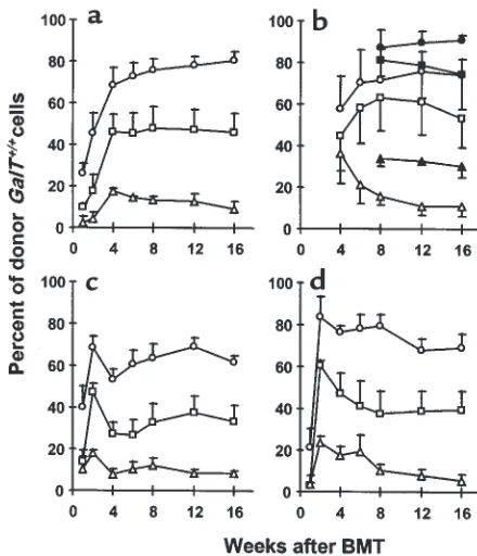
Figure 1
Long-lasting multilineage GalT+/+→GalT–/–mixed chimerism in
recip-ients prepared with a nonmyeloablative conditioning regimen. Peripheral WBCs were stained with FITC-conjugated CD4, anti-CD8, B220, and anti-MAC1 mAb’s, together with biotinylated anti–donor mouse H-2Kb5F1 mAb and PE-streptavidin. Cells
stain-ing with 5F1 were identified as donor-derived cells. Kinetics of donor reconstitution (mean ±SEM) among B cells (a), T cells (b; open sym-bols: CD4+cells; closed symbols: CD8+cells), monocytes (c), and
granulocytes (d) in WBCs. GalT–/–(H-2d) mice received anti-CD4 and
anti-CD8 mAb’s on day –5, followed by whole-body irradiation (3 Gy), thymic irradiation (7 Gy), and T cell–depleted GalT+/+(H-2bxd)
BMCs (circles: 20 ×106BMCs [n= 5]; squares: 7.5 ×10⁶BMCs [n=
immunoglobulin (250 ng/mL; Southern Biotechnolo-gy Associates Inc.). Color development was achieved using 0.1 mg/mL o-phenylenediamine dihydrochloride (OPD; Sigma Chemical Co., St. Louis, Missouri, USA) in substrate buffer. The OPD reaction was stopped using 3 M NH2SO4, and absorbance at 492 nm was measured.
Enzyme-linked immunospot for detecting anti-Gal–produc-ing cells.The enzyme-linked immunospot (ELISPOT) assay was performed as described previously (19). Briefly, nitrocellulose membranes from a 96-well fil-tration plate (Millipore Corp., Bedford, Massachusetts, USA) were coated with 5 µg/mL of synthetic Galα 1-3Galβ1-4GlcNAc conjugated to BSA (Gal-BSA; Alber-ta Research Council, AlberAlber-ta, AlberAlber-ta, Canada). Non-specific binding sites were blocked with 0.4% BSA in culture medium. Serial dilutions of spleen, bone mar-row, or peritoneal cell suspension were added to tripli-cate wells. After a 24-hour culture at 37°C, bound anti bodies were detected using horseradish peroxi-dase–conjugated goat mouse IgM plus IgG anti-bodies (Southern Biotechnology Associates Inc.), fol-lowed by color development with 3-amino-9-ethyl carbazole (Sigma Chemical Co.).
FCM analysis and cell sorting of B cells bearing receptors for Gal.One million cells per 100 µL were incubated with 0.5 µg/100 µL FITC-conjugated Gal-BSA (Alberta Research Council) or 0.5 µg/100 µL control FITC-con-jugated BSA (Fisher Scientific Co., Pittsburgh,
Penn-sylvania, USA) in FCM medium for 2 hours at 4°C, together with biotinylated anti-mouse IgM mAb (PharMingen) (for 2-color FCM) or PE-conjugated anti-CD19 (PharMingen) and biotinylated anti–donor mouse H-2Kb5F1 mAb’s (for 3-color FCM). FITC
con-jugation of Gal-BSA and BSA was performed with the QuickTag FITC Conjugation Kit (Boehringer Mannheim Biochemicals, Indianapolis, Indiana, USA) according to the manufacturer’s instructions. Biotiny-lated mAb’s were viewed with PE-streptavidin (for 2-color FCM) or CyChrome-streptavidin (for 3-2-color FCM). Based on Gal-BSA binding and IgM expression, splenic Gal-BSA+/IgM+ and Gal-BSA–/IgM+
popula-tions were sorted under sterile condipopula-tions using a MoFlo high-speed cell sorter (Cytomation, Fort Collins, Colorado, USA). Sorted cells were immediate-ly resuspended in culture medium and applied to ELISPOT plates precoated with Gal-BSA to determine the frequency of anti-Gal (IgM)–producing cells.
[image:4.612.83.524.52.273.2]Heterotopic heart transplantation.Cervical heterotopic heart transplantation was performed using the cuff technique modified from a method described previous-ly (27). Briefprevious-ly, the recipients were initialprevious-ly prepared before the donor heart harvest to minimize the graft ischemic time. The right external jugular vein and the right common carotid artery were dissected free, mobi-lized as far as possible, and fixed to the appropriate cuffs. The cuffs were composed of polyethylene tubes
Figure 2
Reduced anti-Gal NAb levels in sera of GalT+/+→GalT–/–mixed chimeras. (a) Representative histograms obtained by FCM analysis show
an absence of anti-Gal NAb’s in GalT+/+→GalT–/–mixed chimeras. C.B.-17 scidmouse (GalT+/+) spleen and BMCs were stained with sera
from normal GalT+/+, from control conditioned GalT–/–mice, or from BMT recipients; NAb’s were detected using rat anti-mouse
IgM-FITC as secondary mAb. Representative histogram appearances from sera obtained at 4 weeks after conditioning/BMT are shown (10
µL of undiluted serum per 1 ×10⁶C.B.-17 scidcells was used). (b) Kinetics of serum anti-Gal NAb levels measured by FCM analysis. The
anti-Gal NAb levels are presented as median fluorescence intensity (MFI). Average values ±SEM for the individual groups are shown. Number of animals in each group: normal GalT–/–mice, n= 6; conditioned GalT–/–mice, n= 5; chimeras receiving 20 ×106BMCs,
n= 5; chimeras receiving 7.5 ×10⁶BMCs, n= 5; chimeras receiving 1 ×10⁶BMCs, n= 3; and normal GalT+/+mice, n= 5. *P < 0.05
com-pared with similarly conditioned GalT–/–control mice that did not receive BMT. There was no statistical difference between BMT
(2.5F; Portex Ltd., London, United Kingdom) in which diameters were adjusted by the physical extension. The aorta and the main pulmonary artery of the harvested donor heart were drawn over the end of the common carotid artery and the external jugular vein for the anas-tomoses. The ischemic time of the graft hearts was less than 30 minutes. The function of the grafts was
moni-tored by daily inspection and palpation. Rejection was determined by the cessation of beating of the graft and was confirmed by histology.
Histological studies.Formalin-fixed grafted heart tissue sections were stained with hematoxylin and eosin (H&E) and examined microscopically. Fresh frozen grafted heart tissue sections were stained with fluores-cein-labeled anti-mouse IgG and IgM (Sigma Chemical Co.) and C3 (Cappel Research Products, Durham, North Carolina, USA) and were examined by fluores-cence microscopy.
Statistics.The results were statistically analyzed by the unpaired or paired Student’s ttest of means or the log rank test when appropriate. A Pvalue less than 0.05 was considered to be statistically significant.
Results
Lasting multilineage GalT+/+→GalT–/–mixed chimerism can
be induced by nonmyeloablative conditioning.To determine whether GalT+/+pluripotent hematopoietic stem cell
engraftment could be achieved in GalT–/–recipients
treated with a nonmyeloablative conditioning regimen that permits the development of allogeneic chimerism and T-cell tolerance across MHC barriers (23), we administered varying doses (20 ×106, 7.5 ×10⁶, and 1 ×
106) of T cell–depleted GalT+/+BMCs (H2bxd) to GalT–/–
recipients (H-2d) conditioned with depleting anti-CD4
plus anti-CD8 mAb’s on day –5, and with 3 Gy whole-body irradiation and 7 Gy thymic irradiation on day 0. In addition to the Gal epitope, the donor strain expressed a full MHC haplotype (H-2b) not shared by
the recipients. Untreated GalT–/–and GalT+/+mice, and
conditioned GalT–/–and GalT+/+ mice not receiving
BMT, served as controls. In all recipients of 20 ×106
and 7.5 ×10⁶BMCs (n= 5 per group), and in 3 of 5
recipients of 1 ×10⁶BMCs, lasting mixed chimerism
was observed in peripheral blood B cells, CD4+and
CD8+T cells, monocytes, and granulocytes at all times
(Figure 1). Erythrocyte chimerism could not be detect-ed because of low levels of Gal and H-2 expression on erythrocytes. Our data indicate that lasting multilin-eage GalT+/+→GalT–/–chimerism could be induced with
nonmyeloablative conditioning when appropriate doses of BMCs are transplanted.
Loss of anti-Gal NAb’s in mixed GalT+/+→GalT–/–chimeras.
The kinetics of serum anti-Gal NAb levels in each group are shown in Figure 2. In the control untreated
GalT–/–mice, serum anti-Gal NAb levels showed a
grad-ual increase during the observation period, consistent with the age-related increase in mouse NAb described previously (28). In the control non-BMT GalT–/–mice
that received conditioning treatment, serum anti-Gal NAb levels increased further at 2 weeks (P < 0.05 before conditioning vs. 2 weeks after conditioning) and remained high throughout the follow-up period. This increase of anti-Gal NAb levels may be due to loss of regulation by T cells, as similar results were observed in
GalT–/–mice treated only with depleting anti-CD4 plus
anti-CD8 mAb’s (H. Ohdan et al., manuscript in
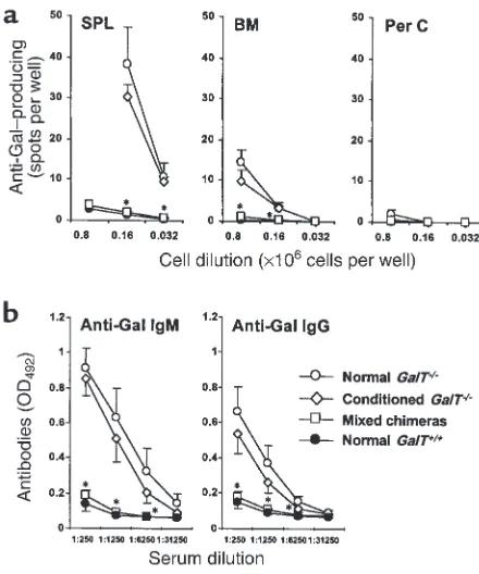
prepa-Figure 3
Absence of anti-Gal–producing cells in GalT+/+→GalT–/– mixed
chimeras (19–20 weeks after BMT). (a) ELISPOT detection of anti-Gal–producing (IgM + IgG) cells. Spleen cells (SPL), BMCs (BM), and peritoneal cavity cells (Per C), prepared from normal and conditioned GalT–/–mice, normal GalT+/+mice, and mixed chimeras 8 days after
immunization with rabbit erythrocytes, were used in ELISPOT assay. The frequency of anti-Gal–producing cells was determined as average of red plaque number in triplicate wells of serially diluted cells. The results shown are the average values ±SEM for the individual groups. Number of animals in each group: normal GalT–/–mice, n= 4; normal GalT+/+mice, n= 4; conditioned GalT–/–mice, n= 5; GalT+/+→GalT–/–
mixed chimeras, n= 7 (results are combined for recipients of 20 ×106,
7.5 ×10⁶, and 1 ×10⁶BMCs). *P < 0.05 compared with normal
con-trol GalT–/–mice and similarly conditioned GalT–/–control mice that
did not receive BMT. There was no statistically significant difference between mixed chimeras and normal GalT+/+control mice in any
tis-sue. (b) Serum levels of anti-Gal antibodies after immunization with Gal-bearing xenogeneic cells. Normal and conditioned GalT–/–mice,
normal GalT+/+mice, and mixed chimeras were immunized with
rab-bit erythrocytes, and serum anti-Gal levels were measured by ELISA assay 8 days after immunization. Average values ±SEM for the indi-vidual groups are shown. Number of animals in each group: normal GalT–/–mice, n= 5; conditioned GalT–/–mice, n= 4; mixed chimeras, n= 13 (results are combined for recipients of 20 ×106, 7.5 ×10⁶, and
1 ×10⁶BMCs); normal GalT+/+mice, n= 5. *P < 0.05 compared with
normal control GalT–/–mice and similarly conditioned GalT–/–control
mice that did not receive BMT. There was no statistical difference between mixed chimeras and normal GalT+/+control mice at any
[image:5.612.66.286.52.316.2]ration). In contrast, GalT+/+→GalT–/–mixed chimeras
had significantly reduced serum levels of anti-Gal NAb by 2 weeks after BMT. These declined further over time, eventually becoming undetectable above the level observed in control GalT+/+mice.
Absence of anti-Gal–producing cells in mixed chimeras.The observation that mixed GalT+/+→GalT–/–chimerism led
to a specific reduction in anti-Gal NAb’s suggested that
GalT+/+hematopoietic chimerism led to tolerance of
anti-Gal NAb–producing cells. However, to rule out the possibility that anti-Gal NAb’s were merely absorbed by the Gal epitope expressed on engrafted GalT+/+cells, the
presence of anti-Gal–producing cells was assessed by ELISPOT assay. Eighteen to 19 weeks after BMT, chimeras were immunized by intraperitoneal injection of 1 × 10⁹ rabbit erythrocytes, which express large
amounts of Gal (29). Eight days after immunization, anti-Gal (IgM and IgG)–producing cells were quanti-fied in recipient spleen cells, BMCs, and peritoneal cav-ity cells. As is shown in Figure 3a, cells producing anti-Gal were undetectable in all 3 tissues of mixed chimeras, whereas large numbers of these cells were detected predominantly in the spleen of both untreat-ed and conditionuntreat-ed GalT–/–mice. The results in mixed
chimeras resembled those from control GalT+/+mice,
which also lacked cells producing anti-Gal. In artificial mixtures of spleen cells from GalT+/+and GalT–/–mice
immunized with rabbit erythrocytes, a linear relation-ship between the frequency of anti-Gal–producing cells and the percentages of GalT–/–cells in the mixtures
(r2= 0.9881) was observed (data not shown), ruling out
absorption of anti-Gal by GalT+/+ cells of mixed
chimeras in the ELISPOT assay.
Consistent with the absence of anti-Gal–producing cells in mixed chimeras, serum levels of anti-Gal (both IgM and IgG) in mixed chimeras were undetectable, even after immunization with rabbit erythrocytes (Fig-ure 3b). In mixed chimeras and conditioned GalT–/–
[image:6.612.116.254.51.231.2] [image:6.612.322.509.227.522.2]mice, total serum IgM and IgG levels were maintained at normal levels at all time points (data not shown). In all mixed chimeras, serum levels of anti-rabbit erythro-cyte IgM antibody were elevated after immunization with rabbit erythrocytes, to levels similar to those detected in normal GalT+/+ control mice (data not
Figure 4
Absence of anti-Gal–producing cells in mixed chimeras 2 weeks after BMT (ELISPOT detection of anti-Gal IgM/IgG–producing cells). Spleen cells from conditioned GalT–/–mice receiving 20 ×106GalT+/+BMCs
(mixed chimeras), from conditioned GalT–/–mice receiving 20 × 10630
Gy–irradiated GalT+/+BMCs (nonchimeras), and from normal GalT+/+
mice were used in ELISPOT assay. The frequency of anti-Gal–producing cells was determined as the average of red plaque numbers in triplicate wells of 8 ×10⁵cells. Average values ±SEM for the individual groups are
shown. Each point represents an individual mouse. *P < 0.05 compared with conditioned GalT–/–mice receiving irradiated BMCs.
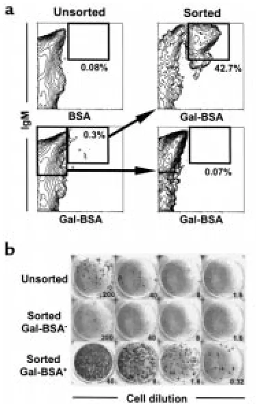
Figure 5
B cells bearing receptors for Gal detected by Gal-BSA comprise the anti-Gal–producing population in the spleens of GalT–/–mice. Spleen
cells were prepared from 5 normal GalT–/–mice (12 weeks of age) 8
days after immunization by intraperitoneal injection of 1 ×10⁹
rab-bit erythrocytes. The pooled cells were stained with FITC-conjugat-ed Gal-BSA or control FITC-conjugatFITC-conjugat-ed BSA, together with biotiny-lated anti-mouse IgM mAb and PE-streptavidin. The populations of Gal-BSA–binding and –nonbinding B cells (IgM+) were sorted as
described in Methods. (a) FCM results of Gal-BSA–binding spleen cells. Sorted cells were reanalyzed for purity; 30,000 cells were ana-lyzed for each contour plot. Percentages given are of total spleen cells. (b) ELISPOT detection of anti-Gal (IgM)–producing cells. The frequencies of anti-Gal–producing cells were determined for unsort-ed and sortunsort-ed cells by ELISPOT assay. Numbers refer to the total cells seeded per well (×103). Anti-Gal–producing cells were greatly
enriched in the sorted Gal-BSA–binding B-cell population. The cal-culated frequencies of anti-Gal–producing cells were 0.1/103,
0.005/103, and 56/103in the unsorted, sorted Gal-BSA–/IgM+, and
shown). Thus, the absence of a response to Gal, and the preserved normal response to other rabbit erythrocyte antigens in mixed GalT+/+→GalT–/–chimeras, confirms
the presence of specific tolerance of Gal-reactive B cells. Tolerance was apparent as early as 2 weeks after BMT, as indicated by the experiment presented in Figure 4, in which conditioned GalT–/–mice received 20 × 106GalT+/+
BMCs that were either untreated or irradiated with 30 Gy. The use of irradiated GalT+/+ BMT provided an
appropriate control in which mixed chimerism could not be induced, but similar antigenic exposure occurred.
As expected, mixed chimerism was observed in all recip-ients of nonirradiated BMCs, but not in the reciprecip-ients of irradiated BMCs. Despite the presence of host B cells in mixed chimeras at 2 weeks (mean: 42.7 ± 4.7% of splenic CD19+cells), cells producing anti-Gal (IgM or IgG) were
undetectable in the spleens of mixed chimeras by ELISPOT assay. In contrast, most conditioned GalT–/–
recipients of irradiated GalT+/+BMCs had measurable
Gal–producing spleen cells. The frequency of anti-Gal–producing cells in the control conditioned GalT–/–
mice receiving irradiated GalT+/+BMCs was similar to
that of untreated normal GalT–/–mice (data not shown).
Thus, mixed hematopoietic chimerism is necessary for the induction of B-cell tolerance, as opposed to antigen exposure alone. Anti-Gal–producing cells were tolerant in mixed chimeras by 2 weeks after BMT.
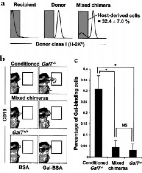
Absence of B cells bearing receptors for Gal in the spleens of mixed chimeras.By surface staining with FITC-conju-gated Gal-BSA, B cells bearing surface IgM (sIgM) receptors that can recognize Gal epitopes could be identified. In normal GalT–/– mice immunized
intraperitoneally with 1 ×10⁹rabbit erythrocytes,
Gal-BSA–binding B cells were detected in the spleen (a pri-mary site of anti-Gal production, as demonstrated in Figure 3a) (Figure 5a). The combined FCM sorting and ELISPOT assay revealed that anti-Gal (IgM)–producing cells were greatly enriched in the sorted Gal-BSA+/IgM+
population, but were undetectable in the sorted Gal-BSA–/IgM+population (Figure 5b), demonstrating that
the Gal-BSA–binding spleen cells included all anti-Gal–producing cells.
To address the question of how the specific toler-ance of Gal-reactive B cells was maintained in mixed chimeras, we looked for the presence of B cells bear-ing receptors for Gal in long-term mixed chimeras. Spleen cells from mixed chimeras were analyzed for Gal-BSA–binding B cells 22 weeks after BMT and 8 days after immunization by intraperitoneal injection of 1 × 10⁹ rabbit erythrocytes. Gal-BSA–binding
spleen cells were detected in control conditioned
GalT–/– mice that did not receive BMT; they were
undetectable in GalT+/+mice (Figure 6b). In mixed
chimeras, the frequency of Gal-BSA–binding splenic B cells among host-derived cells (GalT–/–) was
marked-ly lower than in conditioned GalT–/–mice, and
resem-bled that of GalT+/+ mice (Figure 6c). Thus, B cells
bearing receptors for Gal were absent from the major site of anti-Gal production in mixed chimeras.
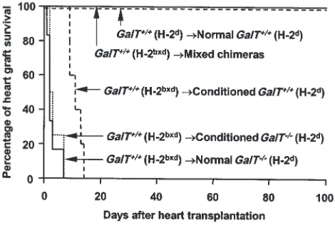
Permanent acceptance of hearts from GalT+/+mice in mixed
chimeras.To evaluate T-cell and antibody tolerance in mixed GalT+/+(H-2bxd)→GalT–/– (H-2d) chimeras, we
transplanted hearts from donor-type GalT+/+(H-2bxd)
mice to chimeras 19–20 weeks after BMT. To enhance anti-Gal production, recipient animals were immunized by intraperitoneal injection of 1 ×10⁹rabbit erythrocytes
8 or 9 days before heart transplantation. All untreated and conditioned control H-2dGalT–/–mice (n= 6 and n
= 4, respectively) rejected H-2bxdGalT+/+hearts within 1–7
days (Figure 7). Upon histological and
immunohisto-Figure 6
Absence of B cells bearing receptors for Gal in the spleens of mixed chimeras (22 weeks after BMT). Spleen cells were prepared from con-ditioned GalT–/–mice (H-2d), normal GalT+/+mice (H-2d), and mixed
chimeras (H-2bxd→H-2d) 8 days after immunization with rabbit
erythrocytes. The cells were stained with FITC-conjugated Gal-BSA or control FITC-conjugated BSA together with PE-conjugated anti-CD19 mAb and biotinylated anti–donor mouse H-2Kb5F1 mAb +
CyChrome-streptavidin. (a) In mixed chimeras, the host-derived H-2Kb–negative cells were selected by gating and analyzed for the
fre-quency of Gal-BSA–binding B cells. The percentage of host-derived cells in the spleens of mixed chimeras is indicated as average value ± SEM. (b) Representative contour plots obtained by FCM analysis show an absence of Gal-BSA–binding B cells in the spleens of mixed chimeras. To ensure statistical significance, data on 100,000 host-derived cells were collected for each sample. (c) The frequency of Gal-BSA–binding B cells was calculated by subtracting the percentage of CD19+cells staining with control FITC-conjugated BSA from the
per-centage of CD19+cells staining with FITC-conjugated Gal-BSA.
Per-centages of total host-derived spleen cells are shown. Average values
±SEM for the individual groups are shown (*P < 0.01; NS = no sta-tistical difference). Number of animals in each group: conditioned GalT–/–mice that did not receive BMT, n= 4; mixed chimeras, n= 4;
[image:7.612.57.290.50.335.2]logical examination, hearts rejected at early time points (1–3 days; 8 of 10 animals) showed massive interstitial hemorrhage, neutrophil infiltration (Figure 8a), and deposition of IgM, IgG, and the C3 component of com-plement on the capillary and vessel endothelia (Figure 9, a–c). These are all major features of humoral rejection. In contrast, the hearts rejected at later time points in this group (7 days; 2 animals) showed diffuse mononuclear cell infiltration, in addition to IgM, IgG, and C3 deposi-tion. All conditioned control H-2dGalT+/+mice (n= 5)
rejected H-2bxdGalT+/+hearts in 9–14 days (Figure 7).
These rejected hearts showed severe diffuse mononu-clear cell infiltration and myocardial necrosis (Figure 8b), which were typical features of cell-mediated rejec-tion, but significant deposits of IgM, IgG, and C3 were not observed. In contrast, in all mixed chimeras (n= 5) — except for one that died in 6 days with a nonrejecting, histologically normal-looking heart — grafted GalT+/+
hearts survived indefinitely (>150 days) (Figure 7). None of these surviving hearts revealed any histological sign of rejection, and they showed a complete absence of immunoglobulin and complement deposition at day 165 (Figure 8c and Figure 9, d–f).
Discussion
We demonstrate here the simultaneous induction of T-cell tolerance and specific tolerance of Gal-reactive B cells in GalT+/+→GalT–/–mixed chimeras prepared with
a relatively nontoxic, nonmyeloablative regimen. These results suggest that the successful induction of mixed hematopoietic chimerism with nonmyeloablative con-ditioning in the pig-to-human (GalT+/+→GalT–/–)
xeno-geneic combination could lead to tolerance among Gal-reactive B cells, as well as T cells recognizing histocompatibility antigens. Although the pathophys-iological consequence of losing anti-Gal antibodies needs to be determined (30), it seems probable that induction of specific tolerance will be associated with less risk of infection than the high level of chronic immunosuppression that would be required to prevent xenograft rejection.
For success to be achieved with this approach, the pos-sible role that preexisting anti-Gal NAb’s may play in resisting GalT+/+xenogeneic marrow engraftment must
first be addressed. In this study, GalT–/–recipient mice
with relatively high levels of preexisting anti-Gal NAb’s before BMT developed reduced GalT+/+hematopoietic
chimerism compared with GalT–/–recipients with lower
levels of preexisting anti-Gal NAb’s (data not shown). However, once GalT+/+→GalT–/–mixed chimerism was
achieved in GalT–/–mice, it was stable, and, even if
pres-ent at low levels (7–10% donor WBCs), it was associated with both B-cell and T-cell tolerance. Our findings sug-gest that anti-Gal NAb’s may have inhibitory effects on the engraftment of GalT+/+BMCs, but that this degree of
resistance can be overcome when sufficient GalT+/+
BMCs are administered. This speculation is also sup-ported by our previous findings that the ability of murine sera to inhibit engraftment of rat BMCs was cor-related with their cytotoxic anti-rat NAb content, and that their inhibitory effect could be overcome by admin-istration of large numbers of rat BMCs (28, 31, 32). Con-sistent with these results, we have confirmed the feasi-bility of inducing mixed chimerism in GalT–/–mice with
high levels of anti-Gal due to immunization with rabbit erythrocytes before conditioning. When high-dose BMT with GalT+/+BMCs was given to such mice, chimerism
and tolerance were achieved (H. Ohdan, K. Swenson, and M. Sykes, manuscript in preparation). If this approach were applied to the pig-to-human combination, previ-ously reported strategies for reducing initial anti-Gal NAb and/or complement levels should be used to facili-tate initial BMC engraftment. Despite such efforts, how-ever, induction of persistent mixed chimerism has not yet been achieved in a pig-to-primate combination. In addition to the vigorous immune response to xenoanti-gens, nonimmune physiological factors (e.g., the failure of crucial growth factors, adhesion molecular interac-tions, and cytokines to function across species barriers) also limit the achievement of chimerism between dis-cordant species (33). Some of these problems might be alleviated by the use of donor-specific growth factors and/or cytokines at the time of BMT (34), and others may require genetic engineering of donor pigs.
[image:8.612.57.292.52.212.2]Our previous results involving induction of mixed chimerism in lethally irradiated mice demonstrated that newly developing anti-Gal–producing B cells could be
Figure 7
Permanent acceptance of GalT+/+ donor-type hearts in
GalT+/+→GalT–/–mixed chimeras. The hearts from donor-type GalT+/+
(H-2bxd) mice were heterotopically transplanted into mixed chimeras
(n= 6; including 2 chimeras transplanted with 20 ×106, 7.5 ×10⁶,
or 1 ×10⁶GalT+/+BMCs) 19–20 weeks after BMT, as well as into
con-ditioned control GalT–/–(H-2d) (n= 6) and GalT+/+mice (H-2d) (n=
5) and untreated control GalT–/–mice (H-2d) (n= 4). As an
H-2–iden-tical control, hearts from GalT+/+(H-2d) mice were transplanted into
GalT+/+(H-2d) mice (n= 3). To enhance anti-Gal NAb production, all
recipient animals were immunized by intraperitoneal injection of 1 × 10⁹rabbit erythrocytes 8 or 9 days before heart transplantation.
Sur-vival curves of the grafted hearts are shown. P < 0.005 normal GalT–/–, conditioned GalT–/–, or conditioned GalT+/+ vs. mixed
chimeras. P < 0.005 normal GalT–/–or conditioned GalT–/–vs.
tolerized by the presence of GalT+/+hematopoietic cells
(19). In contrast to lethal irradiation, the conditioning regimen used here would not be expected to eliminate preexisting anti-Gal–producing B cells (35, 36). Consis-tent with this possibility, we observed increasing anti-Gal NAb levels in sera of GalT–/–mice receiving
non-myeloablative conditioning without BMT. Thus, anti-Gal NAb–producing cells were present at high lev-els after conditioning. In contrast, anti-Gal–producing cells were already undetectable by ELISPOT assay in mixed chimeras as early as 2 weeks after BMT, suggest-ing that mixed chimerism induced tolerance among anti-Gal–producing cells that preexisted at the time of BMT. The cells that produce anti-Gal in GalT–/–mice
have not been defined. However, if it is assumed that their half-life is similar to that of other murine mature B cells (6 weeks) (37) or plasma cells (as long-lived as memory B cells) (36), then it is likely that preexisting anti-Gal NAb–producing mature B or plasma cells are tolerized by the induction of mixed chimerism. Although mature B cells are generally thought to be less sensitive than newly developing cells to tolerance induc-tion by self-antigens or foreign antigens (38), chronic exposure to antigenic determinants present on cell sur-faces has been reported to eliminate mature B cells in immunoglobulin-transgenic mice (39, 40). Based on these results, it is likely that preexisting anti-Gal–pro-ducing cells are tolerized through antigen-receptor cross-linking in mixed chimeras. However, an alterna-tive explanation for the rapid development of tolerance in mixed chimeras is that anti-Gal–producing B cells or plasma cells may have rapid turnover. Studies are in progress to distinguish these possibilities.
In mixed chimeras, Gal epitopes may be recognized as self-antigens during B-cell maturation. The known mechanisms mediating tolerance of self-reactive B cells include clonal deletion (i.e., the physical elimination of
autoreactive B-cell clones) (38, 40–42), anergy (i.e., the functional inactivation of autoreactive B cells) (43, 44), and receptor editing (i.e., the modification of B-cell receptors of autoreactive cells) (45–47). To investigate the possibility that these mechanisms are also involved in maintaining Gal-reactive B-cell tolerance in mixed chimeras, we assayed the presence of B cells with recep-tors (sIgM) recognizing Gal epitopes. Using FITC-con-jugated Gal-BSA, we could directly identify B cells bear-ing anti-Gal receptors in the spleens of normal GalT–/–
mice (Figure 5a). Because anti-Gal–producing cells express sIgM (H. Ohdan and M. Sykes, unpublished data), the specificity of the Gal-BSA ligand for the cor-responding receptors on B cells was demonstrated by showing enrichment of anti-Gal–producing cells among Gal-binding B cells and a complete absence of anti-Gal–producing cells among non–Gal-binding B cells, using combined FCM sorting and ELISPOT assay (Figure 5b). In mixed chimeras, B cells with receptors recognizing Gal were completely absent in the spleens (Figure 6), suggesting the possibility of clonal deletion and/or receptor editing of Gal-reactive B cells as a mechanism of tolerance.
In the present study, indefinite acceptance of vascu-larized GalT+/+ heart grafts was demonstrated in
GalT+/+→GalT–/–mixed chimeras, whereas rapid
vascu-lar rejection was observed in untreated and condi-tioned control GalT–/–mice. Although the kinetics of
[image:9.612.57.295.52.174.2]GalT+/+heart rejection in GalT–/–mice were delayed
Figure 8
Histology of transplanted GalT+/+mouse hearts (H&E staining). (a)
Histological findings of rejected GalT+/+mouse heart 2 days after
transplantation into control conditioned GalT–/–mouse that did not
receive BMT, with interstitial hemorrhage and neutrophil infiltration. (b) Histological findings of rejected GalT+/+mouse heart 12 days after
transplantation into control conditioned GalT+/+mouse, showing
dif-fuse mononuclear cell infiltration. (c) Histological findings of GalT+/+
mouse heart 165 days after transplantation into GalT+/+→GalT–/–
mixed chimera. The graft shows no evidence of any type of rejection.
Figure 9
Immunofluorescence staining for IgM, IgG, and C3 of transplanted GalT+/+mouse hearts. Rejected GalT+/+mouse heart 2 days after
transplantation into control conditioned GalT–/–mouse that did not
receive BMT shows endothelial deposition of IgM (a), IgG (b), and C3 (c). GalT+/+mouse heart 165 days after transplantation into GalT+/+→GalT–/–mixed chimera shows no deposition of IgM (d), IgG
[image:9.612.303.540.406.646.2]compared with the typical HAR that usually occurs within minutes to hours in pig-to-primate combina-tions (12, 48), the histological and immunofluores-cence data are consistent with a role for anti-Gal in rejection. The role of antibodies in this process is fur-ther substantiated by the more delayed (cell-mediated) rejection observed in GalT+/+to GalT+/+mice with
simi-lar histoincompatibilities. The slower time course of
GalT+/+allogeneic mouse heart rejection observed in
GalT–/–mice, compared with pig-to-primate
transplan-tation, may be explained, first, by a lower inherent abil-ity of mice to fix complement by the classical pathway and, second, by the intraspecies compatibility of com-plement regulatory proteins in our model. Using the
GalT+/+to GalT–/–mouse heart transplant model, other
reports also demonstrate the absence of HAR (49) or the presence of DXR-like rejection (with graft survival of 8–12 days) (50). The more rapid rejection of allo-grafts in control mice in our studies may be due to dif-ferences in the anatomical site of heart grafting, to the presence of MHC alloantigens in our donors, and, most probably, to the more advanced age of the mice in our studies, which was associated with high levels of anti-Gal antibodies at the time of heart grafting. Despite this rapid rejection in control mice, GalT+/+
heart allografts in mixed chimeras were free from all types of rejection, indicating the presence of tolerance at the level of both T and B cells. This is, to our knowl-edge, the first direct demonstration that the induction of mixed chimerism can simultaneously prevent both T cell– and antibody-mediated rejection of vascularized solid-organ grafts. Taken together with our previous results demonstrating the induction of T-cell tolerance across rat-to-mouse and pig-to-mouse species barriers induced by a regimen similar to that used in the pres-ent studies (51, 52), it could be expected that the suc-cessful induction of mixed hematopoietic chimerism in the pig-to-human xenogeneic combination would similarly result in T- and B-cell tolerance and accept-ance of vascularized organ xenografts.
Acknowledgments
The authors thank D.K.C. Cooper and A.D. Thall for helpful review of the manuscript; D.H. Sachs for advice and encouragement; H.S. Kruger Gray for cell sorting assistance; and D. Plemenos for expert secretarial assis-tance. This work was supported by the National Insti-tutes of Health (grants POI 18646 and ROI HL-49915) and by a sponsored research agreement between Massachusetts General Hospital and BioTransplant Inc. H. Ohdan was partially supported by Naito Foundation.
1. Platt, J.L. 1996. Xenotransplantation: recent progress and current per-spectives. Curr. Opin. Immunol.8:721–728.
2. Sachs, D.H. 1994. The pig as a xenograft donor. Pathol. Biol.42:217–219. 3. Oriol, R., Ye, Y., Koren, E., and Cooper, D.K.C. 1993. Carbohydrate anti-gens of pig tissues reacting with human natural antibodies as potential targets for hyperacute vascular rejection in pig-to-man organ xeno-transplantation. Transplantation.56:1433–1442.
4. Galili, U. 1993. Interaction of the natural anti-Gal antibody with α -galac-tosyl epitopes: a major obstacle for xenotransplantation in humans.
Immunol. Today.14:480–482.
5. Sandrin, M., Vaughan, H.A., Dabkowski, P.L., and McKenzie, I.F.C. 1993. Anti-pig IgM antibodies in human serum react predominantly with Galα1-3Gal epitopes. Proc. Natl. Acad. Sci. USA.90:11391–11395. 6. Leventhal, J.R., et al. 1993. Prolongation of cardiac xenograft survival by
depletion of complement. Transplantation.55:857–865.
7. Cozzi, E., and White, D.J.G. 1995. The generation of transgenic pigs as potential organ donors for humans. Nat. Med.1:964–967.
8. Sablinski, T., et al. 1995. Xenotransplantation of pig kidneys to nonhu-man primates: I. Development of a model. Xenotransplantation.2:264–270. 9. Simon, P.M., et al. 1998. Intravenous infusion of Galα1-3Gal oligosac-charides in baboons delays hyperacute rejection of porcine heart xenografts. Transplantation.65:346–353.
10. Sandrin, M.S., et al. 1995. Enzymatic remodelling of the carbohydrate surface of a xenogeneic cell substantially reduces human antibody bind-ing and complement-mediated cytolysis. Nat. Med.1:1261–1267. 11. Osman, N., et al. 1998. Combined transgenic expression of α
-galactosi-dase and α1,2-fucosyltransferase leads to optimal reduction in the major xenoepitope Galα(1,3)Gal. Proc. Natl. Acad. Sci. USA.94:14677–14682. 12. Cooper, D.K.C. 1998. Xenoantigens and xenoantibodies.
Xenotransplan-tation.5:6–17.
13. Galili, U., Minanov, O.P., Michler, R.E., and Stone, K.R. 1997. High-affin-ity anti-Gal immunoglobulin G in chronic rejection of xenografts. Xeno-transplantation.4:127–131.
14. Palmetshofer, A., Galili, U., Dalmasso, A.P., Robson, S.C., and Bach, F.H. 1998. α-galactosyl epitope-mediated activation of porcine aortic endothelial cells. Transplantation.65:844–853.
15. Bach, F.H., et al. 1995. Barriers to xenotransplantation. Nat. Med.
1:869–873.
16. Lin, Y., Goebels, J., Xia, G., Vandeputte, M., and Waer, M. 1998. Induc-tion of specific transplantaInduc-tion tolerance across xenogeneic barriers in the T-independent immune compartment. Nat. Med.4:173–180. 17. Lin, Y., Vandeputte, M., and Waer, M. 1998. Accommodation and
T-inde-pendent B cell tolerance in rats with long term surviving hamster heart xenografts. J. Immunol.160:369–375.
18. LaTemple, D.C., and Galili, U. 1998. Adult and neonatal anti-Gal response in knock-out mice for α1,3galactosyltransferase. Xenotrans-plantation.5:191–196.
19. Yang, Y.-G., et al. 1998. Tolerization of anti–Galα1-3Gal natural anti-body-forming B cells by induction of mixed chimerism. J. Exp. Med.
187:1335–1342.
20. Thall, A.D., Maly, P., and Lowe, J.B. 1995. Oocyte Galα1,3Gal epitopes implicated in sperm adhesion to the zona pellucida glycoprotein ZP3 are not required for fertilization in the mouse. J. Biol. Chem.270:21437–21440. 21. Dialynas, D.P., et al. 1983. Characterization of murine T cell surface mol-ecule, designated L3T4, identified by monoclonal antibody GK1.5: sim-ilarity of L3T4 to human Leu3/T4 molecule. J. Immunol.131:2445–2451. 22. Sarmiento, M., Glasebrook, A.L., and Fitch, F.W. 1980. IgG or IgM mon-oclonal antibodies reactive with different determinants on the molecu-lar complex bearing Lyt2 antigen block T cell-mediated cytolysis in the absence of complement. J. Immunol.125:2665–2672.
23. Sharabi, Y., and Sachs, D.H. 1989. Mixed chimerism and permanent spe-cific transplantation tolerance induced by a non-lethal preparative reg-imen. J. Exp. Med.169:493–502.
24. Sykes, M., Abraham, V.S., Harty, M.W., and Pearson, D.A. 1993. IL-2 reduces graft-vs-host disease and preserves a graft-vs-leukemia effect by selectively inhibiting CD4+T cell activity. J. Immunol.150:197–205. 25. Tomita, Y., Khan, A., and Sykes, M. 1994. Role of intrathymic clonal
dele-tion and peripheral anergy in transplantadele-tion tolerance induced by bone marrow transplantation in mice conditioned with a non-myeloablative regimen. J. Immunol.153:1087–1098.
26. Sherman, L.A., and Randolph, C.P. 1981. Monoclonal anti-H-2Kb
anti-bodies detect serological differences between H-2Kbmutants.
Immuno-genetics.12:183–189.
27. Matsuura, A., Abe, T., and Yasuura, K. 1991. Simplified mouse cervical heart transplantation using a cuff technique. Transplantation.51:896–898. 28. Aksentijevich, I., Sachs, D.H., and Sykes, M. 1991. Natural antibody against bone marrow cells of a concordant xenogeneic species. J. Immunol.147:79–85.
29. Galili, U., Rachmilewitz, E.A., Peleg, A., and Flechner, I. 1984. A unique natural human IgG antibody with anti-galactosyl specificity. J. Exp. Med.
16:1519–1531.
30. Weiss, R.A. 1998. Transgenic pigs and virus adaptation. Nature.
391:327–328.
31. Aksentijevich, I., Sachs, D.H., and Sykes, M. 1991. Natural antibodies can inhibit bone marrow engraftment in the rat→mouse species combina-tion. J. Immunol.147:4140–4146.
32. Aksentijevich, I., Sachs, D.H., and Sykes, M. 1992. Humoral tolerance in xenogeneic BMT recipients conditioned with a non-myeloablative regi-men. Transplantation.53:1108–1114.
Transplan-tation.57:906–917.
34. Yang, Y.-G., et al. 1996. Donor-specific growth factors promote swine hematopoiesis in SCID mice. Xenotransplantation.3:92–101.
35. Anderson, R.E., and Warner, N.L. 1975. Radiosensitivity of T and B lym-phocytes. III. Effect of radiation on immunoglobulin production by B cells. J. Immunol.115:161–169.
36. Slifka, M.K., Antia, R., Whitmire, J.K., and Ahmed, R. 1998. Humoral immunity due to long-lived plasma cells. Immunity.8:363–372. 37. Fulcher, D.A., and Basten, A. 1997. Influences on the lifespan of B cell
subpopulations defined by different phenotypes. Eur. J. Immunol.
27:1188–1199.
38. Klinman, N.R. 1996. The “clonal selection hypothesis” and current con-cepts of B cell tolerance. Immunity.5:189–195.
39. Murakami, M., et al. 1992. Antigen-induced apoptotic death of Ly-1 B cells responsible for autoimmune disease in transgenic mice. Nature.
357:77–80.
40. Russell, D.M., et al. 1991. Peripheral deletion of self-reactive B-cells.
Nature.354:308–311.
41. Nemazee, D., and Buerki, K. 1989. Clonal deletion of autoreactive B lym-phocytes in bone marrow chimeras. Proc. Natl. Acad. Sci. USA.86:8039–8043. 42. Hartley, S.B., et al. 1991. Elimination from peripheral lymphoid tissues of self-reactive B lymphocytes recognizing membrane-bound antigens.
Nature.353:765–769.
43. Goodnow, C.C., et al. 1988. Altered immunoglobulin expression and functional silencing of self-reactive B-lymphocytes in transgenic mice.
Nature.334:676–682.
44. Nossal, G.J.V. 1996. Clonal anergy of B cells: a flexible, reversible, and quantitative concept. J. Exp. Med.183:1953–1956.
45. Tiegs, S., Russell, D., and Nemazee, D. 1993. Receptor editing in self-reactive bone marrow B-cells. J. Exp. Med.177:1009–1020.
46. Hertz, M., and Nemazee, D. 1997. BCR ligation induces receptor editing in IgM+IgD-bone marrow B cells in vitro. Immunity.6:429–438. 47. Pelanda, R., et al. 1997. Receptor editing in a transgenic mouse model:
site, efficiency, and role in B cell tolerance and antibody diversification.
Immunity.7:765–775.
48. Cooper, D.K.C., et al. 1988. Effects of cyclosporine and antibody adsorp-tion on pig cardiac xenograft survival in the baboon. J. Heart Transplant.
7:238–246.
49. McKenzie, I.F.C., Li, Y.Q., Patton, K., Thall, A.D., and Sandrin, M.S. 1998. A murine model of antibody-mediated hyperacute rejection by galactose-α(1,3)galactose antibodies in Gal0/0mice. Transplantation. 66:754–763. 50. Pearse, M., et al. 1998. Anti-gal antibody-mediated allograft rejection in α1,3-galactosyltransferase gene knockout mice. A model of delayed xenograft rejection. Transplantation. 66:748–754.
51. Sharabi, Y., Aksentijevich, I., Sundt, T.M., III, Sachs, D.H., and Sykes, M. 1990. Specific tolerance induction across a xenogeneic barrier: produc-tion of mixed rat/mouse lymphohematopoietic chimeras using a non-lethal preparative regimen. J. Exp. Med.172:195–202.