TGF-
bb
antagonists: Why suppress a tumor
suppressor?
Rosemary J. Akhurst
J Clin Invest.
2002;
109(12)
:1533-1536.
https://doi.org/10.1172/JCI15970
.
Tumor metastasis is the major determinant of cancer patient survival. This ultimate phase in
tumorigenesis depends on the ability of a tumor cell to invade the stroma, migrate in and out
of blood or lymphatic vessels, and survive and re-establish itself at a secondary site. A large
number of papers have provided strong evidence for a role of TGF-
b
in tumor invasion
and/or metastasis (1–6). Now, two papers in this issue of the JCI highlight this clinically
significant action of TGF-
b
in tumorigenesis and provide very encouraging results regarding
both the efficacy and the low toxicity of a soluble TGF-
b
receptor antagonist that effectively
reduces tumor spread (7, 8). Positive and negative effects of TGF-
b
signaling in cancer
TGF-
b
is a potent growth inhibitor of all epithelial and hematopoietic cells and can also
induce apoptosis (1–3). For this reason, much emphasis has been placed on elucidating
TGF-
b
signaling pathways, particularly those responsible for growth inhibition (summarized
in Figure 1). After activation of the TGF
b
type II/TGF
b
type I (T
b
RII/T
b
RI) receptor complex,
TGF-
b
s signal predominantly via the Smad pathway, although the activated receptor
complex can also signal independently of Smads, via phosphatidylinositol 3-kinase (PI3K),
protein phosphatase 2A/p70 S6 kinase (PP2A/p70S6K), and various mitogen-activated
protein kinase (MAPK) pathways. There is also interplay between these pathways, such
that activation of the Ras pathway […]
Commentary
Find the latest version:
TGF-
β
antagonists: Why suppress
a tumor suppressor?
Rosemary J. Akhurst
University of California–San Francisco, Mount Zion Cancer Research Institute, Room S231, Box 0875, 2340 Sutter Street, San Francisco, California 94143-0875, USA.
Phone: (415) 514-0215; Fax: (415) 502-6779; E-mail: rakhurst@cc.ucsf.edu.
J. Clin. Invest.109:1533–1536 (2002). doi:10.1172/JCI200215970.
Tumor metastasis is the major deter-minant of cancer patient survival. This ultimate phase in tumorigenesis depends on the ability of a tumor cell to invade the stroma, migrate in and out of blood or lymphatic vessels, and survive and re-establish itself at a sec-ondary site. A large number of papers have provided strong evidence for a role of TGF-β in tumor invasion and/or metastasis (1–6). Now, two papers in this issue of the JCI high-light this clinically significant action of TGF-βin tumorigenesis and pro-vide very encouraging results regard-ing both the efficacy and the low tox-icity of a soluble TGF-β receptor antagonist that effectively reduces tumor spread (7, 8).
Positive and negative effects of TGF-βsignaling in cancer
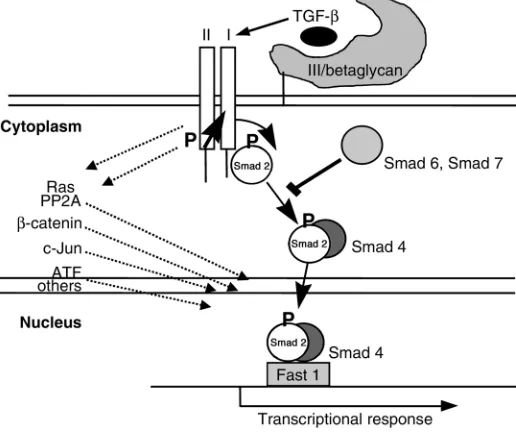
TGF-βis a potent growth inhibitor of all epithelial and hematopoietic cells and can also induce apoptosis (1–3). For this reason, much emphasis has been placed on elucidating TGF-β signaling pathways, particularly those responsible for growth inhibi-tion (summarized in Figure 1). After activation of the TGFβtype II/TGFβ type I (TβRII/TβRI) receptor com-plex, TGF-βs signal predominantly via the Smad pathway, although the activated receptor complex can also signal independently of Smads, via phosphatidylinositol 3-kinase (PI3K), protein phosphatase 2A/p70 S6 kinase (PP2A/p70S6K), and various mitogen-activated protein kinase (MAPK) pathways. There is also inter-play between these pathways, such that activation of the Ras pathway or other non-Smad pathways can mod-ulate signaling via Smads (1–6).
Homozygous mutations or dele-tions in the genes for Smad4, TβRII, or Smad2 are observed in some
human tumors (1–3), suggesting a significant role for TGF-βsignaling in tumor suppression. Nevertheless, only a minority of tumors show this type of genetic aberration, and the most commonly deleted such gene,
MADH4 (encoding Smad4), is not
essential for all TGF-β activities (1–3). Some authors have suggested that the tumor-suppressing function
of MADH4 can be attributed to its
antiangiogenic effect (not necessari-ly mediated by TGF-β), rather than to growth inhibition (9).
The tumor-suppressive effects of TGF-βhave been clearly demonstrat-ed in transgenic mouse models. He-mizygous or homozygous Tgfb1-null animals show an increased incidence of chemically or spontaneously induced tumors, respectively (1–3, 10,
11). Similarly, targeting a dominant negative TβRII to mammary or skin epithelia also enhances tumorigen-icity, whereas TGF-β1–overexpressing mice have a decreased incidence of tumors (1–3). This tumor-suppres-sive function of TGF-β has raised concerns about the use of TGF-β antagonists to treat cancer, des-pite the increasingly strong evidence that TGF-β1 can promote tu-mor metastasis.
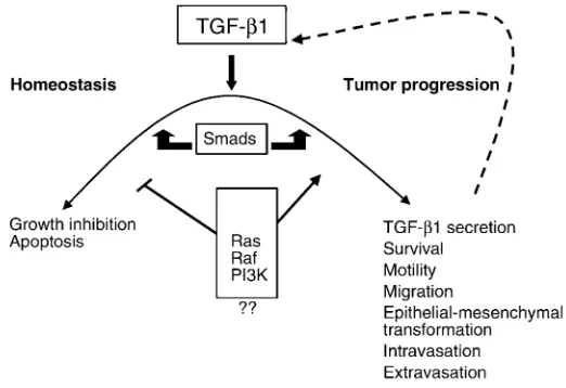
It is widely accepted that during multistage tumorigenesis, TGF-β growth-inhibitory and apoptotic effects are lost, frequently by subver-sion of the normal signaling pathway due to activation of other signaling molecules including PI3K and Ras (1–3). Meanwhile, other TGF-β responses prevail, unrelated to
Commentary
See related articles, pages 1551–1559 [image:2.576.252.510.463.679.2]and pages 1607–1615.
Figure 1
growth inhibition and favoring tumorigenesis (Figure 2; refs. 1–3). Moreover, as tumor cells progress, they secrete ever-increasing quantities of TGF-β1 (1–3, 6). TGF-βactivity is increased partly by autostimulation of the Tgfb1 gene, but also through transcriptional activation by Ras and other effectors, as well as by the action of proteases that activate the latent TGF-βin the ECM (1–3, 6).
In response to elevated TGF-βlevels, the tumor cell becomes more migra-tory and invasive. Indeed, in coopera-tion with activated Ras, TGF-β1 can induce a complete epithelioid-to-fibroblastoid transition in both mam-mary and keratinocyte-derived tumors (1–3, 6), and it can drive metastasis of epithelioid tumors (6–8, 12). TGF-βcan also stimulate tumor angiogenesis, alter the stromal envi-ronment, and cause local and systemic immunosuppression, all of which contribute to tumor progression and metastasis (1–3).
As discussed in the two articles in this issue of the JCI(7, 8), the concept of using soluble protein antagonists that bind and inactivate extracellular TGF-βwas first tested over a decade ago using decorin, a natural inhibitor of TGF-β, in a therapeutic model for fibrosis (8). More recently, the chimeric Fc:TβRII protein used in the current studies has proved attrac-tive because of its high affinity for TGF-β, its ready purification by pro-tein A affinity chromatography, and its effectiveness in a number of mod-els of fibrosis.
Early attempts to demonstrate the efficacy of this approach involved stably transfected glioma (13), thymoma (14), pancreatic (15), or metastatic breast tumor cell lines (16) carrying cDNAs for soluble forms of decorin (13), TβRII (14, 15), or TβRIII (16). Each demon-strated tumor suppression after subse-quent injection of the modified tumor cell line into mice. In the first two cases (13, 14), this was attributed to re-acqui-sition of tumor-specific cellular immu-nity, whereas the effects on the pancreas and breast cancer lines included sup-pression of invasion (15), angiogenesis (15), and lung metastasis (16).
Efficacy and toxicity
The articles in this issue of the JCI(7, 8) have pushed the story two steps further, firstly by applying soluble
Fc:TβRII as an injectable drug to prove efficacy in suppression of breast tumor metastasis in vivo (7), and secondly by screening for any adverse effects on the mice after life-time exposure to high-level circulat-ing Fc:TβRII (8). Muraoka et al. (7), using the MMTV-PyV mT transgenic model of mammary tumorigenesis, show that twice-weekly intraperi-toneal injection of Fc:TβRII reduces lung metastasis tenfold. Fc:TβRII treatment also inhibits metastasis of two metastatic mammary cell lines. In all three cases, Fc:TβRII has no effect on proliferative rate of the pri-mary tumor cells. Yang et al. (8) take a different approach, focusing on possible adverse effects in transgenic mice that stably express soluble Fc:TβRII. Circulating Fc:TβRII, which is found at about 1 mg/ml in the blood, not only reduces metasta-sis formation of melanoma cells injected into the tail vein of the mice but also reduces metastasis to the lung from endogenous mammary tumors that arise when the mice are crossed onto the MMTV-Neu trans-genic model of mammary carcino-genesis. Both groups find that Fc:TβRII leads to no changes in tumor latency, yield, or size.
Taken together, the two papers (7, 8) show that soluble Fc:TβRII is effi-cacious in reducing tumor metastasis, whether delivered genetically from within the neoplastic cell or adminis-tered as an injectable circulating
drug. Both groups also addressed the mechanisms of action of Fc:TβRII in attenuating metastatic spread. In the MMTV-PyV mT model, Muraoka et al. (7) specifically exclude an effect on TGF-β–induced angiogenesis. In their model, Fc:TβRII appears to decrease tumor cell intravasation and/or decrease survival of tumor cells in the circulation, since the number of cir-culating tumor cells is lower in the Fc:TβRII-treated mice than in con-trols (7). In support of this mecha-nism, Smad2 activation has recently been shown to drive tumor cell extravasation in a skin tumor model (6). Consistent with an effect on tumor intravasation, the Fc:Tβ RII-treated mammary tumor cells are altered toward a more differentiated, less motile/migratory phenotype than is seen in untreated tumor cells. Production of active matrix metallo-proteinase 2 (MMP2) and MMP9, proteases thought to be important in tumor invasion, migration, and intravasation, is diminished and apoptosis is elevated in response to Fc:TβRII (7).
[image:3.576.252.513.501.680.2]In the injectable melanoma model, Yang et al. (8) argue, the effect of Fc:TβRII on metastasis is likely indi-rect, possibly including decreased angiogenesis and/or elevated immunosuppression. Although me-tastasis is diminished in the Fc:TβRII transgenic mice following tail vein injections of melanoma cells, the ini-tial appearance of micro-metastases
Figure 2
is no different from that seen in wild-type mice. Since TGF-βhas multiple actions that can drive tumor metas-tasis, the exact mechanisms involved are probably context-specific, de-pending on the tumor type, genetic constitution, and the exact stage of carcinogenesis of the tumor. Howev-er, the exciting take-home message is that soluble TGF-βantagonists can significantly decrease metastasis in models of breast cancer and melanoma, as previously suggested for thymoma (13), glioma (14), and pancreatic (15) and mammary carcinoma (16).
The complete absence of TGF-β1 in mice leads to death resulting from uncontrolled systemic inflammation, and even a T cell–specific deficit in TGF-βcauses lethal immune system defects (discussed in ref. 8). There-fore, the apparent absence of side effects, even after lifetime exposure to approximately 1 mg/ml circulat-ing Fc:TβRII (8), is particularly encouraging. No immune phenotype was seen in the study of Yang et al. (8), apart from a minimal increase in memory T cells, and a nonsignificant increase in lymphocytic infiltration into organs of aged mice. The authors employed several techniques to demonstrate that Fc:TβRII at this level does not completely block all TGF-β1 bioactivity in vivo (8). Indeed, although circulating TGF-β levels are reduced in the Fc:TβRII transgenics to probably ≤10% of the wild-type level, these animals still thrive in the laboratory. It would be interesting to examine how the mice fare when challenged with other envi-ronmental hazards such as foreign antigens and pathogens.
Also heartening is the finding by both groups that soluble Fc:TβRII had no tumor-promoting action in vivo. Conversely, mice in which TGF-β activity is diminished globally, as a result of hemizygosity forTgfb1, are tumor-prone (10, 11), as are animals in which this factor is ablated in a tissue-specific manner using dominant neg-ative (DN) TβRII. The basis of this dis-crepancy is uncertain, but it may be that Fc:TβRII preferentially targets cir-culating TGF-β1 because it is too bulky to gain access to the more func-tionally important TGF-β tightly bound to the cell surface or ECM. In addition, different thresholds of
TGF-β activity required for the growth-suppressing and the metasta-sis-promoting effects of TGF-βcould help account for the tumor inci-dence seen in DN-TβRII transgenic strains. Growth inhibition, for exam-ple, is more sensitive than other TGF-β responses to decreases in the level of TβRII (3, 17). Moreover, recent studies in a skin carcinogene-sis model do indeed show that a high threshold of Smad2 activation must be surpassed in order to drive metastatic spread (6).
Nevertheless, since Tgfb1+/–animals
have an increased incidence of chem-ically induced tumorigenesis (10), one might expect a similar phenotype in Fc:TβRII mice (8), but this is not the case. The explanation probably lies in the different models used. The tumors and cell lines studied by Muraoka et al. (7) have already lost growth sensitivity to TGF-β, as assessed by BrdU incorporation, so tumor-suppressive effects of TGF-β would not be expected. The TGF-β growth sensitivity of MMTV-Neu tumor cells has not been studied, but transfection of normal differentiated thyroid cells with ErbB2 (Neu) atten-uates the growth-inhibitory response to TGF-β, suggesting that Neu does indeed attenuate growth sensitivity to TGF-β(18). In this context, chem-ical carcinogenesis studies on Fc:TβRII transgenic mice are war-ranted to uncover any tumor-pro-moting effects of Fc:TβRII, especially in view of the fact that a soluble TβRII transfected into a hepatoma cell line has been shown to promote tumor development (19).
Despite these reservations, Fc:TRII clearly is highly efficacious in reducing metastasis and is of exceptionally low toxicity in mice. Indeed, many drugs for treatment of both malignant and nonmalignant conditions, such as cyclosporin, have tumor-promoting activity (12), and most cancer drugs show general cytotoxicity levels orders of magnitude higher than does this soluble TβRII receptor.
Future developments in anti–TGF-βdrug design
Pharmaceutical companies have avoided TGF-βagonists or antago-nists, partly because of fear of non-specificity and consequent side effects. The articles in this issue of
the JCI (7, 8) might cause them to reconsider this decision. TGF-β antagonists such as Fc:TβRII could prove as useful clinically as Herceptin (20), an anti-Neu antibody used for the treatment of Neu-positive breast tumors. They would also be expected to have a wider range of applications, since metastasis of many tumor types may be inhibited by their use.
Small-molecule inhibitors of TGF-βaction could also be of value and should offer better drug speci-ficity than the fusion protein described here. Their design will depend on a greater understanding of the cross-talk between the intra-cellular signaling pathways that propagate TGF-βmetastatic versus homeostatic signals in different cell and tumor types (Figure 2). However, it should be possible to design and select small-molecule drugs that specifically inhibit the invasion/ metastasis branch of TGF-βaction, while leaving growth-inhibitory and apoptotic pathways intact. Inhibition of the Ras/Raf and/or PI3K path-ways, in addition to blocking the cell survival and mitogenic effects of these pathways, might also attenuate the adverse effects of TGF-β (1). Hence, combination therapies using metastasis inhibitors that target TGF-β, as well as specific Ras/Raf and/or PI3K inhibitors, might be particularly efficacious and safe.
Acknowledgments
The work in the author’s laboratory is funded by the NIH, the American Heart Association, and the March of Dimes.
1. Derynck, R., Akhurst, R.J., and Balmain, A. 2001. TGF-βsignaling in tumor suppression and can-cer progression. Nat. Genet. 29:117–129. 2. Akhurst, R.J., and Derynck, R. 2001. TGF-β
sig-naling in cancer: a double-edged sword. Trends Cell Biol. 11:S44–S51.
3. Wakefield, L.M., and Roberts, A.B. 2002. TGF-β
signaling: positive and negative effects on tumorigenesis. Curr. Opin. Genet. Dev. 12:22–29. 4. Janda, E., et al. 2002. Ras and TGFβ cooperative-ly regulate epithelial cell plasticity and metasta-sis: dissection of Ras signaling pathways. J. Cell Biol. 156:299–313.
5. Kakonen, S.M., et al. 2002. TGFβstimulates parathyroid hormone-related protein and oste-olytic metastases via Smad and mitogen-activat-ed protein kinase signaling pathways. J. Biol. Chem. In press.
6. Oft, M., Akhurst, R.J., and Balmain, A. 2002. Ele-vated levels of actiEle-vated Smad2 and H-ras con-trol epithelial-mesenchymal transformation, tumor cell extravasation and metastasis. Nat. Cell Biol. In press.
7. Muraoka, R.S., et al. 2002. Blockade of TGF-β
migration, and metastases. J. Clin. Invest.
109:1551–1559. doi:10.1172/JCI200215234. 8. Yang, Y., et al. 2002. Lifetime exposure to a soluble TGF-βantagonist protects mice against metasta-sis without adverse side effects. J. Clin. Invest.
109:1607–1615. doi:10.1172/JCI200215333. 9. Schwarte-Waldhaff, I., et al. 2000.
Smad4/DPC4-mediated tumor suppression through suppres-sion of angiogenesis.Proc. Natl. Acad. Sci. USA.
97:9624–9629.
10. Tang, B., et al. 1998. Transforming growth
factor-β1 is a new form of tumor suppressor with true haploid insufficiency. Nat. Med. 4:802–807. 11. Engle, S.J., et al. 1999. Transforming growth
fac-tor β1 suppresses nonmetastatic colon cancer at an early stage of tumorigenesis.Cancer Res.
59:3379–3386.
12. Hojo, M., et al. 1999. Cyclosporine induces
can-cer progression by a cell-autonomous mecha-nism. Nature. 397:530–534.
13. Stander, M., et al. 1998. Decorin gene transfer-mediated suppression of TGF-βsynthesis abro-gates experimental malignant glioma growth in vivo. Gene Ther. 5:1187–1194.
14. Won, J., et al. 1999. Tumorigenicity of mouse thy-moma is suppressed by soluble type II trans-forming growth factor βreceptor therapy. Cancer Res. 59:1273–1277.
15. Rowland-Goldsmith, M.A., et al. 2001. Soluble type II transforming growth factor-β(TGF-β) receptor inhibits TGF-βsignaling in COLO-357 pancreatic cancer cells in vitro and attenuates tumor formation. Clin. Cancer Res. 7:2931–2940. 16. Bandyopadhyay, A., et al. 1999. A soluble trans-forming growth factor βtype III receptor sup-presses tumorigenicity and metastasis of human
breast cancer MDA-MB-231 cells. Cancer Res.
59:5041–5046.
17. Portella, G., et al. 1998. Transforming growth fac-tor βis essential for spindle cell conversion of mouse skin carcinoma in vivo: implications for tumor invasion. Cell Growth Differ. 9:393–404. 18. Mincione, G., et al. 1993. Loss of thyrotropin
reg-ulation and transforming growth factor β -induced growth arrest in erbB-2 overexpressing rat thyroid cells. Cancer Res. 53:5548–5553. 19. Kim, K.-Y., Jeong, S.-Y., Won, J., Ryu, P.-D., and
Nam M.-J. 2001. Induction of angiogenesis by expression of soluble type II transforming growth factor-βreceptor in mouse hepatoma. J. Biol. Chem. 276:38781–38786.
20. de Bono, J.S., and Rowinsky, E.K. 2002. The ErbB receptor family: a therapeutic target for cancer.