CD4
+
T cells specific to a glomerular basement
membrane antigen mediate glomerulonephritis
Jean Wu, … , William F. Glass II, Ya-Huan Lou
J Clin Invest.
2002;
109(4)
:517-524.
https://doi.org/10.1172/JCI13876
.
Ab-mediated mechanisms have been considered the major causes of glomerulonephritis
(GN). However, recent studies suggest that T cells may be more important in mediating GN.
To investigate the effects of antigen-specific CD4
+T cells, we generated Th1 cell lines
specific for this antigen from rats that had been immunized with a recombinant form of the
glomerular basement membrane (GBM) antigen, Col4
a
3NC1. Upon the transfer of in vitro–
activated T cell lines to pertussis toxin-primed, naive syngeneic rats, the recipients
developed severe proteinuria/albuminuria, which plateaued after
~
35 days. Although no IgG
binding to GBM or C3 deposition could be detected by immunofluorescence, five out of
eleven rats exhibited severe GN, as judged by the formation of characteristic
crescent-shaped lesions in the glomerluli, whereas the others exhibited modest GN. Thus
Col4
a
3NC1-specific T cells directly initiated glomerular injury in the recipients. One notable
difference from GN induced by active immunization was a T cell infiltration in the renal
interstitium, which affected some tubules. We therefore injected fluorescence-labeled
Col4
a
3NC1-specific into naive rats, and we found that they were enriched 4.5-fold in the
kidney cortex relative to nonspecific control T cells 24 hours later. Many of the T cells were
located in the Bowman’s space and had a flattened shape, suggesting that the primary
target for the T cells was in or adjacent to the Bowman’s […]
Article
Immunology
Find the latest version:
http://jci.me/13876/pdf
Introduction
Glomerulonephritis (GN), a common cause of end-stage renal failure worldwide, is believed to result from immunologically induced inflammatory injury in the glomerulus (1). Some cases result from autoimmune responses to glomerular antigens, such as glomerular basement membrane (GBM), and thus are autoim-mune diseases in nature (2, 3).
Historically, the involvement of the immune system in GN was first shown by the demonstration of deposits of Ab within glomeruli (2, 4). This influenced early ideas to such an extent that it was thought that all types of GN could be explained by Ab–mediated mechanisms, either by a direct reaction with intrinsic glomerular antigen, such as anti-GBM Ab, or by the deposition of circulating immune complexes as seen in systemic lupus erythematosus (SLE) (2–6). Howev-er, the participation of Ab and associated pathways alone cannot fully explain many human GN cases or some aspects of the pathways in experimental and human GN (7, 8).
T cell–mediated cellular immunity has long been sus-pected to be potentially the most important mediator of GN (8). Besides participating as helper cells in T cell–dependent Ab response to renal autoantigens, T cells may be directly involved in glomerular damage. Early evidence showed T cells were actually present in the
glomerulus in a variety of human and experimental GN (9–11). Those glomerular-infiltrating T cells may have been activated, as revealed by their expression of IL-2 receptor and various T cell cytokines (12). The contribu-tion of T cells to GN, especially the proliferative/cres-centic type, has been investigated in animal models either lacking T cells or with interrupted B7/CD28 cos-timulation pathway (13–15). In a mouse lupus model, the mice without secreted Ab developed GN, suggesting the direct involvement of T and/or B cells (16).
One of the most significant questions for the T cell involvement in GN, however, has been whether anti-gen-specific CD4+ T cells per se could initiate
glomerular injury. A weak T cell proliferative response specific to a GBM antigen has been found in the peripheral blood in anti-GBM disease patients (17). Whether such a weak response is a true reflection of a T cell–mediated attack in the glomerulus remains unclear. In a chicken model, mononuclear cells from GBM-primed chicks transferred GN in naive animals (18). Collagen 4α3 chains (Col4α3), which is a unique component of GBM and alveolar basement mem-brane, has been identified as autoantigen for Good-pasture syndrome (3, 19, 20). Recently, several excel-lent rodent models have been developed based on this antigen (21, 22). Susceptibility to anti-GBM GN induced by Col4α3 has been linked to the MHC class
CD4
+T cells specific to a glomerular basement membrane
antigen mediate glomerulonephritis
Jean Wu,
1John Hicks,
2Jason Borillo,
1William F. Glass II,
3and Ya-Huan Lou
11Department of Basic Science, Dental Branch, University of Texas Houston Health Science Center, Houston, Texas, USA 2Department of Pathology, Texas Children’s Hospital, Baylor College of Medicine, Houston, Texas, USA
3Department of Pathology and Anatomy, Eastern Virginia Medical School, Norfolk, Virginia, USA
Address correspondence to: Ya-Huan Lou, Department of Basic Science, Dental Branch, University of Texas Houston Health Science Center, Houston, Texas 77030, USA. Phone: (713) 500-4059; Fax: (713) 500-4500; E-mail: ylou@mail.db.uth.tmc.edu.
Received for publication August 1, 2001, and accepted in revised form January 7, 2002.
Ab-mediated mechanisms have been considered the major causes of glomerulonephritis (GN). How-ever, recent studies suggest that T cells may be more important in mediating GN. To investigate the effects of antigen-specific CD4+T cells, we generated Th1 cell lines specific for this antigen from rats that had been immunized with a recombinant form of the glomerular basement membrane (GBM) antigen, Col4α3NC1. Upon the transfer of in vitro–activated T cell lines to pertussis toxin-primed, naive syngeneic rats, the recipients developed severe proteinuria/albuminuria, which plateaued after
∼35 days. Although no IgG binding to GBM or C3 deposition could be detected by immunofluores-cence, five out of eleven rats exhibited severe GN, as judged by the formation of characteristic cres-cent-shaped lesions in the glomerluli, whereas the others exhibited modest GN. Thus Col4α 3NC1-specific T cells directly initiated glomerular injury in the recipients. One notable difference from GN induced by active immunization was a T cell infiltration in the renal interstitium, which affected some tubules. We therefore injected fluorescence-labeled Col4α3NC1-specific into naive rats, and we found that they were enriched 4.5-fold in the kidney cortex relative to nonspecific control T cells 24 hours later. Many of the T cells were located in the Bowman’s space and had a flattened shape, sug-gesting that the primary target for the T cells was in or adjacent to the Bowman’s capsule.
II gene in a murine model (23). However, it is unclear whether T cells are merely helper cells in Ab respons-es, or if they directly initiate the glomerular injury.
To address this question, we have developed a rat model for anti-GBM GN based on noncollagen domain 1 of Col4α3 (Col4α3NC1). We have shown previously the disassociation between anti-GBM Ab and disease severity (24). In the present study, we were able to demonstrate that GN could be induced by transferring Col4α3NC1-specific CD4+T cells. Thus,
antigen-specific CD4+T cells per se are sufficient to
cause glomerular injury.
Methods
Ab’s. Biotin-labeled anti-rat CD3 (G.4.18), biotin-labeled anti-rat CD11b/c, phycoerythrin-biotin-labeled (PE-labeled) rat CD4 (OX35), FITC-labeled anti-rat IFN-γ(DB-1), PE-labeled anti-rat IL-4 (OX-81) Ab’s, FITC-labeled rat IgG-1, and PE-labeled rat IgG-2 iso-type controls were obtained from PharMingen (San Diego, California, USA). FITC-labeled anti-rat IgG, FITC-labeled anti-rat IgM, peroxidase-labeled anti-rat IgG Ab’s, and purified rat IgG were obtained from Southern Biotechnology Associates (Birmingham, Alabama, USA), and FITC–anti-rat C3 Ab was obtained from ICN Radiochemicals Inc. (Costa Mesa, California, USA). Biotin- or FITC-labeled Ab SR-13 and SR-6 were kind gifts from Y. Sado, Okayama Uni-versity, Okayama City, Japan. SR-13 reacts mainly with GBM, whereas SR-6 reacts with both GBM and tubu-lar basement membrane (TBM).
Immunization and GN evaluation. Female Wistar-Kyoto rats (4–6 weeks of age) were purchased from Harlan (Indianapolis, Indiana, USA). The rats were maintained in the animal facility at the University of Texas Houston Health Science Center and allowed to acclimate for 3 days. Rats were immunized with 300 µg of recombinant Col4α3NC1 emulsified in CFA in one hind footpad and at the base of the tail (24). Lymphocytes were isolated from the immunized rat for generating T cell lines. Rats immunized with CFA alone served as controls.
GN was evaluated by proteinuria and renal histopathology. Random urine samples were moni-tored daily by Multstix (Bayer Corp., Pittsburgh, Penn-sylvania, USA), and the selected samples were analyzed in the Clinical Chemistry Laboratory, Texas Children’s Hospital, Baylor College of Medicine starting at day 14. Urine albumin was semiquantitated by 12% SDS-PAGE (2 µl urine/lane) using BSA as a standard.
The experimental animals were sacrificed as indicat-ed. Renal tissues fixed in Bouin’s fixative were used for hematoxylin and eosin staining and fixed in 10% for-malin for periodic acid-Schiff, trichrome, or Jones’s staining. Part of the renal tissue was snap-frozen in liq-uid nitrogen for direct immunofluorescence staining. Based on the percentage of glomeruli with crescent for-mation, GN was graded as severe (>50%), modest (<50% but >5%), or normal (<5%). Infiltrating T cells were identified by immunoperoxidase staining with anti-rat
CD3 mAb, monocytes/macrophages by anti-rat CD11b/c mAb, and B cells by immunofluorescent staining with anti-rat IgM/IgG.
Lymphocyte proliferation assay and T cell line generation. Lymphocyte proliferation assay (LPA) was carried out as described previously (24). Briefly, T cells and irradi-ated thymocytes (at 1:1 ratio) were cultured in 96-well plates at 4 ×10⁵cells/well in 200 µl of complete T cell
medium. Digested rat GBM proteins (24) or rCol4α3NC1 (0.1–10 µg/ml) were added to each well in triplicate, and purified protein derivative (PPD) was used as positive control. The cells were incubated at 37°C in a humidified, 5% CO2 atmosphere for 72
hours, pulsed with 3H-thymidine, 0.5 µCi/well, for 18
hours (ICN Radiochemicals Inc.), and harvested onto glass fiber filters using a semiautomatic cell harvester. The incorporated radioactivity was measured by a liquid scintillation counter (Beckman Instruments Inc., Fullerton, California, USA). The results were expressed as mean triplicate cpm with antigen minus mean trip-licate cpm without antigen (∆cpm).
A similar protocol was used to stimulate T cells for generating T cell lines. Lymphocytes isolated from the immunized rats were stimulated by rCol4α3NC1 for 4 days and harvested by two gradient centrifugations with Histopaque (Sigma Chemical Co., St. Louis, Mis-souri, USA) to completely remove dead cells and remaining rCol4α3NC1. The cells were allowed to rest for 7 days in the presence of 2.5% supernatant of Con-A–stimulated (Sigma Chemical Co.) normal rat splenic cells. Based on our experience, addition of irra-diated syngeneic thymocytes during rest was necessary to exhaust potential self-reactive T cells. After the first stimulation, the T cells were stimulated in the presence of irradiated syngeneic thymocytes.
Cellular composition of T cell lines was determined by two methods. First, the cells were stained by anti–CD4-PE and anti-rat IgM/IgG–FITC Ab’s fol-lowed by flow cytometry analyses (FACSCalibur; Bec-ton Dickinson Immunocytometry Systems, San Jose, California, USA). Second, supernatants after each stim-ulation were collected for detecting Ab activities by Western blot analysis and rat IgG by ELISA using puri-fied rat IgG as a standard as described (24).
After more than three cycles of stimulation, T cell lines were used for transfer. The naive recipient received 100 µl of pertussis toxin (10 µg/rat, a kind gift from K. Tung, University of Virginia, Charlottesville, Virginia, USA) 24 hours before experiments. T cells (60 ×106in HBSS) were transferred into naive
recipi-ents by intravenous injection.
staining with FITC-labeled anti-rat INF-γ and PE-labeled anti-rat IL-4 Ab’s. A small portion of unfixed T cells was stained by PE-labeled anti-rat CD4 Ab. The samples were analyzed by flow cytome-try. Cytokines were also determined by RT-PCR. One million T cells at rest or harvested after antigen-specific stimulation were used to isolate total RNA using a kit from Ambion Inc. (Austin, Texas, USA), and cDNA was synthesized by using an RT-PCR kit (Sigma Chemical Co.). PCR was carried out to detect expression of IL-4 and INF-γfollowing a published method (25). PCR products were visualized by ethid-ium bromide staining after rapid agarose gel elec-trophoresis (RAGE) elecelec-trophoresis.
T cell labeling experiments. Activated T cells were labeled by CFDA-SE (5/6-carboxyfluorescein diacetate succinimidyl ester; 37.5 µM in DMSO) (Molecular Probes Inc., Eugene, Oregon, USA) as described previ-ously (26). The labeled cells were injected into pertus-sis toxin–primed rats (60 ×106cells/rat). As a control,
T cells from CFA-immunized rats were activated by Con-A, labeled, and transferred to pertussis-primed rats. The recipients were sacrificed 24 hours later. Peripheral white blood cells were isolated. Different organs including lung, gut, liver, pancreas, spleen, lymph nodes, thymus, and kidney were dissected out for (a) isolation of cells for flow cytometry by digestion with collagenase D (Roche Molecular Biochemicals, Indianapolis, Indiana, USA) following a published method (27) and (b) observation of frozen sections using a fluorescent microscope. Some cells remained in the culture as a positive control. Labeled cells in the kidney cortex were counted in more than ten serial frozen sections (5 µm). The areas of the section were calculated by a computer image-analyzing program (Alpha Innotech, San Leandro, California, USA). The density of the labeled cells in the cortex was calculated as follows: cell density (cells/mm3) = labeled cell
num-ber/section area (mm2) ×0.005 mm ×section number.
The data were analyzed by ttest. Some sections were stained by an anti-GBM Ab, SR-13, to reveal the loca-tion of the labeled T cells.
Results
Characterization of Col4α3NC1-specific T cell lines. We have shown previously that T cells from rCol4α 3NC1-immunized rats responded not only to rCol4α3NC1, but also to the digested GBM proteins (24). The antigen specificity of the T cell lines (three in total) gen-erated from rCol4α3NC1-immunized rats was deter-mined after each stimulation cycle. It was not surpris-ing to find that T cell lines reacted with both rCol4α3NC1 and digested GBM proteins (Figure 1). Flow cytometry showed that the T cell lines after the third stimulation cycle were composed of more than 98% CD4+T cells with essentially no IgG/IgM–positive
cells (Figure 2a). Supernatants collected after each stimulation were analyzed for rat IgG concentration and Ab to rCol4α3NC1. Rat IgG, as well as Ab to rCol4α3NC1, could be well detected in the super-natants after the first stimulation, but quickly became undetectable after third stimulation (Figure 2b). The undetectable rat IgG and Ab activity in the super-natants confirmed the absence of antigen-specific B cells in the T cell lines.
[image:4.576.71.259.53.180.2]Cytokine phenotype of the T cell lines after the third stimulation was characterized by intracellular cytokine staining. The cytokine phenotype was first determined by RT-PCR after stimulation by rCol4α3NC1. Only IFN-γmRNA was detected in the rCol4α 3NC1-specif-ic T cell lines after stimulation (Figure 3a). As shown in Figure 3b, a T cell population that secreted IFN-γ, but not IL-4, was detected in the T cell lines after activation by mitogens, indicating a Th1 phenotype.
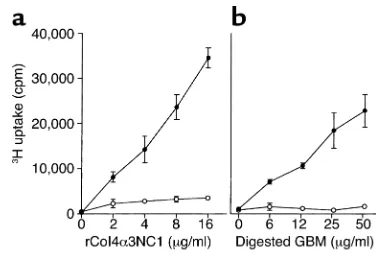
Figure 1
[image:4.576.312.528.482.637.2]Antigen specificities of a T cell line generated from rCol4α 3NC1-immu-nized rat. (a) An rCol4α3NC1 T cell line (filled circles) or T cells from CFA-immunized rat (open circles) were incubated with different con-centrations of rCol4α3NC1 (xaxis) in the presence of irradiated syn-geneic thymocytes. The proliferation of T cells was measured as uptake of 3H (∆cpm, yaxis). (b) T cells’ responses to digested rat GBM.
Figure 2
Transfer of rCol4α3NC1-specific CD4+T cells induces
pro-teinuria and GN in naive recipients. Three characterized rCol4α3NC1-specific, CD4+T cell lines of Th1 type
were transferred into 11 syngeneic rats that had received pertussis toxin 24 hours earlier. Mild protein-uria became detectable in the T cell recipients around 10 to 12 days after cell transfer. Two out of 11 returned to normal without further development, while, in the other nine, proteinuria persisted and gradually reached a plateau, ranging from 1,100 to over 2,500 mg/dl around day 35 to 40 (Figure 4a). SDS-PAGE analyses of the urine samples revealed severe albuminuria (Figure 4b). The proteinuria was accompanied by severe hema-turia. Severe proteinuria/albuminuria indicated the glomerular damage in the T cell recipients. However, the development of proteinuria/hematuria in the T cell recipients was much slower as compared with the rats actively immunized with rCol4α3NC1 in which pro-teinuria rapidly plateaued around 24 days after immu-nization (24). No T cell recipients died during the experimental period, as compared with 30% mortality in rCol4α3NC1-immunized rats around 35 days (24). Rats were sacrificed around 65 days after the T cell transfer. Kidneys from five rats with proteinuria above 1,500 mg/dl appeared pale and enlarged, while the kid-neys from the other six had normal size and color. Histopathology of the renal tissues demonstrated sim-ilar glomerular damage as seen in rCol4α 3NC1-immu-nized rats (24) (Figure 5a). The severity of GN was cor-related with the amount of proteinuria and abnormal gross appearance. Five kidneys with abnormal appear-ance showed severe GN (50–65% of glomeruli with cres-cent formation). In contrast, the kidneys from the four rats with proteinuria lower than 1,500 mg/dl had mod-est GN in which crescent formation affected less than 25% of glomeruli. Kidneys from the two without sig-nificant proteinuria had no sign of GN. In severe and
modest GN, glomeruli with crescents, as well as some without, demonstrated segmental fibrin deposition and necrosis. Jones’s staining demonstrated deposition of the matrix proteins around glomeruli and irregular, thickened GBM (data are not shown). Electron micro-scope (EM) studies especially revealed significant loss of podocyte processes (Figure 6). Examination of the other organs (except for the lung, which is currently under pathological evaluation) in two T cell recipients with severe GN did not reveal pathological changes. As controls, T cells isolated from CFA-immunized rats were either stimulated with Con-A or PPD. Transfer of those T cells did not cause any pathological changes or proteinuria in six rats. Occasionally, clusters of T cells were found to surround blood vessels. We concluded that transfer of Col4α3NC1-specific T cells induced GN in syngeneic naive recipients.
Infiltration of mononuclear cells in the interstitial tissue in the T cell recipients. Widely distributed leukocyte infiltration in the interstitial tissues, though also seen in rats immu-nized with rCol4α3NC1, was significant in the T cell recipients (Figure 5a). The cellular composition of the infiltrates was investigated. Morphologically, the infil-trates consisted of mononuclear cells essentially free of polymorphonuclear leukocytes (Figure 5a). Immuno-histochemistry demonstrated that the infiltrating cells were mainly CD3+T cells (56.1% ± 5.9%) (Figure 5b).
[image:5.576.93.270.54.202.2]Infiltration of monocytes in both the glomeruli and interstitium was confirmed by anti-CD11b/c staining. Interestingly, glomerular infiltrating cells were mainly monocytes/macrophages (CD11b/c+) and clustered Figure 3
[image:5.576.334.498.428.640.2]Cytokine profiles in an rCol4α3NC1-specific T cell line. (a) RT-PCR detection of rat INF-γ/IL-4 expressions at rest (R) or after rCol4α3NC1 stimulation (S). DNA size markers are shown at the left. (b) Flow cytometry analyses on cells from a Col4α3NC1-specific T cell line at rest (left panel) or stimulated by mitogens (right panel) after intracellular stain with anti–rat IFN-γand IL-4 Ab’s.
Figure 4
within the Bowman’s capsule (Figure 5c). Surface IgG/M+B cells were not found in any leukocyte
infil-trates. The infiltrates were frequently centered around glomeruli. Tubular injury was also present. Interstitial infiltration by mononuclear cells was not observed in the control rats that received CFA T cells.
Absence of IgG binding/C3 deposition in GBM of the T cell recipients. That activation of self-reactive T cells may trigger autoantibody production through T to B cell epitope spreading is well recognized (28). It was possi-ble that transfer of Col4α3NC1-specific T cells might have led to production of anti-GBM Ab. To determine whether anti-GBM Ab existed in the T cell recipients, direct immunofluorescence on the recipients’ renal tis-sues was used to detect IgG/IgM bound to the GBM or deposition of C3 (Figure 7). Neither binding of IgG nor deposition of C3 was observed in any T cell recipients (Figure 7b). The IgM staining, with an obvious “cres-cent” shape, was localized within the crescentic lesions of affected glomeruli; however, linear staining was not observed (Figure 7a). We concluded that the detected IgM was not anti-GBM Ab, but serum IgM nonspecifi-cally trapped in the fibrin deposits of crescentic lesions. Since there was no evidence of linear staining for IgG or IgM, it was unlikely that anti-GBM Ab was produced in the T cell recipients. Thus the antigen-specific T cells were responsible for glomerular injury by direct target-ing of glomerular antigen.
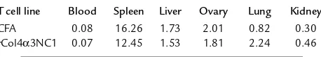
Tissue distribution of transferred T cells. We next asked whether antigen-specific T cells were able to target glomeruli and initiate glomerular inflammation. Col4α3NC1-specific T cells were activated in vitro and labeled by CFDA-SE before transfer. Another T cell line from CFA-immunized rats was stimulated by Con-A and labeled as a control. The recipients, having received either labeled Col4α3NC1-specific or control T cells, were sacrificed 24 hours later. Distributions of the labeled cells in the tissues of recipients were investigat-ed. Flow cytometry analyses did not reveal significant
differences in densities (percent-age) of labeled cells between rCol4α3 T cells and CFA T cells in most locations (Table 1). One exception was in the lung, in which rCol4α3 T cells were twice as dense as CFA T cells. Although the densi-ty of rCol4α3NC1 T cells in the whole kidney was slightly higher than that of CFA T cells, there was no significant difference. However, fluorescence microscopy showed that rCol4α3NC1 T cells in the kidney cortex were enriched 4.5-fold in comparison with CFA T cells (Figure 8a). More significant-ly, the two cell lines differed great-ly in cell shape and distribution. CFA T cells were distributed evenly in the interstitial tissue, as well as within the glomerulus. Most of the infiltrating cells were round, similar to the cells found in other organs or in the culture. On the other hand, most of the rCol4α3NC1-specific T cells (65%) were found within the Bowman’s space or surrounding the base of tubules. These T cells became elongated and/or flat-tened (Figure 8, b and c).
Discussion
The results from the present study demonstrate that transfer of GBM-specific CD4+T cells was sufficient to
[image:6.576.61.365.56.159.2]induce GN in naive animals. We further showed that pathogenic features of GN induced by T cell transfer were similar to that induced by active immunization with rCol4α3NC1 (24). First, severe proteinuria/albu-minuria developed in both cases. It is widely accepted that albuminuria is one of the most critical clinical indices for glomerular damage in both human disease and animal models. Second, histopathology revealed glomerular damage, characterized mainly by crescent formation, in the kidney of the T cell recipients.
Figure 5
Development of GN in the rCol4α3NC1-specific T cell recipients. (a) Histology shows a glomerulus with fibrin deposition and crescent formation. Adjacent to the glomerulus is a mononuclear infiltrate. Focal tubules are dilated and contain intensely eosinophilic mate-rial consistent with protein. (b) Immunoperoxidase staining shows CD3+T cells (brown in color) in mononuclear cell infiltrate surrounding a glomerulus with crescentic lesion. (c) Immunofluorescence stain show CD11b/c+monocytes/macrophages (red) clustered in the Bowman’s space of an affected glomerulus. GBM and TBM are counterstained green by FITC-labeled SR-6. Arrowheads outline the basement membrane of the Bowman’s capsule.
Figure 6
[image:6.576.347.489.547.685.2]Traditionally, Ab and its associated pathways have been considered the most important cause of GN (1). To our knowledge, this study is the first direct evi-dence to support the hypothesis that GBM antigen-specific CD4+T cells per se are able to initiate
glomeru-lar injury that leads to GN.
The next question is whether B cells are involved in GN pathogenesis in our passive-transfer model. B cells might be involved in several ways. First, antigen-specif-ic B cells may produce anti-GBM Ab, whantigen-specif-ich in turn causes glomerular injury. Since we did not detect any anti-GBM Ab in the T cell recipients with severe GN, it seems unlikely that B cells might be involved in this way. Second, B cells might participate in GN pathogenesis as antigen-presenting cells (APCs). This has been docu-mented in a few T cell–mediated autoimmune disease models (29–31). In general, some believe that B cells may act as APCs in the initial activation of naive autoreactive T cells. Thus it is important to test whether B cells may play roles as APCs if an autoimmune disease is induced by active immunization. The situation in the disease induced by transferring T cells, however, is very differ-ent. Because the transferred T cells are not naive and have been activated in vitro, they have rendered the abil-ity to target the autoantigen without further activation by the recipients’ endogenous APCs, including B cells. Third, B cells might participate in GN pathogenesis as infiltrating leukocytes (31). However, no B cells were observed in any leukocyte infiltrates in the kidney of the T cell recipients in our study. Therefore, it is unlikely that endogenous B cells might have been involved in GN pathogenesis in our model.
T cell–mediated immunity has long been suspected to be the most important mediator of GN (8). T cells may be involved in the pathogenesis of GN at three dif-ferent levels. First, T cells may participate as helper cells in T cell–dependent Ab response to renal autoantigens. It has been shown that several autoantibodies to glomerular antigens are T cell independent (32). Second, T cells may cause glomerular damage as infil-trating leukocytes. Several studies have demonstrated that CD4+ and CD8+ T cell populations may be
required in Ab-mediated GN (14). In a mouse lupus model, the mice without secreted Ab still developed GN, suggesting direct involvement of T and/or B cells (16). Third, antigen-specific T cells may directly target glomerular antigen to initiate damage. The potential involvement of T cells in GN has been a subject of sig-nificant interest. Although this mechanism has been
suspected, evidence to support it, despite development of several excellent animal models, has been lacking (21–23, 33). In those models, the antigens were either purified native GBM proteins or recombinant proteins expressed in mammalian cells that also closely resem-bled the native GBM protein. Immunization with those antigens always led to production of autoantibodies to host’s GBM. There was little analysis of the T cell responses in those models. Therefore, it was difficult to determine which mechanism, Ab, T cells, or both, mediated the glomerular injury in those models.
Some studies have focused more on the role of anti-gen-specific T cells in anti-GBM GN. Disease suscepti-bility has been strongly linked to MHC class II, sug-gesting the potential roles of the antigen-specific T cells in glomerular damage (23). Transfer of unstimulated splenocytes from the immunized animals into naive recipients induced a mild GN, as well as anti-GBM Ab (23). An early study also showed that transfer of mononuclear cells from chicks immunized with isolat-ed GBM inducisolat-ed GN in naive animals (18). Although these models used cell transfer to induce GN, it was dif-ficult to interpret data due to the following reasons. First, transferred cells might have included antigen-specific B cells, which may produce significant autoan-tibody in vivo. Actually, our study showed that antigen-specific B cells probably could survive and secrete Ab for up to two cycles of stimulation. Second, specificity of T cells were never determined. Thus, the cell transfer experiments in those studies might suggest, but did not prove, that antigen-specific T cells per se could induce glomerular damage. The most unique feature of our model, therefore, was successful generation of CD4+T cell lines, which recognized not only
recombi-nant Col4α3NC1, but also digested rat GBM proteins. With this unique tool, we were able to address the ques-tion of whether antigen-specific CD4+T cells could
[image:7.576.59.367.54.150.2]transfer the diseases in the syngeneic naive animals. Recently, we have identified two peptides derived from Col4α3NC1 that encode T cell epitopes. Immunization with those short peptides also induced similar
Figure 7
Direct immunofluorescence of Ab on serial frozen sections of kidney from an rCol4α3NC1 T cell recipient. (a) Section stained with FITC-labeled anti-rat IgM Ab. Crescent-shaped fluo-rescence staining is visible. (b) Section stained with Ab to rat IgG (negative). (c) Counter-staining by methylene blue illustrates fibrin in a crescentic lesion in glomerulus.
Table 1
Density of the labeled T cells in different tissues, expressed as percentage
T cell line Blood Spleen Liver Ovary Lung Kidney
CFA 0.08 16.26 1.73 2.01 0.82 0.30
[image:7.576.306.535.689.730.2]proteinuria and GN. Those results further support the conclusion that antigen-specific CD4+T cells alone
may be sufficient to induce GN.
Despite the similarity between GN induced by T cell transfer and active immunization, several differences were significant. First, there was much more widely dis-tributed mononuclear infiltration, mainly T cells, in the renal interstitial tissue of the T cell recipients. Such infiltrates may cause damage to tubules. Second, it took a much longer period for the T cell recipients to develop severe proteinuria than it did in those immu-nized with rCol4α3NC1. Currently, it is not under-stood what causes these differences. One explanation may lie in the source of antigen-specific T cells. Active immunization may provide a continuous supply of antigen-specific T cells. Thus nearly all glomeruli could be attacked by the T cells over a certain period. While in the case of T cell transfer only very few antigen-specific T cells may have a chance to infiltrate into the kidney to reach the target, most of them die out in other tissues. Thus T cell transfer may induce inflam-mation in a limited number of glomeruli. Unlike some autoimmune disease models, such as experimental allergic encephalomyelitis in which a localized injury to CNS may still lead to significant clinical manifesta-tions, damage to a limited number of glomeruli in GN may not result in clinically significant manifestations. If transferred T cells were able to target only a few glomeruli, the next question is why the T cell recipients eventually develop severe proteinuria and glomerular damage. Occurrence of “T cell epitope spreading” dur-ing GN pathogenesis may be an explanation (34). The endogenous self-reactive T cells may be activated with-in the with-inflamed glomeruli and/or with-in the with-interstitial infiltrates. Proteinuria and a large-scale glomerular damage might be caused by the second-wave attack by endogenously activated T cells. However, this mecha-nism needs to be determined in the future.
The observation that GBM-specific CD4+T cells can
directly cause glomerular injury gives rise to the ques-tion, which types of cells may serve as APCs in glomeruli for antigen-specific CD4+T cells? Several
early studies have investigated the MHC class II (Ia) expression in the rat glomerulus and found that the cell population expressing MHC class II was localized to the renal mesangium (35, 36). Such MHC class II expression is enhanced by various reagents such as IFN-γ(37). Cultured mesangial cells were able to stim-ulate sensitized lymphocytes to proliferate, though the nature of proliferation is unclear (38). By tracing labeled antigen-specific T cells, however, we found that the T cells may invade glomeruli through the Bow-man’s capsule. At present, it is not clear to which types of cells the T cells adhere; however, mesangial cells may not necessarily be the first target for T cells. This infer-ence is partially supported by our observation that monocytes/macrophages clustered in the Bowman’s space of the affected glomerulus (Figure 5c). Our next goal is to identify the cells in glomeruli, which may serve as APCs for T cells.
What is the significance of the finding that antigen-specific CD4+T cells per se also cause GN?
Accumulat-ing data showed that Ab-mediated mechanisms alone do not explain many aspects or cases of GN, either for human disease or animal models. For example, in rare cases of crescentic GN, neither immune complex, nor Ab to GBM, nor antineutrophil cytoplasm Ab’s were found in patients (39). Even in GN with the Ab’s, it has been found that autoantibody titers failed to predict the severity of GN in many human patients (40). Our finding could provide another explanation of those cases. Fully understanding the mechanism of T cell–mediated glomerular injury will be essential in designing new therapies for this human disease. Our model may serve as an excellent tool to explore T cell mechanisms in GN.
Acknowledgments
We thank Chin-nan Ou, Estella Tam, S. Zhu, and J. Bar-rish from Baylor Medical College for their excellent technical support. This study was supported by NIH grant RO1 HD-35993 (Y.-H. Lou) and internal funding from Dental Branch, University of Texas Houston Health Science Center (Y.-H. Lou).
1. Wilson, C.B. 1996. Renal response to immunologic glomerular injury. In The kidney. Volume 2. B.M. Brennen, editor. W.B. Saunders Co. Philadelphia, Pennsylvania, USA. 1263–1349.
2. Unaue, E.R., and Dixon, F.J. 1967. Experimental allergic glomeru-lonephritis induced in the rabbit with heterologous renal antigens. J. Exp. Med. 125:149–163.
3. Kalluri, R., Gattone, V.H., Jr., Noelken, M.E., and Hudson, B.G. 1994. The
α3(IV) chain of type IV collagen induces autoimmune Goodpasture syn-drome. Proc. Natl. Acad. Sci. USA. 91:6201–6205.
4. Theofilopoulos, A.N., and Dixon, F.J. 1980. Immune complexes in human diseases: a review. Am. J. Pathol. 100:529–594.
[image:8.576.58.294.54.161.2]5. Wang, Y., et al. 1966. Amelioration of lupus-like autoimmune disease in NZB/W F1 mice after treatment with a blocking monoclonal antibody spe-cific for complement component C5. Proc. Natl. Acad. Sci. USA. 93:8563–8568. 6. Clynes, R., Dumitru, C., and Ravetch, J.V. 1998. Uncoupling of immune complex formation and kidney damage in autoimmune glomeru-lonephritis. Science. 279:1052–1054.
Figure 8
7. Cunningham, M.A., Huang, X.R., Dowling, J.P., Tipping, P.G., and Holdsworth, S.R. 1999. Prominence of cell-mediated immunity effectors in “pauci-immune” glomerulonephritis.J. Am. Soc. Nephrol. 10:499–506. 8. Rovin, B.H., and Schreiner, G.F. 1991. Cell-mediated immunity in
glomerular disease.Annu. Rev. Med. 42:25–33.
9. Nolasco, F.E., et al. 1987. Intraglomerular T cells and monocytes in nephritis: study with monoclonal antibodies. Kidney Int. 31:1160–1166. 10. Bolton, W.K., Innes, D.J., Sturgill, B.C., and Kaiser, D.L. 1987. T-cells and macrophages in rapidly progressive glomerulonephritis: clinicopatho-logic correlations. Kidney Int. 32:869–876.
11. Bhan, A.K., Schneeberger, E.E., Collins, A.B., and McCluskey, R.T. 1982. Evidence for a pathogenic role of a cell-mediated immune mechanism in experimental glomerulonephritis. J. Exp. Med. 148:246–260. 12. Li, H.L., Hancock, W.W., Dowling, J.P., and Atkins, R.C. 1991. Activated
(IL-2R+) intraglomerular mononuclear cells in crescentic glomeru-lonephritis. Kidney Int. 39:793–798.
13. Li, S., Holdsworth, R., and Tipping, P.G. 1997. Antibody independent crescentic glomerulonephritis in µ chain deficient mice. Kidney Int.
51:672–678.
14. Tipping, P.G., Huang, X.R., Qi, M., Van, G.Y., and Tang, W.W. 1998. Cres-centic glomerulonephritis in CD4- and CD8-deficient mice.Am. J. Pathol.
152:1541–1548.
15. Reynolds, J., et al. 2000. CD28-B7 blockage prevents the development of experimental autoimmune glomerulonephritis. J. Clin. Invest.
105:643–651.
16. Chan, O.T., Hannum, L.G., Haberman, A.M., Madaio, M.P., and Shlom-chik, M.J. 1999. A novel mouse with B cells but lacking serum antibody reveals an antibody-independent role for B cells in murine lupus. J. Exp. Med. 189:1639–1648.
17. Derry, C.J., et al. 1995. Analysis of T cell response to the autoantigen in Goodpasture’s disease. Clin. Exp. Immunol. 100:262–268.
18. Bolton, W.K., Tucker, F.L., and Sturgill, B.C. 1984. New avian model of experimental glomerulonephritis consistent with mediation by cellular immunity. J. Clin. Invest. 73:1263–1276.
19. Miner, J.H., and Sanes, J.R. 1994. Collagen IV α3, α4 and α5 chains in rodent basal laminae: sequence, distribution, association with laminins, and developmental switches. J. Cell Biol. 127:879–891.
20. Neilson, E.G., et al. 1993. Specificity of Goodpasture autoantibodies for the recombinant noncollagenous domains of human type IV collagen. J. Biol. Chem. 268:8402–8405.
21. Abbate, M., et al. 1998. Experimental Goodpasture’s syndrome in Wis-tar-Kyoto rats immunized with α3 chain of type IV collagen. Kidney Int.
54:1550–1561.
22. Sado, Y., et al. 1998. Induction of anti-GBM nephritis in rats by recom-binant α3(IV)NC1 and α4(IV)NC1 of type IV collagen. Kidney Int.
53:664–671.
23. Kalluri, R., Daniff, T.M., Okada, H., and Neilson, E.G. 1997. Suscepti-bility to anti-glomerular basement membrane disease and Goodpasture syndrome is linked to MHC class II genes and the emergence of T cell-mediated immunity in mice. J. Clin. Invest. 100:2263–2275.
24. Wu, J., et al. 2001. Glomerulonephritis induced by recombinant Col4a3NC1 is not associated with GBM antibody: a potential T cell mediated mechanism. J. Immunol. 167:2388–2395.
25. Montgomery, R.A., and Dallman, M.J. 1991. Analysis of cytokine gene expression during fetal thymic ontogeny using polymerase chain reac-tion. J. Immunol. 147:554–560.
26. Enelow, R.I., et al. 1998. Structural and functional consequences of alve-olar cell recognition by CD8(+) T lymphocytes in experimental lung dis-ease. J. Clin. Invest. 102:1653–1661.
27. Sung, S., Rose, C.E., and Fu, S.M. 2001. Intratracheal priming with oval-bumin- and ovalbumin 323-339 peptide-pulsed dendritic cells induces airway hyperresponsiveness, lung eosinophilia, goblet cell hyperplasia, and inflammation. J. Immunol. 166:1261–1271.
28. Tung, K.S., Lou, Y.-H., Garza, K.M., and Teuscher, C. 1998. Autoimmune ovarian disease: mechanism of disease induction and prevention.Curr. Opin. Immunol. 9:839–845.
29. Lin, R.H., Mamula, M.J., Hardin, J.A., and Janeway, C.A. 1991. Induction of autoreactive B cells allows priming of autoreactive T cells. J. Exp. Med.
173:1433–1439.
30. Serreze, D.V., et al. 1996. B lymphocytes are essential for the initiation of T cell-mediated autoimmune diabetes: analysis of a new “speed con-genic” stock of NOD.Igµnullmice. J. Exp. Med.184:2049–2053.
31. Holmdahl, R., et al. 1995. Chronicity of tissue-specific experimental autoimmune disease: a role for B cells? Immunol. Rev. 144:111–135. 32. Diamond, B., et al. 1992. The role of somatic mutation in the pathogenic
anti-DNA response.Annu. Rev. Immunol. 10:731–757.
33. Sugihara, K., Sado, Y., Ninomiya, Y., and Wada, H. 1996. Experimental anti-GBM glomerulonephritis induced in rats by immunization with synthetic peptides based on six αchains of human type IV collagen. J. Pathol. 178:352–358.
34. Lehmann, P.V., Forsthuber, T., Miller, A., and Sercarz, E.E. 1992. Spread-ing of T-cell autoimmunity to cryptic determinants of an autoantigen. Nature. 358:155–157.
35. Schreiner, G.F., and Unanue, E.R. 1984. Origin of the rat mesangial phagocyte and its expression of the leukocyte common antigen. Lab. Invest. 51:515–523.
36. Gurner, A., Smith, J., and Cattell, V. 1986. In vivo induction of Ia antigen in resident leukocytes in the normal rat renal glomerulus. Lab. Invest.
55:546–550.
37. Martin, M., Schwinzer, R., Schellekens, H., and Resch, K. 1989. Glomeru-lar mesangial cells in local inflammation. Induction of the expression of MHC class II antigens by IFN-gamma. J. Immunol. 142:1887–1894. 38. Radeke, H.H., et al. 1994. Activation of autoreactive T-lymphocytes by
cultured syngeneic glomerular mesangial cells. Kidney Int. 45:763–774. 39. Angangco, R., et al. 1994. Does truly ‘idiopathic’ crescentic
glomeru-lonephritis exist?Nephrol. Dial. Transplant. 9:630–636.