Expression of the AT2 receptor
developmentally programs extracellular
signal-regulated kinase activity and influences fetal
vascular growth
Masahiro Akishita, … , Victor J. Dzau, Masatsugu Horiuchi
J Clin Invest.
1999;
103(1)
:63-71.
https://doi.org/10.1172/JCI5182
.
Angiotensin II type 2 (AT2) receptor is abundantly expressed in vascular smooth muscle
cells (VSMC) of the fetal vasculature during late gestation (embryonic day 15–20), during
which the blood vessels undergo remodeling. To examine directly the influence of AT2
receptor expression in the developmental biology of VSMC, we studied cultures of VSMC
from fetal and postnatal wild-type (
Agtr2
+) and AT2 receptor null (
Agtr2
–) mice. Consistent
with
in vivo
data, AT2 receptor binding in cultured
Agtr2
+VSMC increased by age, peaking
at embryonic day 20, and decreased dramatically after birth. Angiotensin II–induced growth
in
Agtr2
+VSMC (embryonic day 20) was increased by the AT2 receptor blocker PD123319,
indicating that the AT2 receptors are functional and exert an antigrowth effect in
Agtr2
+VSMC. Growth of VSMC in response to serum decreased age dependently and was higher
in
Agtr2
–than in
Agtr2
+, inversely correlating with AT2 receptor expression. However,
serum-induced growth in
Agtr2
+and
Agtr2
–VSMC and the exaggerated
Agtr2
–VSMC
growth was maintained even in the presence of PD123319 or losartan, an AT1 receptor
blocker. Moreover,
Agtr2
–VSMC showed greater growth responses to platelet-derived
growth factor and basic fibroblast growth factor, indicating that
Agtr2
–cells exhibit a
generalized exaggerated growth phenotype. We studied the mechanism responsible for this
phenotype and observed that extracellular signal-regulated kinase (ERK) activity was
higher in
Agtr2
–VSMC […]
Article
Introduction
Angiotensin II (Ang II), a key regulator of cardiovascu-lar homeostasis, exerts various actions in its diverse tar-get tissues that control vascular tone, hormone secre-tion, tissue growth, and neuronal activity (1). To date, at least two Ang II receptor subtypes are defined: type 1 (AT1) and type 2 (AT2). Most of the actions of Ang II are mediated by the well-characterized AT1 receptor, while less is known about the functions of the recently cloned AT2 receptor (2, 3).
In vitrostudies have shown that the AT2 receptor medi-ates growth inhibition in vascular smooth muscle cells (VSMC) (4, 5), coronary endothelial cells (6), PC12W cells, which is a rat pheochromocytoma cell line (7), and car-diomyocytes (8). It mediates differentiation in neuronal cells (7, 9) and/or apoptosis in vascular smooth muscle cells (VSMC) (10), PC12W cells (11, 12), cardiomyocytes (13), R3T3 mouse fibroblasts (11, 14), and endothelial cells (15). Signal transduction pathways of these AT2
receptor actions include extracellular signal-regulated kinase (ERK)-1 and -2, also known as p44 and p42 mito-gen-activated protein (MAP) kinases, which are critical to cell proliferation, differentiation, and, in some cells, hypertrophy (16). AT2 receptor stimulation causes the inactivation of ERK, which may be mediated by the acti-vation of protein tyrosine phosphatases such as SHP-1 (17) and MAP kinase phosphatase-1 (MKP-1) (11, 12).
The AT2 receptor is abundantly and widely expressed in fetal tissues, but is present only at low levels in adult tis-sues, including tissues of the adrenal gland, brain, heart, uterine myometrium, and atretic ovarian follicles (18, 19). The AT2 receptor expression is upregulated in some dis-ease states such as vascular injury (4, 20), myocardial infarction (21), and failing heart (22). The AT2 receptor expression in the aorta also exhibits a developmental pat-tern in that it is very low (or undetectable) during early embryonic development, but is very high during the later stages of embryonic development and in the neonate (23,
Expression of the AT2 receptor developmentally programs
extracellular signal-regulated kinase activity
and influences fetal vascular growth
Masahiro Akishita, Masaaki Ito, Jukka Y.A. Lehtonen, Laurent Daviet, Victor J. Dzau,
and Masatsugu Horiuchi
Cardiovascular Research, Department of Medicine, Brigham and Women’s Hospital, Harvard Medical School, Boston, Massachusetts 02115, USA
Address correspondence to: Masatsugu Horiuchi, Cardiovascular Research, Department of Medicine, Brigham and Women’s Hospital, Harvard Medical School, 75 Francis Street, Thorn-13, Boston, Massachusetts 02115, USA. Phone: (617) 732-8917; Fax: (617) 975-0995; E-mail: [email protected]
Received for publication September 10, 1998, and accepted in revised form November 10, 1998.
Angiotensin II type 2 (AT2) receptor is abundantly expressed in vascular smooth muscle cells (VSMC) of the fetal vasculature during late gestation (embryonic day 15–20), during which the blood vessels undergo remodeling. To examine directly the influence of AT2 receptor expression in the developmen-tal biology of VSMC, we studied cultures of VSMC from fedevelopmen-tal and postnadevelopmen-tal wild-type (Agtr2+) and AT2
receptor null (Agtr2–) mice. Consistent with in vivodata, AT2 receptor binding in cultured Agtr2+VSMC
increased by age, peaking at embryonic day 20, and decreased dramatically after birth. Angiotensin II–induced growth in Agtr2+VSMC (embryonic day 20) was increased by the AT2 receptor blocker
PD123319, indicating that the AT2 receptors are functional and exert an antigrowth effect in Agtr2+
VSMC. Growth of VSMC in response to serum decreased age dependently and was higher in Agtr2–than
in Agtr2+, inversely correlating with AT2 receptor expression. However, serum-induced growth in Agtr2+
and Agtr2–VSMC and the exaggerated Agtr2–VSMC growth was maintained even in the presence of
PD123319 or losartan, an AT1 receptor blocker. Moreover, Agtr2–VSMC showed greater growth
respons-es to platelet-derived growth factor and basic fibroblast growth factor, indicating that Agtr2–cells
exhib-it a generalized exaggerated growth phenotype. We studied the mechanism responsible for this pheno-type and observed that extracellular signal-regulated kinase (ERK) activity was higher in Agtr2–VSMC
at baseline and also in response to serum. ERK kinase inhibitor PD98059 inhibited both growth and ERK phosphorylation dose–dependently, while the regression lines between growth and ERK phos-phorylation were identical in Agtr2+and Agtr2–VSMC, suggesting that increased ERK activity in Agtr2–
VSMC is pivotal in the growth enhancement. Furthermore, the difference in ERK phosphorylation between Agtr2+and Agtr2–was abolished by vanadate but not by okadaic acid, implicating tyrosine
phos-phatase in the difference in ERK activity. These results suggest that the AT2 receptor expression during the fetal vasculogenesis influences the growth phenotype of VSMC via the modulation of ERK cascade.
24). After birth, the level of this receptor declines rapidly. The transient expression of the AT2 receptor suggests that it plays a role in fetal vascular development, growth, and/or differentiation. Indeed, in uteroadministration of AT2 blocker could inhibit the developmental reduction of the DNA synthesis in fetal rat aorta (4), supporting the notion that this receptor is implicated in vasculogenesis. In this study, to further investigate the role of the AT2 receptor in the developmental biology of VSMC, we stud-ied cultures of VSMC from wild-type (Agtr2+) and AT2
receptor null (Agtr2–) mice (25). We analyzed the receptor
expression, growth, and signaling in these cells. We demonstrated that Agtr2–VSMC, compared with Agtr2+
VSMC, showed developmentally regulated growth enhancement attributed to a higher ERK activity that is regulated by the tyrosine phosphatase pathway. We pos-tulate that developmental expression of VSMC AT2 receptor programs the subsequent level of ERK activity and influences fetal VSMC growth.
Methods
Animals. Female mice heterozygous for the AT2 receptor mutant allele (25) were mated with Agtr2+FVB/N male mice
mice were sacrificed by overdose anesthesia, and the fetuses were dissected from uterine decidua. The day when a vaginal plug was observed was considered to be embryonic day 1 (E1). Fetuses at the ages of E15, E18, and E20 were used in this study. In addition, postnatal day 7 and day 28 male mice, which were obtained by the same mating as the fetuses, were used. These fetuses and postnatal mice, backcrossed for six generations into the FVB/N background, had 98% FVB/N and 2% 129/SV background. Animal genotyping was performed as described previously (25), using body or tail genomic DNA samples. The animals were housed in a room where lighting was controlled (12 h on, 12 h off) and room temperature was kept at 22°C. They were given standard diet and water ad libi-tum. All experimental procedures were approved and carried out in accordance with the guidelines of the Harvard Medical Area Standing Committee on Animals.
Culture of VSMC.Thoracic aortae were removed from fetus-es and postnatal animals and micro dissected free from the surrounding adventitial tissues. The aortae from individual animals were placed into separate individual test tubes and then digested in collagenase (1 mg/ml, type II; Sigma Chem-ical Co., St. Louis, Missouri, USA) in DMEM (Life Technolo-gies Inc., Rockville, Maryland, USA) for 30 min at 37°C. After triturating and centrifuging twice, the cells were seeded in 10-cm culture dishes and cultivated in DMEM supplement-ed with 10% FBS. Thus, a culture line was isolatsupplement-ed from each animal. Culture lines obtained from littermates were main-tained exactly in the same manner. Culture medium was changed every other day. At 10–14 days after primary culture, the cells were trypsinized and seeded for the following exper-iments. Accordingly, all the experiments were performed using the cells at the second passage, except the experiment in which the effect of passages was examined. Comparisons between Agtr2+and Agtr2–cells were performed using litter-mate culture lines.
[image:3.612.99.245.54.346.2]In addition to the morphological observation, characterization of VSMC was examined by indirect immunofluorescence using a monoclonal antibody specific for the α-smooth muscle actin (clone 1A4; Sigma Chemical Co.) (26).
Figure 1
AT1 and AT2 receptor binding densities in wild-type (Agtr2+) and AT2
receptor null (Agtr2–) VSMC. Radioligand binding assay was performed
using subconfluent Agtr2+and Agtr2–VSMC derived from embryonic day
15 (E15), day 18 (E18), and day 20 (E20) fetuses, and from mice of post-natal day 7 (7d) and day 28 (28d). Similar results were obtained in at least three different culture lines. The values are expressed as mean ± SEM (n= 4). §P< 0.05 vs. E15; *, **P< 0.05, 0.01 vs. 28d. AT1, angiotensin II type
[image:3.612.317.500.466.616.2]1; AT2, angiotensin II type 2; VSMC, vascular smooth muscle cells.
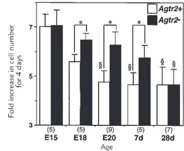
Figure 2
Developmental change in wild-type (Agtr2+) and AT2 receptor null (Agtr2–)
Receptor binding assay.AT1 and AT2 receptor binding was meas-ured using subconfluent cells grown in 24-well plates (Becton Dickinson and Co., Franklin Lakes, New Jersey, USA). After wash-ing twice with PBS containwash-ing 0.1% BSA, the cells were incubat-ed for 1 h at 37°C with 0.2 nM 125I-[Sar1,Ile8]Ang II (Du Pont Nen Research Products, Boston, Massachusetts, USA) in the absence (for the total count) or presence of 1 µM losartan (Merck Research Laboratories, Rahway, New Jersey, USA) or 1 µM PD123319 (Research Biochemicals International, Natick, Mass-achusetts, USA). The cells were then washed twice with ice-cold PBS containing 0.1% BSA and were lysed in 0.5 N NaOH. The raw radioactivity count of the lysate was measured by gamma count-er. AT1 receptor binding was calculated as the difference between the total count and the count from samples incubated with losar-tan. AT2 receptor binding was determined by subtracting the count of samples incubated with PD123319 from the total count. The net radioactivity count was converted to molar value by using specific activity of the ligand and was normalized by the cell number, which was measured at the same time.
Developmental change in VSMC growth.To investigate the devel-opmental change in the growth of Agtr2+and Agtr2– cells, VSMC were seeded in 24-well plates at a density of 3 ×10⁴cells per well and maintained in DMEM containing 10% FBS. The medium was replaced every other day. The number of cells were deter-mined using a Coulter Counter (model ZM; Coulter Electronics Ltd., Hialeah, Florida, USA) at 1 day and 5 days after seeding and were averaged for 4 wells. The growth of each culture line was expressed as the fold increase in cell number from 1 day to 5 days. Thus, 5 to 9 independent experiments, each of which con-sisted of several Agtr2+and Agtr2–littermate culture lines, were performed for each age.
Effects of Ang II receptor blockers on Ang II- or FBS-induced prolif-eration.VSMC were seeded in 24-well plates at a density of 3 × 10⁴cells per well in DMEM containing 10% FBS. The follow-ing day, the medium was replaced with the defined serum-free (DSF) DMEM containing 0.5 µM insulin, 5 µg/ml transferrin, and 0.2 mM ascorbate, and was incubated for 48 h to induce quiescence. Then the cells were treated for 3 days with DSF containing various agents. For Ang II-induced proliferation, vehicle or Ang II (0.3 µM) with or without PD123319 (10 µM) or losartan (10 µM) was added to DSF. For FBS-induced pro-liferation, vehicle or 10% FBS with or without PD123319 (10
µM) or losartan (10 µM) was added to DSF. Each medium was
changed every day. Cell numbers were counted before treat-ment (baseline) and at 3 days after treattreat-ment.
Effects of growth factors on DNA synthesis.DNA synthesis was assayed by measuring 3H-thymidine incorporation. VSMC (E20) were seeded in 24-well plates at a density of 5 ×10⁴cells per well in DMEM containing 10% FBS. The following day, the medium was replaced with DSF and incubated for 48 h to induce quies-cence. The cells were treated with a DSF-containing vehicle, 10% FBS, 10 ng/ml platelet-derived growth factor (PDGF)-BB (Life Technologies Inc.), or 10 ng/ml basic fibroblast growth factor (bFGF) (Life Technologies Inc.) for 20 h and were pulsed with 1
µCi/ml 3H-thymidine (Du Pont Nen Research Products) for an additional 4 h. The cells were washed twice with ice-cold PBS and subsequently incubated with ice-cold 5% TCA for 20 min at 4°C. The cells were washed twice with ice-cold 5% TCA, then with ice-cold PBS, and were lysed with 0.5 N NaOH. The radioactivity of the cell lysate was determined using a liquid scintillation counter and was normalized by the cell number. Determination of ERK activity, phosphorylation, and protein level. Subconfluent Agtr2+and Agtr2–VSMC (E20) were incubated in serum-free DMEM for 16 h and were treated with 10% FBS for 0–30 min. After the medium was completely removed, the cells were washed quickly with HEPES-buffered saline twice and frozen in liquid nitrogen. The cells were lysed in the lysis buffer (25 mM Tris-HCl [pH 7.5], 25 mM NaCl, 0.5 mM EGTA, 10 mM NaF, 20 mM β-glycerophosphate, 1 mM Na3VO4, 1 mM PMSF, and 10 µg/ml aprotinin). These lysates were used for kinase assay and immunoblot for phospho-ERK and ERK.
ERK activity was assayed by its ability to phosphorylate myelin basic protein as described previously (5). The lysate (100 µg) was precipitated with 5 µg of the anti–ERK-1/2 antibody (Upstate Biotechnology, Lake Placid, New York, USA) and a 40-µg suspen-sion of protein G Sepharose (Pharmacia Biotech Inc., Piscataway, New Jersey, USA). The immunoprecipitates were incubated for 10 min at room temperature with 24 µl of reaction buffer, including 1 mg/ml myelin basic protein and 40 µM γ-32P-ATP (Du Pont Nen Research Products) as substrates. The kinase reaction was termi-nated by adding Laemmli sample buffer. The samples were run on 14% SDS-PAGE, and the proteins were stained with Coomassie brilliant blue, dried, and analyzed by autoradiography. The bands corresponding to myelin basic protein were cut, and their radioac-tivity was measured by scintillation counting.
[image:4.612.64.537.493.620.2]For immunoblotting, the cell lysate was separated on 12%
Figure 3
(a–c) Growth of wild-type (Agtr2+) and AT2 receptor null (Agtr2–) VSMC derived from the aortae of embryonic day 20 fetuses, in response to angiotensin
II (Ang II) (a), FBS (b), or other growth factors (c). (a) Subconfluent, quiescent second-passage cells were treated with vehicle, Ang II (0.3 µM), Ang II plus PD123319 (10 µM), or Ang II plus losartan (10 µM) for 3 days. The cell number is expressed as percent of the number before the treatment (base-line). Similar results were obtained in three different culture lines. The values are expressed as mean ± SEM (n= 6). *P< 0.05 vs. vehicle, Ang II, and Ang II plus losartan; † P< 0.05 vs. vehicle and Ang II plus losartan. (b) Subconfluent, quiescent second-passage cells were treated with vehicle, 10% FBS, 10% FBS plus PD123319 (10 µM), or 10% FBS plus losartan (10 µM) for 3 days. The cell number is expressed as percent of the number before the treatment (baseline). Similar results were obtained in three different culture lines. The values are expressed as mean ± SEM (n= 4). (c) DNA syn-thesis was assayed by measuring 3H-thymidine incorporation. Subconfluent, quiescent second-passage cells were treated with vehicle, 10% FBS,
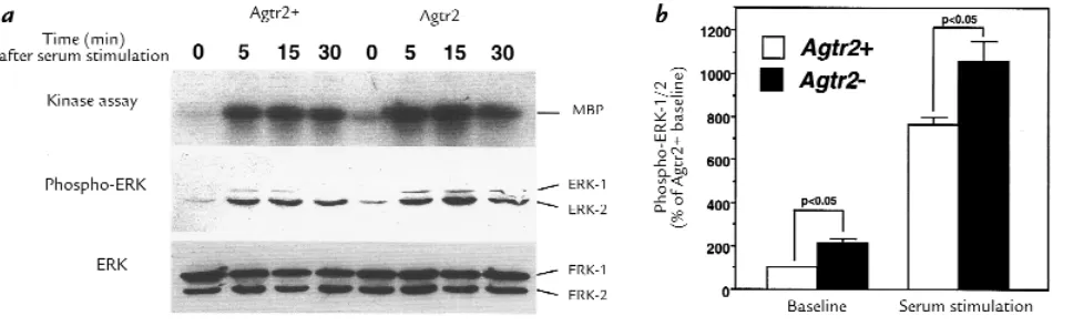
Figure 4
ERK activity at baseline and in response to serum in wild-type (Agtr2+) and AT2 receptor null (Agtr2–) VSMC derived from the aortae of embryonic
day 20 fetuses. (a) Subconfluent, quiescent second-passage cells were treated with 10% FBS for the indicated time. The cell lysate was applied to the kinase assay for ERK and immunoblots for phospho-ERK and ERK. (b) Densitometric measurements of phospho-ERK (ERK-1 and ERK-2) at base-line and 15 min after serum stimulation. The values are expressed as mean ± SEM of four different pair of culture base-lines. ERK, extracellular signal-reg-ulated kinase; MBP, myelin basic protein.
SDS-PAGE, electroblotted onto nitrocellulose membrane, and immunoblotted with phospho-specific p44/42 MAP kinase polyclonal antibody (New England Biolabs Inc., Beverly, Mass-achusetts, USA) or anti–ERK-1/2 antibody (Upstate Biotech-nology, Inc.). Antibodies were detected by horseradish peroxi-dase–linked secondary antibody using an enhanced chemiluminescence system (Amersham Life Sciences Inc., Arlington Heights, Illinois, USA). Densitometric analysis was performed using an image scanner (Arcus II; Agfa Graphic Sys-tems, Wilmington, Massachusetts, USA) and National Insti-tutes of Health (Bethesda, Maryland, USA) image software. Densities of the bands corresponding to ERK-1 and ERK-2 were added to represent total ERK.
Effects of ERK kinase inhibitor on growth and ERK phosphorylation. The same pair of Agtr2+and Agtr2–culture lines (E20) were used for DNA synthesis and phospho-ERK immunoblotting. Sub-confluent, quiescent cells were treated with 10% FBS plus 0–100
µM ERK kinase (MEK) inhibitor PD98059 (New England Bio-labs Inc.). DNA synthesis, 20 h stimulation followed by 4 h pulse labeling, was assayed as described above. ERK phospho-rylation was measured after treating for 15 min.
Determination of ERK dephosphorylation and MKP-1 level. ERK dephosphorylation was assayed according to the method described previously (11), with some modification. Sample lysates were prepared from subconfluent, quiescent cells (E20) in lysis buffer (20 mM HEPES [pH 7.5], 0.27 M sucrose, 2 mM EDTA, 2 mM EGTA, 1% Triton X-100, 1 mM β -glycerophos-phate, 0.1% 2-mercaptoethanol, 1 mM benzamidine, 1 mM PMSF, 10 µg/ml aprotinin). The lysates (100 µg) were precipi-tated with the anti–ERK-1/2 antibody (5 µg) to remove endoge-nous ERK. Consequently, the lysate supernatants were used for the dephosphorylation assay. Activated/phosphorylated ERK was immunoprecipitated with the anti–ERK-1/2 antibody using lysates of serum-stimulated Agtr2+VSMC (E20). It was then incubated in dephosphorylation buffer (40 mM HEPES [pH 7.5], 0.01% BSA, 2 mM dithiothreitol) for 1 h at room tempera-ture with or without the sample lysates. ERK phosphorylation was detected by immunoblotting with phospho-specific p44/42 MAP kinase monoclonal antibody (New England Biolabs Inc.). The density of the band was compared with that treated with-out the sample lysate; then, dephosphorylation was calculated as percent decrease in ERK phosphorylation. Protein levels of MKP-1 in the lysates were determined by immunoblotting using the anti–MKP-1 antibody (Santa Cruz Biotechnology Inc., Santa
Effects of vanadate and okadaic acid on ERK phosphorylation. Sub-confluent VSMC (E20) were treated with the tyrosine phos-phatase inhibitor vanadate (0–20 µM; Sigma Chemical Co.) or with the serine/threonine phosphatase inhibitor okadaic acid (0 or 100 nM; Life Technologies Inc.), in serum-free DMEM for 16 h. Phosphorylation and protein level of ERK were deter-mined as described above.
Data analysis.The values in the text and figures are expressed as mean ± SEM. The data were analyzed using one-factor ANOVA. If a statistically significant effect was found, Newman-Keuls’ test was performed to isolate the difference between the groups. In some experiments using several different pair of Agtr2+and Agtr2–culture lines, paired ttests were also performed to clarify the difference, which might be invisible because of the variance among the culture lines. In interpreting results, P< 0.05 was considered to be statistically significant.
Results
Developmental change of Ang II receptors and growth in VSMC. All of our culture lines (fetal through postnatal day 28) exhibited the “hill-and-valley” appearance characteristic of adult VSMC. The cells were uniformly positive for α -smooth muscle actin by immunofluorescence. These results indicate that the cells are characteristic of VSMC, although the possible contamination of fibroblasts and endothelial cells cannot be avoided.
AT2 receptor expression in rat vasculature is reported to be abundant at the late gestational age and to decrease rapidly after birth (23, 24). Consistent with these results, reverse transcription-PCR analysis using mouse aorta revealed that AT2 receptor started to increase at E15, peaked at E20, and disappeared after seven days, where-as AT1 receptor wwhere-as constantly expressed during the development (27). To examine whether the cultured VSMC retain the expression pattern for Ang II receptors, we determined AT1 and AT2 receptor expression by receptor binding assay. In Agtr2+VSMC, AT2 receptor
binding density increased by age, peaking at E20, and dramatically decreased after birth (Fig. 1), whereas Agtr2–
difference between Agtr2+and Agtr2–(Fig. 1). AT2
recep-tor binding density in Agtr2+cells was significantly lower
than AT1 binding density at each age (one-third of the AT1 receptor binding at E20).
By using these cells, we examined the developmental change in VSMC growth. The rate of growth, determined by the cell number increase in response to 10% fetal bovine serum (FBS) for four days, decreased according to age (Fig. 2). Importantly, VSMC growth at E18, E20, and postnatal day 7 was higher in Agtr2– than in Agtr2+,
inversely correlating with the presence of the AT2 receptor. No difference between Agtr2+and Agtr2–was
observed at E15 or postnatal day 28, when the AT2 receptor is minimally or not expressed. Thus, develop-mental decline in VSMC growth was delayed in cells derived from Agtr2–mice.
Ang II-dependent and -independent growth of VSMC.To exam-ine the mechanism of the growth difference, we studied VSMC prepared from E20 fetuses (which express abundant vascular AT2 receptor) and used these cells in the follow-ing experiments. The growth difference in response to 10% FBS between Agtr2+and Agtr2–VSMC persisted for up to
eight days of culture at the second passage (data not shown). Third-passage cells exhibited a growth phenotype similar to second-passage cells, while fourth-passage cells lost the growth ability and the morphological characteris-tics of VSMC (data not shown), suggesting that these cells reach terminal senescence at the fourth passage.
Because Agtr2+VSMC express the significant amount
of AT2 receptor, and we have reported that the AT2 receptor mediates growth inhibition in VSMC (4, 5), we investigated whether the growth difference between Agtr2+and Agtr2–VSMC was Ang II–dependent
or–inde-pendent. Second-passage cultured VSMC were stimu-lated with Ang II or 10% FBS, and the effects of Ang II receptor blockers were examined (Fig. 3, aand b). In Agtr2+VSMC, Ang II did not increase the cell number,
but Ang II plus AT2 blocker PD123319 enhanced the growth, suggesting that the AT1 receptor stimulates cell growth and the AT2 receptor antagonizes it. Consistent with this, we observed that Ang II stimulated cell growth in Agtr2– VSMC. Interestingly, significant
growth difference between Agtr2+and Agtr2–VSMC was
found even in the absence of Ang II and 10% FBS (Fig. 3, vehicle). Moreover, the growth difference in response to 10% FBS was not affected by AT2 blocker in either the Agtr2+or Agtr2–cells (Fig. 3b), indicating that this
dif-ference did not depend on a direct activation of the cell-surface AT2 receptor by its peptide ligand, but rather by a possible programming effect on growth response by the AT2 receptor in vivoduring development.
The effects of growth factors on DNA synthesis in VSMC (E20) were examined. DNA synthesis in second-passage Agtr2–VSMC was also higher than that in
cor-responding Agtr2+ VSMC, in response to 10% FBS,
PDGF-BB, and bFGF, as well as vehicle (Fig. 3c), sup-porting the notion that AT2 receptor null cells have the exaggerated growth phenotype.
[image:6.612.317.523.49.591.2]ERK and its link with the growth phenotype.It is well known that ERK plays a pivotal role in regulating cell growth (16). Previous studies have shown that AT2 receptor stimulation inactivates ERK via the activation of tyrosine
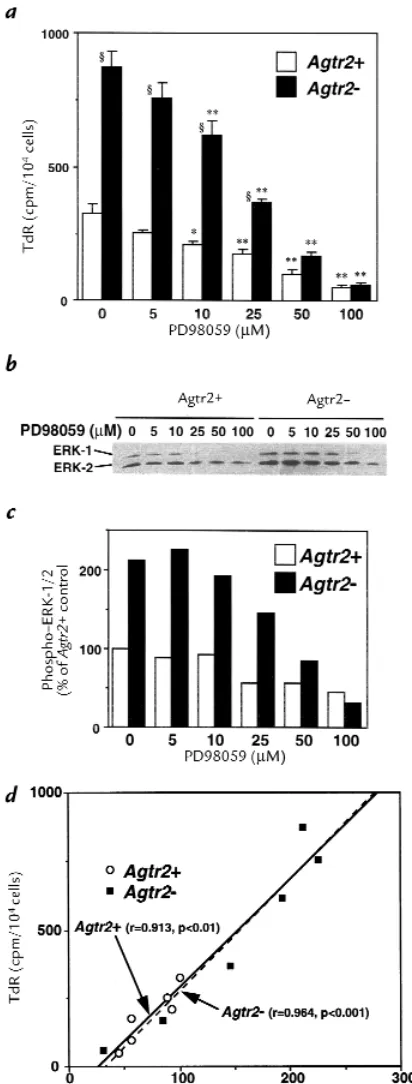
Figure 5
Effects of MEK inhibitor on DNA synthesis and ERK phosphorylation in wild-type (Agtr2+) and AT2 receptor null (Agtr2–) VSMC derived from the
aortae of embryonic day 20 fetuses. Subconfluent, quiescent cells were treated with 10% FBS plus 0–100 µM MEK inhibitor PD98059. (a) DNA synthesis was assayed as 3H-thymidine incorporation (TdR). The values
are expressed as mean ± SEM (n= 6). *, **P< 0.05, 0.01 vs. PD98059 (–); §P< 0.05 vs. Agtr2+. (band c) Immunoblotting for phospho-ERK and
phosphatases (5, 11, 12, 17). Consistent with this, we have demonstrated the attenuated ERK activity in vivoin AT2 receptor in transgenic mouse hearts (28). Therefore, we hypothesized that ERK activity would be higher in AT2 receptor null VSMC, resulting in a higher growth rate compared with Agtr2+cells.
As shown in Fig. 4a, both kinase assay and immunoblot using anti–phospho-ERK antibody documented that ERK activity was higher in Agtr2– VSMC at baseline
(before serum stimulation) and also in response to 10% FBS, whereas the protein levels of ERK were similar. Because densitometric measurements of immunoblot-ting for phospho-ERK correlated very well with ERK activities determined by kinase assay (r= 0.979, P< 0.001, n= 8), we used phospho-ERK immunoblotting to
exam-sitometric analysis using four different pair of culture lines showed that phospho-ERK was 200% higher at base-line and 140% higher 15 minutes after serum stimulation in Agtr2– than in Agtr2+ VSMC (Fig. 4b). In contrast,
VSMC from E15 fetuses or postnatal day 28 animals did not show the difference in ERK phosphorylation between Agtr2+and Agtr2–(data not shown).
To examine whether ERK activity is linked to growth response, we treated the cells with MEK inhibitor PD98059. PD98059 inhibited both growth as meas-ured by DNA synthesis and ERK phosphorylation, dose dependently (Fig. 5, a–c). Interestingly, the regression lines between VSMC thymidine incorporation and ERK phosphorylation were identical in Agtr2+and Agtr2–
cells (Fig. 5d), suggesting that the increased growth in Agtr2–cells is attributable to a higher level of
intracel-lular ERK activity.
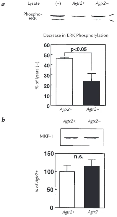
Because it has been reported that protein tyrosine phosphatases play a critical role in AT2 receptor–medi-ated ERK inactivation (5, 11, 12, 17), we studied the involvement of phosphatases in ERK activity in these cells. We examined phosphatase activity that deter-mined ERK activity in Agtr2+and Agtr2–VSMC by
meas-uring ERK dephosphorylation (Fig. 6a). The phos-phatase activity of the Agtr2+cells was approximately
twofold higher than that of Agtr2–cells, showing a
rea-sonable difference that would explain the difference in ERK activity. These results suggest that the activity of some specific phosphatase(s) that directly regulates ERK activity is enhanced in Agtr2–cells. Next, we
exam-ined the protein level of MKP-1, which is a dual speci-ficity phosphatase that dephosphorylates ERK, and demonstrated that MKP-1 protein levels were similar in Agtr2+and Agtr2–VSMC. As shown in Fig. 7, aand b,
vanadate increased ERK phosphorylation predomi-nantly in Agtr2+ cells and abolished the difference
between Agtr2+and Agtr2–cells. In contrast, 100 nM of
okadaic acid, a concentration enough to inhibit protein phosphatases 1 and 2A (29) and exhibit a small increase in ERK phosphorylation in both cells, did not influence the difference between Agtr2+and Agtr2–cells. The
pro-tein levels of ERK were not affected by the treatments. These results suggest that tyrosine phosphatase, rather than serine/threonine phosphatase, is a primary deter-minant in the quantitative difference of ERK activities between the Agtr2+and Agtr2–VSMC.
Discussion
[image:7.612.57.255.53.421.2]The adult intact artery, which is fully developed and dif-ferentiated, is a quiescent tissue. Indeed, replication rate of VSMC in the normal aorta is less than 0.05% per day (30). In contrast, VSMC in the developing aorta undergo remarkable proliferation (31–33). However, this replica-tion rate exhibits a dramatic reducreplica-tion in the late gesta-tional stage (4, 33), when the AT2 receptor is abundantly expressed (23, 24). We have demonstrated that in utero pharmacologic blockade of the fetal AT2 receptor attenu-ated this decline in the rate of aortic DNA synthesis (4), indicating that the AT2 receptor modulates growth of the developing blood vessel and thus contributes to vascular remodeling in late gestation. The AT2 receptor null
Figure 6
ERK dephosphorylation and MKP-1 protein level in wild-type (Agtr2+) and
AT2 receptor null (Agtr2–) VSMC. (a) Phosphorylated ERK was
incubat-ed with the lysates preparincubat-ed from Agtr2+and Agtr2–VSMC and was
the role of this receptor in vascular development. The AT2 receptor null mouse exhibits no gross abnormalities; how-ever, the basal blood pressure and the acute response to Ang II are enhanced in the adult (three to five months old) AT2 receptor null mouse (25, 34). Because the vascular AT2 receptor is not expressed at this age, the data suggest that the transient and developmentally regulated AT2 receptor expression in late gestation exerts a long-term effect on blood pressure, possibly via its influence on vas-cular structure. However, the structural consequences of fetal AT2 receptor expression in vascular development in the AT2 receptor null mouse await detailed analysis.
To study the influence of AT2 receptor expression on the developmental biology of VSMC, we studied cultures of VSMC derived from wild-type and AT2 receptor null mice. In this study we demonstrated that VSMC derived from the aorta of fetal AT2 receptor null mice exhibited the exaggerated growth phenotype compared with age-matched wild-type VSMC. ERK activity, which was close-ly associated with growth, was programmed to be high-er in AT2 receptor null VSMC. This high ERK activity in AT2 receptor null cells may be attributable to the low tyrosine phosphatase activity.
Cook et al. (33) reported that cultured VSMC derived from rat aorta exhibited the developmentally regulated growth, mimicking the in vivopattern. They showed that the high growth potential of fetal VSMC was lost by E20. These data suggest that the significant change in VSMC phenotype occurs during the development and that
cul-tured VSMC reflect the in vivophenotype. Consistent with their data, we observed that the growth rate of wild-type VSMC declined after E18. In contrast, the growth rate of AT2 receptor null VSMC remained increased up to post-natal day 7. These results suggest that AT2 receptor expres-sion during the fetal vasculogenesis influences the growth phenotype of VSMC. However, the finding that the growth rate decreased developmentally, even in AT2 receptor null VSMC and in postnatal wild-type VSMC in which AT2 receptor was minimally expressed, indicates that some other factors are also implicated in this process. Little is known about the factors that control the growth pheno-type of VSMC during development. Majesky et al. (35, 36) have reported that developmentally regulated expression of endogenous PDGF and its receptor influences rat VSMC growth. In our preliminary study, however, condi-tioned medium derived from cultured AT2 receptor null VSMC did not contain higher growth-promoting activity than that from wild-type VSMC (data not shown). Fur-thermore, AT2 receptor null cells showed greater growth responses to serum, PDGF, and bFGF, indicating that AT2 receptor null VSMC exhibit a generalized exaggerated growth phenotype. Thus, an alteration in signaling path-ways in AT2 receptor null VSMC is likely to be responsible for the exaggerated growth phenotype, rather than alter-ations in growth factors or their receptor expressions.
[image:8.612.74.510.62.334.2]In this study, we focused on the ERK pathway because this cascade is crucial in proliferation, differentiation, and hypertrophy of VSMC (16), and is implicated in the AT2
Figure 7
Effects of vanadate and okadaic acid on ERK phosphorylation in wild-type (Agtr2+) and AT2 receptor null (Agtr2–) VSMC derived from the aortae of
receptor–mediated antigrowth and proapoptotic actions on VSMC (4, 5, 10). ERK is also involved in migration (37) and contraction (38, 39) of VSMC, denoting that ERK plays pivotal roles in the regulation of VSMC biology. We observed that ERK activity was significantly higher in AT2 receptor null VSMC than in wild-type VSMC at baseline and in response to serum, whereas protein levels of ERK were similar, suggesting that the difference is due to acti-vation/phosphorylation, not to transcription or the trans-lation of ERK. We demonstrated that phosphatase activi-ty, which directly regulated ERK, was higher in wild-type VSMC than in AT2 receptor null VSMC.or vanadate, but not serine/threonine phosphatase inhibitor okadaic acid, abolished the difference in the basal ERK phosphoryla-tion between wild-type and AT2 receptor null VSMC with-out influence on ERK protein levels supports the notion that tyrosine phosphatases are involved in programming the level of ERK activity in these cells. To investigate the upstream events in the ERK cascade, we determined MEK phosphorylation in the cells. We could not detect a sig-nificant difference in MEK phosphorylation between wild-type and AT2 receptor null VSMC (data not shown), suggesting that the kinase activity in AT2 receptor null cells is increased at the level of ERK. However, possible reg-ulation of other signaling molecules by decreased tyrosine phosphatase activities in AT2 receptor null VSMC still remains to be defined. Some tyrosine phosphatases are shown to be critical to embryonic development (40–42) and regulation of VSMC function (43, 44). Tyrosine phos-phatases can regulate the development and function of VSMC by dephosphorylating their target substrates, including ERK. Given that stimulation of the AT2 recep-tor activates tyrosine phosphatases in vivo, the precise mechanism by which multiple-passaged cultured VSMC maintain this phenotype in vitroin the absence of Ang II, has to be explained. One possible mechanism is mental programming. We hypothesize that the develop-mental expression and activation of vascular AT2 recep-tor stimulate tyrosine phosphatase activity, whereas the lack of this receptor in AT2 receptor null vessels results in lower enzyme activity. This developmental regulation pro-grams the subsequent level of local tyrosine phosphatase activity even in the absence of AT2 receptor activation or expression. Indeed, the finding that cultured VSMC derived from genetic disease animals such as sponta-neously hypertensive rats (45) and spontasponta-neously diabet-ic rats (46) exhibit the exaggerated growth phenotype sup-ports this possibility. The exact molecular mechanism of genetic programming of intracellular regulatory pathways is currently unknown.
The results of this study may be relevant to vascular pathobiology. It has been shown that neointimal VSMC in vascular lesions such as atherosclerosis and restenosis after balloon angioplasty resemble fetal VSMC, rather than adult medial VSMC, in terms of morphology, expres-sion of smooth–muscle specific markers, and growth abil-ity (36, 47). Furthermore, ERK is activated in neointimal VSMC (48), and the downregulation of MKP-1 is associ-ated with the activation of ERK after vascular injury (44). Our data may provide an explanation for why these altered growth phenotypes persist in culture. We
hypoth-of specific growth regulatory receptors and their signaling pathways for a specific time period can have a genetic pro-gramming effect that subsequently influences the pheno-type, at least for several cell cycles. In the case of AT2 recep-tor, it is reexpressed in injured blood vessels and attenuates neointima formation (4, 20). The expression pattern may play a role in the reversal of the phenotypic shift, thereby participating in the vascular remodeling in these pathophysiologic states.
Acknowledgments
This work was supported by National Institutes of Health (NIH) grants 46631, 35252, 35610, 48638, HL-07708, and HL-58616, and by a grant from the Longwood Foundation for Translational Research. V.J. Dzau is a recipient of NIH Merit Award HL-35610.
1. Dzau, V.J., Gibbons, G.H., and Pratt, R.E. 1991. Molecular mechanisms of vascular renin-angiotensin system in myointimal hyperplasia. Hyper-tension. 18(Suppl II):100–105.
2. Mukoyama, M., et al. 1993. Expression cloning of type 2 angiotensin II receptor reveals a unique class of seven-transmembrane receptors. J. Biol. Chem. 268:24539–24542.
3. Kambayashi, Y., et al. 1993. Molecular cloning of novel angiotensin II receptor isoform involved in phosphotyrosine phosphatase inhibition. J. Biol. Chem. 268:24543–24546.
4. Nakajima, M., et al. 1995. The angiotensin II type 2 (AT2) receptor antag-onizes the growth effects of the AT1 receptor: gain of function study using in vivo gene transfer. Proc. Natl. Acad. Sci. USA.92:10663–10667. 5. Hayashida, W., Horiuchi, M., and Dzau, V.J. 1996. Intracellular third loop domain of angiotensin II type 2 receptor: role in mediating signal transduction and cellular function. J. Biol. Chem. 271:21985–21992. 6. Stoll, M., et al. 1995. The angiotensin AT2-receptor mediates inhibition
of cell proliferation in coronary endothelial cells. J. Clin. Invest. 95:651–657.
7. Meffert, S., Stoll, M., Steckelings, U.M., Bottari, S.P., and Unger, T. 1996. The angiotensin II AT2 receptor inhibits proliferation and promotes dif-ferentiation in PC12W cells. Mol. Cell. Endocrinol. 122:59–67. 8. Booz, G.W., and Baker, K.M. 1996. Role of type 1 and type 2 angiotensin
receptors in angiotensin II-induced cardiomyocyte hypertrophy. Hyper-tension. 28:635–640.
9. Laflamme, L., Gasparo, M., Gallo, J.M., Payet, M.D., and Gallo-Payet, N. 1996. Angiotensin II induction of neurite outgrowth by AT2 receptors in NG108-15 cells. Effect counteracted by the AT1 receptors. J. Biol. Chem. 271:22729–22735.
10. Yamada, T., et al.1998. Angiotensin II type 2 receptor mediates vascular smooth muscle cell apoptosis and antagonizes angiotensin II type 1 receptor action: an in vitro gene transfer study. Life Sci. 63:PL289-PL295. 11. Yamada, T., Horiuchi, M., and Dzau, V.J. 1996. Angiotensin II type 2
receptor mediates programmed cell death. Proc. Natl. Acad. Sci. USA. 93:156–160.
12. Horiuchi, M., Hayashida, W., Kambe, T., Yamada, T., and Dzau, V.J. 1997. Angiotensin II type 2 receptor dephosphorylates Bcl-2 by activating mitogen-activated protein kinase phosphatase-1 and induces apoptosis. J. Biol. Chem. 272:19022–19026.
13. Hayashida, W., Horiuchi, M., Grandchamp, J., and Dzau, V.J. 1996. Antagonistic action of angiotensin II type-1 and type-2 receptors on apoptosis in cultured neonatal rat ventricular myocytes. Hypertension. 28:535. (Abstr.)
14. Horiuchi, M., Yamada, T., Hayashida, W., and Dzau, V.J. 1997. Interfer-on regulatory factor-1 upregulates angiotensin type 2 receptor and induces apoptosis. J. Biol. Chem. 272:11952–11958.
15. Dimmeler, S., Rippmann, V., Weiland, U., Haendeler, J., and Zeiher, A.M. 1997. Angiotensin II induces apoptosis of human endothelial cells. Pro-tective effect of nitric oxide. Circ. Res. 81:970–976.
16. Force, T., and Bonventre, J.V. 1998. Growth factors and mitogen-acti-vated protein kinases. Hypertension. 31:152–161.
17. Bedecs, K., et al. 1997. Angiotensin II type 2 receptors mediate inhibition of mitogen-activated protein kinase cascade and functional activation of SHP-1 tyrosine phosphatase. Biochem. J.325:449–454.
18. Daud, A.I., Bumpus, F.M., and Husain, A. 1988. Evidence for selective expression of angiotensin II receptors on atretic follicles in the rat ovary: an autoradiographic study. Endocrinology. 122:2727–2734.
20. Akishita, M., Horiuchi, M., Yamada, H., Zhang, L., and Dzau, V.J. 1997. Accentuated vascular proliferation and altered remodeling after injury in mice lacking angiotensin II type 2 receptor. Circulation. 96:1-547. (Abstr.)
21. Nio, Y., Matsubara, H., Murasawa, S., Kanasaki, M., and Inada, M. 1995. Regulation of gene transcription of angiotensin II receptor subtypes in myocardial infarction. J. Clin. Invest.95:46–54.
22. Wharton, J., et al. 1998. Differential distribution of angiotensin AT2 receptors in the normal and failing human heart.J. Pharmacol. Exp. Ther. 284:323–336.
23. Viswanathan, M., Tsutsumi, K., Correa, F.M., and Saavedra, J.M. 1991. Changes in expression of angiotensin receptor subtypes in the rat aorta during development. Biochem. Biophys. Res. Commun. 179:1361–1367. 24. Shanmugam, S., Corvol, P., and Gasc, J-M. 1996. Angiotensin II type 2
receptor mRNA expression in the developing cardiopulmonary system of the rat. Hypertension. 28:91–97.
25. Hein, L., Barsh, G.S., Pratt, R.E., Dzau, V.J., and Kobilka, B.K. 1995. Behavioural and cardiovascular effects of disrupting the angiotensin II type-2 receptor gene in mice. Nature. 377:744–747.
26. Skalli, O., et al. 1986. A monoclonal antibody against α-smooth muscle actin: a new probe for smooth muscle differentiation. J. Cell Biol. 103:2787–2796.
27. Yamada, H., Horiuchi, M., Akishita, M., and Dzau, V.J. 1997. Regulation of vascular development and differentiation by angiotensin II type 2 receptor. Hypertension. 30:470. (Abstr.)
28. Masaki, H., et al. 1998. Cardiac-specific overexpression of angiotensin II AT2 receptor causes attenuated response to AT1 receptor-mediated pres-sor and chronotropic effects. J. Clin. Invest. 101:527–535.
29. Cohen, P., Holmes, C.F., and Tsukitani, Y. 1990. Okadaic acid: a new probe for the study of cellular regulation. Trends Biochem. Sci. 15:98–102. 30. Lombardi, D.M., Reidy, M.A., and Schwartz, S.M. 1991. Methodologic considerations important in the accurate quantitation of aortic smooth muscle cell replication in the normal rat. Am. J. Pathol. 138:441–446. 31. Owens, G.K., and Thompson, M.M. 1986. Developmental changes in
isoactin expression in rat aortic smooth muscle cells in vivo. Relationship between growth and cytodifferentiation. J. Biol. Chem. 261:13373–13380. 32. Bochaton-Piallat, M.L., Gabbiani, F., Ropraz, P., and Gabbiani, G. 1993. Age influences the replicative activity and the differentiation features of cultured rat aortic smooth muscle cell populations and clones. Arte-rioscler. Thromb. 13:1449–1455.
33. Cook, C.L., Weiser, M.C., Schwartz, P.E., Jones, C.L., and Majack, R.A. 1994. Developmentally timed expression of an embryonic growth phe-notype in vascular smooth muscle cells. Circ. Res. 74:189–196. 34. Ichiki, T., et al. 1995. Effects on blood pressure and exploratory
behav-iour of mice lacking angiotensin II type-2 receptor. Nature. 377:748–750. 35. Majesky, M.W., Benditt, E.P., and Schwartz, S.M. 1988. Expression and developmental control of platelet-derived growth factor A-chain and B-chain/Sis genes in rat aortic smooth muscle cells. Proc. Natl. Acad. Sci. USA.85:1524–1528.
36. Majesky, M.W., Giachelli, C.M., Reidy, M.A., and Schwartz, S.M. 1992. Rat carotid neointimal smooth muscle cells reexpress a developmental-ly regulated mRNA phenotype during repair of arterial injury. Circ. Res. 71:759–768.
37. Graf, K., et al. 1997. Mitogen-activated protein kinase activation is involved in platelet-derived growth factor-directed migration by vascu-lar smooth muscle cells. Hypertension. 29:334-339.
38. Khalil, R.A., Menice, C.B., Wang, C.L., and Morgan, K.G. 1995. Phos-photyrosine-dependent targeting of mitogen-activated protein kinase in differentiated contractile vascular cells. Circ. Res. 76:1101–1108. 39. Epstein, A.M., Throckmorton, D., and Brophy, C.M. 1997.
Mitogen-acti-vated protein kinase activation: an alternate signaling pathway for sus-tained vascular smooth muscle contraction. J. Vasc. Surg. 26:327–332. 40. Tang, T.L., Freeman, R.M. Jr., O’Reilly, A.M., Neel, B.G., and Sokol, S.Y. 1995. The SH2-containing protein-tyrosine phosphatase SH-PTP2 is required upstream of MAP kinase for early Xenopus development.Cell. 80:473–483.
41. Saxton, T.M., et al. Abnormal mesoderm patterning in mouse embryos mutant for the SH2 tyrosine phosphatase Shp-2. EMBO J.16:2352–2364. 42. Fuchs, M., Wang, H., Ciossek, T., Chen, Z., and Ullrich, A. 1998. Differ-ential expression of MAM-subfamily protein tyrosine phosphatases dur-ing mouse development. Mech. Dev.70:91–109.
43. Duff, J.L., et al.1993. Angiotensin II induces 3CH134, a protein-tyrosine phosphatase, in vascular smooth muscle cells. J. Biol. Chem. 268:26037–26040.
44. Lai, K., et al. 1996. Mitogen-activated protein kinase phosphatase-1 in rat arterial smooth muscle cell proliferation. J. Clin. Invest. 98:1560–1567. 45. Hadrava, V., et al. 1991. Vascular smooth muscle cell proliferation and its therapeutic modulation in hypertension. Am. Heart J.122:1198–1203. 46. Yoo, H.J., et al. 1997. Augmented Ca2+ influx is involved in the mecha-nism of enhanced proliferation of cultured vascular smooth muscle cells from spontaneously diabetic Goto-Kakizaki rats. Atherosclerosis. 131:167–175.
47. Schwartz, S.M., Campbell, G.R., and Campbell, J.H. 1986. Replication of smooth muscle cells in vascular disease. Circ. Res. 58:427–444. 48. Hu, Y., Cheng, L., Hochleitner, B.W., and Xu, Q. 1997. Activation of