Bone resorption induced by parathyroid
hormone is strikingly diminished in
collagenase-resistant mutant mice
Weiguang Zhao, … , Brendan F. Boyce, Stephen M. Krane
J Clin Invest. 1999;
103(4)
:517-524.
https://doi.org/10.1172/JCI5481
.
Parathyroid hormone (PTH) stimulates bone resorption by acting directly on
osteoblasts/stromal cells and then indirectly to increase differentiation and function of
osteoclasts. PTH acting on osteoblasts/stromal cells increases collagenase gene
transcription and synthesis. To assess the role of collagenase in the bone resorptive actions
of PTH, we used mice homozygous (r/r) for a targeted mutation (r) in Col1a1 that are
resistant to collagenase cleavage of type I collagen. Human PTH(1–34) was injected
subcutaneously over the hemicalvariae in wild-type (+/+) or r/r mice four times daily for three
days. Osteoclast numbers, the size of the bone marrow spaces and periosteal proliferation
were increased in calvariae from PTH-treated +/+ mice, whereas in r/r mice, PTH-induced
bone resorption responses were minimal. The r/r mice were not resistant to other skeletal
effects of PTH because abundant interstitial collagenase mRNA was detected in the
calvarial periosteum of PTH-treated, but not vehicle-treated, r/r and +/+ mice. Calcemic
responses, 0.5–10 hours after intraperitoneal injection of PTH, were blunted in r/r mice
versus +/+ mice. Thus, collagenase cleavage of type I collagen is necessary for PTH
induction of osteoclastic bone resorption.
Article
Introduction
Parathyroid hormone (PTH) induces hypercalcemia in part through increasing bone resorption mediated by osteo-clasts. Receptors for PTH are present not on osteoclasts, however, but on mesenchymal cells of the osteoblast line-age and stromal cells in the bone marrow (1). PTH must therefore act directly on these mesenchymal cells, which then — through direct cell–cell contact mediated by cell-bound ligands such as osteoclast differentiation factor (ODF) and/or production of soluble ligands — modulates the activity of existing osteoclasts and the differentiation of osteoclasts from precursor cells (2, 3).
Shortly after the discovery of the animal collagenases by Gross and Lapière (4), Walker et al. (5) found that bones removed from mice injected with doses of PTH sufficient to elevate serum calcium levels by ∼4 mg/dl produce collagenase activity at levels much higher than in bones removed from uninjected mice. The time course of the effects on inducing hypercalcemia and collagenase production was different in that increased collagenase production was not detected until several hours after serum calcium levels reached their peak (6). It had also been shown that the sustained hypercalcemia induced by PTH was obviated by inhibitors of mRNA synthesis (acti-nomycin D) (7, 8) or protein synthesis (puromycin) (9), suggesting that, at least in part, PTH-induced hypercal-cemia is dependent upon the synthesis of a protein in bone; collagenase was a candidate. In subsequent stud-ies from several laboratorstud-ies it was shown that in organ cultures of bone fragments, collagenase was released into
the ambient medium and collagenase production could be stimulated by PTH (10–14). Later, it was possible to clone a collagenase gene from a cDNA library prepared from a rat osteogenic sarcoma cell line stimulated with PTH (15). A mouse collagenase cDNA was subsequently cloned (16) using the rat cDNA probe (15), and it was determined that the rodent enzymes had only ∼50% amino acid sequence similarity to human collagenase-1 (matrix metalloproteinase-1 [MMP-1]) but had high similarity to what was later identified as collagenase-3 (MMP-13) in humans (17). Osteoclasts, in addition to producing cysteine proteinases, also produce MMPs such as the 92-kDa gelatinase (gelatinase B) (18) and one of the membrane-bound (MT) MMPs, MT1-MMP (19). In all but a few reported studies (e.g., ref. 20), however, it has not been possible to identify the expression of spe-cific collagenases in osteoclasts using cDNA or cRNA probes. In contrast, several investigators have been read-ily able to measure collagenase produced by osteoblasts or stromal fibroblasts from different species, as described above, either constitutively or induced by sev-eral ligands, including PTH. When osteoblasts are exposed to PTH, they start producing collagenase and stop synthesizing collagen (21).
During normal embryonic development, collagenase is expressed in cells (osteoblasts, stromal cells) in the bone shaft and in hypertrophic chondrocytes of the distal growth plate (22–25). More proximal hypertrophic chondrocytes that characteristically express type X collagen do not express collagenase. Furthermore, the expression of collagenase (26)
Bone resorption induced by parathyroid
hormone is strikingly diminished
in collagenase-resistant mutant mice
Weiguang Zhao,
1Michael H. Byrne,
1Brendan F. Boyce,
2and Stephen M. Krane
11Department of Medicine, Harvard Medical School, Medical Services (Arthritis Unit), Massachusetts General Hospital, Boston,
Massachusetts 02114, USA
2Department of Pathology, University of Texas Health Science Center at San Antonio, San Antonio, Texas 78284, USA
Address correspondence to: Stephen M. Krane, Arthritis Unit/Bulf 165, Massachusetts General Hospital, 55 Fruit Street, Boston, Massachusetts 02114, USA. Phone: (617) 726-2870; Fax (617) 726-2872; E-mail: krane.stephen@mgh.harvard.edu
Received for publication October 12, 1998, and accepted in revised form January 5, 1999.
Parathyroid hormone (PTH) stimulates bone resorption by acting directly on osteoblasts/stromal cells and then indirectly to increase differentiation and function of osteoclasts. PTH acting on osteoblasts/stro-mal cells increases collagenase gene transcription and synthesis. To assess the role of collagenase in the bone resorptive actions of PTH, we used mice homozygous (r/r) for a targeted mutation (r) in Col1a1that are resistant to collagenase cleavage of type I collagen. Human PTH(1–34) was injected subcutaneously over the hemicalvariae in wild-type (+/+) or r/r mice four times daily for three days. Osteoclast numbers, the size of the bone marrow spaces and periosteal proliferation were increased in calvariae from PTH-treat-ed +/+ mice, whereas in r/r mice, PTH-inducPTH-treat-ed bone resorption responses were minimal. The r/r mice were not resistant to other skeletal effects of PTH because abundant interstitial collagenase mRNA was detect-ed in the calvarial periosteum of PTH-treatdetect-ed, but not vehicle-treatdetect-ed, r/r and +/+ mice. Calcemic respons-es, 0.5–10 hours after intraperitoneal injection of PTH, were blunted in r/r mice versus +/+ mice. Thus, col-lagenase cleavage of type I collagen is necessary for PTH induction of osteoclastic bone resorption.
assayed by in situ hybridization is reduced, although not absent, in the distal growth plate and midshafts of bones from PTH/PTHrP receptor–null (–/–) mouse embryos (27). These observations imply that PTH and/or PTHrP could be important in regulation of collagenase expression, and therefore of resorption of the extracellular matrix (ECM) of distal growth plate calcified cartilage and bone during development. It has been suggested, based on assessments of osteoclast attachment and pit formation in in vitro assays, that collagenase produced by osteoblasts in remodeling bone acts on the layer of hypomineralized collagen on bone surfaces to permit osteoclasts to attach and then resorb bone by other mechanisms as described previously (28). Thus, chondrocytes and osteoblasts (or possibly related mesenchymal cells), which at some stage function to syn-thesize the ECM of cartilage and bone, also could function at another stage (in bone resorption) by preparing the matrix for osteoclastic attachment and subsequent degra-dation by cathepsins that act at acid pH.
To understand the role of collagenase in remodeling of type I collagen–containing tissues, we have generated mice (Col1a1tml Jae) that have a targeted mutation(r) in the Col1a1gene that encodes amino acid substitutions in the α1(I) chains that induce resistance to cleavage in the hel-ical domain by collagenases (29, 30). In this study, we have used these mice to further define mechanisms underlying the osteoclastic bone resorption induced by PTH. Here, we have used the models of Boyce et al. (31) and Yates et al. (32), in which PTH is injected over one-half of the calvariae four times daily for three days, and the calvariae are removed and examined histologically. In the calvariae of 4-, 6-, and 40-week-old wild-type (+/+) mice injected with PTH, we observed marked bone resorption and enlarged marrow spaces with prominent osteoclasts. In contrast, in homozygous (r/r) mice of similar age, resorption inside the calvariae in response to PTH was strikingly decreased compared with +/+ mice, and few
osteoclasts were identified. In both groups of animals, however, collagenase was induced in osteoblasts and fibroblastic cells in the periosteum as assessed by in situ
hybridization with collagenase riboprobes. Collagenase must therefore have a critical role in mediating the acute osteoclastic bone resorption induced by PTH, in vivo.
Methods
Mice.To obtain sufficient r/r mice for the experiments to be described, we chose to use the progeny of r/r breeding pairs of the Col1a1tml Jaemice. Homozygous offspring were genotyped
using a PCR-based method (see below) rather than by the more tedious method of Southern analysis reported previously (30). Wild-type (+/+) controls comprised C57BL/6, 129 strain, from which the J1 embryonic stem cells were derived, as well as the +/+ progeny of r/+ (heterozygous) breeding pairs.
Genotyping. We developed a rapid, nested PCR method with genomic DNA as template for genotyping. The DNA Thermal Cycler from Perkin-Elmer Corp. (Norwalk, Connecticut, USA) was used. We took advantage of changes in DNA sequence in the mutant IV construct (29) used for gene targeting in the Col1a1tml Jaemice. In the mutant construct, a downstream KpnI
site (within an intron) was destroyed but the KpnI site in exon 40, where the amino acids surrounding the collagenase cleavage site are encoded in Col1a1, was retained and a new SphI site was introduced in exon 40. First-round PCR was carried out using 1
µg of genomic DNA, a forward oligonucleotide primer in intron 35 (5′-TGAGACACGAGGCATGGGACC-3′) and a reverse primer in intron 43 (5′-GCATGTCTGAAGAAGAGGTCT-3′) to obtain sufficient amplified DNA. Second-round PCR was car-ried out using the first-round product as template with a for-ward oligonucleotide primer in intron 38 (5′ -GTGAGTATCT-GTGGTTCTGGA-3′) and a reverse primer in intron 41 (5′-CAGGGGGACTGGCTAGGAGGT-3′). Conditions were as follows: genomic DNA (∼1 µg/ml), 1.0 µl, as template for the first round; primers (50 mM), 0.5 µl of each; 5×PCR buffer (Tris-HCl [pH 8.5, 300 mM] and [NH2]SO4, [75 mM]), 10.0 µl; MgCl2
(25 mM), 4.0 µl; dNTP mixture (2.5 mM), 5.0 µl; sterile water to a total volume of 50 µl. Template for the second round was 1 µl of first-round product. The above samples were denatured for 2 min at 97°C. After reducing block temperature to 90°C, 1 µl of
Taqpolymerase (5 U/µl; Fisher Scientific, Pittsburgh, Pennsyl-vania, USA) was added to achieve a “hot start.” Temperatures were then 94°C for 30 s, 62°C for 40 s, 72°C for 2 min for 35 cycles, followed by a final extension time of 5 min at 72°C. Restriction enzymes were obtained from Promega Corp. (Madi-son, Wisconsin, USA). For restriction enzyme (Promega Corp.) digestion, 6.0 µl of the second-round PCR product was incu-bated with 0.5 µl of either SphI or KpnI at 10 U/µl, with 2.0 µl of the appropriate 10×buffer in a total volume of 20.0 µl. The pre-dicted size, in bp, of the digestion products is: SphI +/+, 986; SphI r/r, 562,428; KpnI +/+, 409, 360, 217; KpnI r/r, 581, 409.
When-Figure 1
[image:3.612.81.269.470.736.2]ever sufficient genomic DNA was amplified, +/+, r/+, and r/r ani-mals could readily be distinguished (data not shown).
Effects of PTH on bone resorption in vivo. To measure responses of calvarial bone to PTH, we followed the procedures developed by Boyce and coworkers (31, 32). Synthetic human PTH(1–34) was obtained from A. Khatri (Endocrine Unit, Massachusetts General Hospital, Boston, Massachusetts, USA) and was dis-solved in vehicle (1 mM HCl, 0.1% BSA). PTH, at 10 µg in 25 µl or 2 µg in 10 µl, or vehicle in the same volume, were injected subcutaneously four times daily for 3 days, using a Hamilton syringe, into the subcutaneous tissue overlying the right side of the calvariae. After sacrifice by CO2narcosis, calvariae were
removed intact, soft tissues gently dissected, and the calvariae were fixed in 10% phosphate-buffered formalin for 24 h for fur-ther processing and analysis.
Tissue processing and analysis.After fixation, calvariae were decal-cified in 14% EDTA for 7–8 days and then dehydrated in graded alcohol. Calvariae were then bisected perpendicular to the sagit-tal suture through the central portion of the pariesagit-tal bone par-allel to the lambdoidal and coronal sutures and paraffin embed-ded. Four to six 5-µm-thick representative, nonconsecutive step sections were cut. The sections were routinely stained with hema-toxylin and eosin (H&E). The noninjected side was shortened after dissection to distinguish the two sides. To facilitate histo-morphometric measurements, a standard length of 5 = mm of each section from the edge of the sagittal suture to the muscle insertion at the lateral border of each bone was used.
Small cavities filled with bone marrow are present within the calvariae of normal mice, and most of these are adjacent to sutures in the midline, anterior, posterior, and lateral aspects of the calvariae. After treatment with PTH and other resorp-tion-stimulating agents, osteoclasts enlarge these cavities and extend them towards the middle portion of the parietal bones. In untreated mice, the middle of the parietal bone is typically composed of solid bone matrix, with few, if any marrow cavi-ties. Thus, the resorptive activity after PTH treatment can be assessed quantitatively by measuring the area of these bone marrow spaces at multiple sites in the calvarial bones and com-paring mean values with those in controls. The total areas of bone and marrow cavities (i.e., bone and marrow between the inner and outer periosteal surfaces within the 5 mm length from the sagittal suture) were measured on digitized images using the National Institutes of Health (NIH; Bethesda, Mary-land, USA) Image program for the Macintosh computer (Apple Computer Inc., Cupertino, California, USA). The amount of resorptive activity was estimated by expressing the bone mar-row area as a percentage of the total bone area to accommodate variations in the absolute amount of bone resorption among animals (total resorption area % = Σareas of bone marrow cav-ity / Σtotal bone areas ×100). Active bone resorption was also estimated by measuring the number of osteoclasts along the interface between bone and bone marrow, expressed as a per-centage of the total length of this interface (active resorption surface % = Σrough crenated surface with associated osteoclasts / total length of interface between bone and marrow cavity × 100). In some samples, the number of osteoclasts was counted within the marrow cavity and expressed as number/mm2of the
total bone area within the 5 = mm standard length). Osteoclasts were identified by their morphology and location in scalloped lacunae, and their presence was confirmed by staining for tar-trate-resistant acid phosphatase (TRAP) (33).
To reduce measurement errors, at least two of the most cen-tral sections among all sections from each sample were exam-ined and quantified. Each area of bone resorption was digitized twice using the NIH Image program, and two readings were averaged. All measured areas of bone resorption within each section were then summed. Statistical significance was deter-mined using ANOVA.
Calcemic responses to PTH. To measure calcemic responses, syn-thetic human PTH(1–34) (15 µg) or vehicle was injected once intraperitoneally in groups of +/+ and r/r mice of different ages, and blood ionized calcium concentration, [Ca2+], was
deter-mined with the 634 ISC Ca2+/pH Analyzer from Ciba-Corning
Diagnostics Corp. (Medfield, Massachusetts, USA) at intervals from 0.5 up to 10 h after injection. Statistical significance was determined using Student’s ttest.
In situ hybridization. Paraffin sections were used. Osteoclasts were further identified by in situ hybridization using a [35S]α-UTP 5′
antisense riboprobe composed of sequences in the 3′ untranslat-ed region of the mouse 92-kDa gelatinase cDNA (18), a gift from K. Tryggvason (Karolinska Institute, Stockholm, Sweden). For the mouse collagenase, we prepared an antisense [35S]α-UTP 5′
ribo-probe composed of the NcoI/AvrII sequences that cover the open reading frame through the zinc-binding site in the cDNA (16), a gift from Y. Eeckhout (University of Louvain Medical School, Brussels, Belgium). Sense riboprobes were used as controls.
Results
[image:4.612.321.523.47.314.2]Effects of PTH on resorption of calvarial bone.PTH injected subcutaneously over hemicalvariae induced marked increases in bone resorption in the +/+ mice of different ages and different genetic background (i.e., +/+ mice of the C57BL and/or 129 strains and the +/+ progeny of C57BL ×129 matings). In contrast, there was little or no bone resorption in the calvariae of the r/r mice treated with PTH. The histological appearance of representative calvarial bone sections stained with H&E from mice in the different treatment groups is shown in Fig. 1.
Figure 2
Presence of osteoclast-like cells, identified by TRAP staining, in calvarial bone from +/+ mice injected subcutaneously over the hemicalvariae with PTH. The identification of osteoclasts in the PTH-treated +/+ calvariae was confirmed using TRAP staining. (a) The section stained with H&E. (b) An adjacent section stained with TRAP. Arrows indicate multinucle-ated osteoclasts in intracalvarial resorption spaces. Scale bar, 0.05 mm.
Resorption spaces containing blood vessels, multinucle-ated cells resembling osteoclasts, as well as mononucle-ated cells were seen in samples from +/+ mice injected with PTH (∆). In contrast, the bones from the r/r animals treated with PTH contained few resorption spaces and only rare osteoclasts, mostly located in the suture areas. Identification of the multinucleated cells in the PTH-treated +/+ mouse calvariae as osteoclasts was further
supported by positive staining for TRAP, as shown in Fig. 2. Because TRAP can be found in cells other than osteoclasts, we also used in situhybridization with the 92-kDa gelatinase riboprobe (see below).
The resorption area as a function of total bone area was first quantitated in an experiment involving four-week-old +/+ and r/r mice. As shown in Fig. 3, there was a marked increase of approximately fivefold in resorp-tion space areas in bones from +/+ animals treated with PTH (P < 0.005) and an approximately parallel increase in osteoclast number. In contrast, in the calvariae from r/r mice, the baseline resorption area was significantly decreased (P < 0.005) compared with the calvariae from +/+ mice, and there was no significant increase after injection of PTH. The changes in calvarial bone resorp-tion area in response to PTH injecresorp-tion in four separate experiments involving +/+ and r/r mice of different ages are summarized in Fig. 4. In all experiments, there were significant bone resorptive responses to PTH in +/+ mice but no detectable responses in r/r mice.
MMP gene expression using in situ hybridization.Because there was no detectable osteoclastic bone resorption in response to PTH in the r/r mice, it was important to demonstrate that other cellular responses to PTH were intact, particularly enhancement of collagenase (MMP-13) gene expression (14, 21). Using in situhybridization in +/+ mice injected with PTH in vivo, we found that in the intracalvarial resorption spaces, the nonosteoclastic cells strongly expressed the collagenase gene, whereas there was no detectable expression of collagenase in osteoclasts (Fig. 5). The most intense expression of the collagenase gene in the resorption spaces was in those mononucleated cells adjacent to the osteoclasts and close to the bone surfaces. The latter TRAP-negative cells were not further identified but, based on their location within the resorption areas, they could be osteoblasts, stromal fibroblasts, or monocyte/macrophages. In con-trast, the 92-kDa gelatinase was expressed most intense-ly in the multinucleated, TRAP-positive osteoclasts. Because the area of these resorption spaces was small in bones from the r/r mice and osteoclasts were rare in the basal state, and their number was not significantly increased by PTH, we examined the response to PTH injection of cells in the periosteum (Fig. 6). In calvariae from +/+ as well as r/r mice injected with PTH, periosteal cells, particularly in the osteoblastic layer, strongly expressed collagenase, as analyzed by in situ hybridiza-tion. There was little expression of collagenase, however, in periosteal cells in calvariae from +/+ or r/r mice inject-ed only with vehicle. Thus, although the r/r mice were resistant to the effects of PTH in inducing osteoclastic bone resorption, effects of PTH in stimulating collage-nase expression in osteoblasts were intact in the r/r mice.
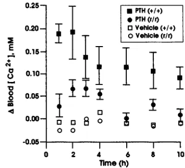
[image:5.612.77.266.151.296.2]Calcemic responses top PTH in +/+ and r/r mice.Because PTH did not stimulate osteoclastic bone resorption in the r/r mice, we asked whether PTH could nevertheless increase circulating calcium levels in these animals. PTH was given intraperitoneally in a single dose of 15 µg/30 g body weight to r/r and +/+ mice, and blood ionized calci-um concentration, [Ca2+], was measured at intervals as shown for the first calcemia experiment in Fig. 7. In the +/+ mice, PTH increased blood [Ca2+] by ∼0.2 mM by one
Figure 3
Changes in calvarial bone area and osteoclast number in 4-week-old +/+ and r/r mice injected subcutaneously over the hemicalvariae with vehicle (Veh) or PTH. Black bars (means ± SEM) indicate the bone mar-row space area (total resorption area), expressed as a percentage of the total bone area in a standard length of calvarial sections. Clear bars (means ± SEM) indicate the number of osteoclasts in the total bone area (i.e., per mm2). The number of mice examined are indicated in
parentheses. The bone resorption area and the number of osteo-clasts/mm2were both significantly increased after PTH injection in the
[image:5.612.57.287.444.629.2]+/+ mice (P < 0.005), but not in the r/r mice. In contrast, resorption in response to PTH was markedly reduced, and only occasional osteo-clasts were identified in calvarial bone from PTH-treated r/r mice.
Figure 4
to two hours after injection, and [Ca2+] was still elevated at 10 hours after injection. In contrast, as is also shown in Fig. 7, in the r/r mice, the peak rise in [Ca2+] was signifi-cantly blunted compared with the +/+ mice (only ∼0.08 mM above baseline) and was at control levels by three hours after injection. In the r/r mice, the peak ∆ [Ca2+] at one to three hours after injection of PTH was 0.056 ± 0.010 (SEM) mM compared with 0.169 ± 0.025 mM in +/+ mice (P < 0.001). In the second calcemia experiment (not shown), the peak ∆[Ca2+] at one to three hours after injec-tion of PTH in r/r mice was 0.061 ± 0.009 mM compared with 0.186 ± 0.044 mM in +/+ mice (P < 0.001). Because both +/+ C57BL and 129 strain mice responded to PTH with an increase in [Ca2+] of similar magnitude to that shown in Fig. 7 (data not shown), it is unlikely that differ-ent genetic backgrounds account for the differences in cal-cemic responses of r/r and +/+ mice observed here.
Discussion
We report here that the homozygous Col1a1tml Jaemice (30) that are resistant to collagenase cleavage of type I collagen have a markedly blunted response to the bone-resorptive effects of PTH. These mice have a targeted
mutation (29, 30) in Col1a1that encodes amino acid sub-stitutions in the helical region of the α1(I) chain around the collagenase cleavage site from P2 through P8′ (residues 774–783). We chose to target the substrate rather than the enzyme(s) because of the potential redundancy in collagenase genes in the mouse analo-gous to that in humans, although at the present time, only the genes homologous to human collagenase-2 (MMP-8) (33) and human collagenase-3 (MMP-13) (17) have been identified in the mouse. Neither the α1(I) chain nor the α2(I) chain of the heterotrimeric type I col-lagen extracted from skin, bone, and tendon of the Col1a1tml Jaemice that carry the mutation (r) on both alleles (r/r) was cleaved by rat or mouse collagenase (now called MMP-13) or human fibroblast collagenase (MMP-1) at what was previously considered to be the single cleavage site at Gly775/Ile776 (P1/P1′) in the helical domain in the α1(I) chain (34, 35).
[image:6.612.96.504.49.392.2]Although embryonic development in the r/r mice appeared to be normal, we have now observed that after as early as two to four weeks of age, the r/r mice begin to display abnormalities such as thickened skin with patchy hair loss, joint contractures, and nodules containing
Figure 5
Figure 6
In situ hybridization, using a collagenase riboprobe, of calvarial bone from +/+ and r/r mice injected subcutaneously over the hemicalvariae with vehicle or PTH. The outer (subcutaneous) side is shown. Sections were hybridized with the antisense collagenase riboprobe and were visualized using bright field (top) and dark field (bottom). Arrows indicate the periosteum. No hybridization with the collagenase riboprobe was detected in the periosteum in calvariae from vehicle-treated +/+ mice or vehicle-treated r/r mice. In calvariae from both +/+ and r/r mice, levels of collagenase mRNA in periosteal osteoblasts were marked-ly increased after PTH injection. No signal was observed in samples hybridized with the collagenase sense riboprobe (not shown). Scale bar, 0.05 mm.
type I collagen in the uterus of postpregnant females (30). In skin from r/r mice, histological examination with H&E and trichrome staining shows a remarkable increase in collagen extending through to the deep der-mis (30). We also observed bony deformities, particular-ly of long bones, with increased deposition of woven bone; these lesions could possibly have resulted from healing fractures as well (Zhao, W., and Krane, S.M., manuscript in preparation). Subsequently, we observed that highly purified rat collagenase (97% amino acid sequence identity with the mouse; ref. 16), which did not cleave at the helical site in the r/r collagen, did cleave at an additional site between a Gly/Val in the NH2
-telopep-tide (30). We later determined (36) that the capacity to cleave type I collagen in the NH2-telopeptide is a
proper-ty of these mouse (16) and rat collagenases, which have
∼97% amino acid sequence identity with each other but
only ∼50% amino acid sequence identity with human
MMP-1 (16). We proposed (30, 36) that the NH2
-telopep-tide cleavage activity of the rodent-type collagenases that are homologous with human MMP-13 might be suffi-cient for type I collagen cleavage during embryonic devel-opment, but that later in life during phases of intense resorption and/or after the collagen is covalently cross-linked, helical cleavage may be required.
The decrease in bone resorption and the paucity of osteoclasts in response to PTH in the r/r mice whose col-lagen cannot be cleaved at the helical locus could have several possible explanations. First, as mentioned previ-ously, there are indications that osteoblasts, when exposed to concentrations of PTH sufficient to induce bone resorption, start to produce collagenase and decrease synthesis of collagen (21). It has been suggest-ed, based on assessments of osteoclast attachment and bone pit formation in in vitro assays, that collagenase produced by osteoblasts in remodeling bone acts on a
layer of hypomineralized collagen on bone surfaces (28) to permit osteoclasts to attach and then resorb bone by mechanisms utilizing other proteinases (37–40). Thus, osteoblasts and/or related mesenchymal cells, such as marrow stromal fibroblasts, that at some stage function to synthesize the ECM of bone, also could function at another stage (in bone resorption) by preparing the matrix for osteoclastic attachment and subsequent degradation by proteinases such as cathepsins that act at the acid pH in the unique environment adjacent to the ruffled border (37–40). Second, it has been shown by
Pfeilschifter and Mundy (41) and Bonewald et al. (42)
osteoclasts to bone surfaces prepared by collagenase itself generates an anti-apoptotic signal. Failure of colla-genase to cleave bone type I collagen in the r/r mice might therefore indirectly abrogate this antiapoptotic signal. Some evidence to support this hypothesis follows. It has been shown that cells can bind to type I collagen through the α2β1 integrin (46) and that “liganding” α2β1 induces collagenase gene transcription followed by secretion and activation of latent (pro)enzyme (47, 48). Proteolytic attack on type I collagen can then result in unwinding of the cleaved ends to reveal a cryptic bind-ing site(s) for the αvβ3 integrin (48). Liganding this cryp-tic binding site by the αvβ3 integrin then promotes a phosphatidylinositol 3-kinase (PI-3 kinase)–mediated adhesion-dependent survival signal necessary for cells to normally progress through the cell cycle. Failure to bind through αvβ3 results in apoptosis, mediated by p53 and p21WAF1/CPI1 (49, 50). Thus, αvβ3 integrin antagonists suppress p53 activity and block p21WAF1/CPI1expression and increase the ratio of bcl-2/bax, whereas αvβ3 antag-onists activate p53 and increase p21WAF1/CPI1expression and decrease the ratio of bcl-2/bax (49, 50). Other obser-vations pertain to these considerations. Keratinocytes bind to type I collagen through the α2β1 integrin result-ing in activation of MMP-1 expression (47). Collagenase is then secreted and activated and the type I collagen substrate at least partially cleaved, events essential for migration of the keratinocytes (47). These cells also acti-vate collagenase expression using a type I collagen sub-strate prepared from the r/r mice, but they do not migrate, presumably because the collagen cannot be cleaved at the helical locus. Osteoclasts adhere to unde-natured type I collagen through the α2β1 integrin, but to denatured collagen through αvβ3 (51). Recognition by αvβ3 of sites in denatured collagen could be equiva-lent to a site generated by collagenase cleavage in the hel-ical region that then results in unwinding (denaturation) of the cut ends, and collagenase-generated type I colla-gen fragments can activate osteoclasts to resorb bone (52). Furthermore, synthetic peptidomimetic antago-nists of αvβ3 integrin inhibit bone resorption in vivo (53). One of the mechanisms of this inhibition of osteoclast activity, as well as osteoclast generation, could involve induction of osteoclast apoptosis, an effect observed by one of us when murine osteoclasts were treated with RGD (Arg-Gly-Asp) peptide–based inhibitors (54).
Whatever the mechanism, it is clear from these studies that induction of bone resorption by PTH is markedly inhibited in the r/r mice. Collagenase cleavage at the heli-cal locus in type I collagen appears to be essential for osteo-clastic resorption. Because embryonic development is nor-mal in the r/r mice, implying that bone resorption during the early phase of life can still occur, it is possible that cleav-age by collcleav-agenase at the NH2-telopeptide site before for-mation of stable intermolecular cross-links could be suffi-cient at this stage, although NH2-telopeptide cleavage has only been demonstrated in vitro (30, 36). Alternatively, bone trabeculae, formed in the primary and secondary spon-giosa during intrauterine and neonatal development, may be laid down and removed so rapidly that they do not become coated with the layer of hypomineralized matrix observed on bone surfaces of mature animals.
Acknowledgments
This work was supported by grants AR-03564, AR-07258, and AR-44855 from the National Institutes of Health.
1. Potts, J.T., Jr., and Jüppner, H. 1997. Parathyroid hormone and parathy-roid hormone-related peptide in calcium homeostasis, bone metabo-lism, and bone development: the proteins, their genes, and receptors. In
Metabolic bone disease and clinically related disorders. L.V. Avioli and S.M. Krane, editors. Academic Press. San Diego, CA. 51–94.
2. Suda, T., Takahashi, N., and Martin, T.J. 1992. Modulation of osteoclast differentiation. Endocr. Rev. 13:66–80.
3. Yasuda, H., et al. 1998. Osteoclast differentiation factor is a ligand for osteoprotogerin/osteoclastogenesis-inhibitory factor and is identical to TRANCE/RANKL. Proc. Natl. Acad. Sci. USA. 95:3597–3602.
4. Gross, J., and Lapière, C.M. 1962. Collagenolytic activity in amphibian tissues: a tissue culture assay. Proc. Natl. Acad. Sci. USA. 48:1014–1022. 5. Walker, D.G., Lapière, C.M., and Gross, J. 1964. A collagenolytic factor in rat bone promoted by parathyroid extract. Biochem. Biophys. Res. Com-mun.15:397–402.
6. Krane, S.M. 1975. Skeletal remodeling and metabolic bone disease. In
Calcium-regulating hormones. R.V. Talmage, M. Owen, and J.A. Parsons, edi-tors. Excerpta Medica. Amsterdam, The Netherlands. 57–65. 7. Rasmussen, H., Arnaud, C., and Hawker, C. 1964. Actinomycin D and
the response to parathyroid hormone. Science. 144:1019–1021. 8. Tashjian, A.H., Jr., Ontjes, D.A., and Goodfriend, T.L. 1964. Mechanism
of parathyroid hormone action. Effects of actinomycin D on hormone-stimulated ion movement in vivo and in vitro. Biochem. Biophys. Res. Commun.16:209–215.
9. Kunin, A.S., and Krane, S.M. 1965. Inhibition by puromycin of the cal-cium-mobilizing activity of parathyroid extract. Endocrinology.
76:343–344.
10. Sakamoto, S., Sakamoto, M., Goldhaber, P., and Glimcher, M.J. 1975. Collagenase and bone resorption: isolation of collagenase from culture medium containing serum after stimulation of bone resorption by addi-tion of parathyroid hormone extract. Biochem.Biophys. Res. Commun.
63:172–178.
11. Sakamoto, S., Sakamoto, M., Goldhaber, P., and Glimcher, M.J. 1975. Studies on the interaction between heparin and mouse bone collagenase.
Biochim. Biophys. Acta. 385:41–50.
12. Sakamoto, S., Sakamoto, M., Goldhaber, P., and Glimcher, M.J. 1978. Mouse bone collagenase. Purification of the enzyme by heparin- substi-tuted Sepharose-4B affinity chromatography and preparation of specif-ic antibody to the enzyme. Arch. Biochem. Biophys.188:438–449. 13. Sakamoto, S., Sakamoto, M., and Horton, J.E. 1984. Evidence that
col-Figure 7
Changes in blood [Ca2+] in +/+ and r/r mice injected intraperitoneally with
vehicle or PTH. The calcemic response after a single intraperitoneal injection of vehicle or 15 µg PTH in +/+ and r/r mice is shown as described for the first calcemia experiment. The change in blood ionized calcium levels, ∆blood [Ca2+], at intervals from 0.5 to 10 h after PTH injection (shown as means ±
SEM) is depicted on the vertical axis. Peak levels of ∆blood [Ca2+] were
observed within the first 1–3 h after PTH injection in the +/+ mice (filled cir-cles). As described in the text, peak levels of ∆blood [Ca2+] after PTH
[image:8.612.325.506.52.216.2]lagenase is involved in the mechanisms of bone resorption stimulated with parathyroid hormone: a study in two different bone culture sys-tems. InEndocrine control of bone and calcium metabolism. D.V. Cohn, T. Fujita, J.T. Potts, Jr., and R.V. Talmage, editors. Excerpta Medica. Ams-terdam, The Netherlands. 140–143.
14. Partridge, N.C., et al. 1987. Hormonal regulation of the production of col-lagenase and a colcol-lagenase inhibitor activity by rat osteogenic sarcoma cells.
Endocrinology. 120:1956–1962.
15. Quinn, C.O., et al. 1990. Rat collagenase. Cloning, amino acid sequence comparison, and parathyroid hormone regulation in osteoblastic cells.
J. Biol. Chem.265:22342–22347.
16. Henriet, P., Rousseau, G.G., and Eeckhout, Y. 1992. Cloning and sequencing of mouse collagenase cDNA. Divergence of mouse and rat collagenases from the other mammalian collagenases. FEBS Lett.
310:175–178.
17. Freije, J.M.P., et al. 1994. Molecular cloning and expression of collage-nase-3, a novel human matrix metalloproteinase produced by breast car-cinomas. J. Biol. Chem.269:16766–16773.
18. Reponen, P., Sahlberg, C., Munaut, C., Thesleff, I., and Tryggvason, K. 1994. High expression of 92-kD type IV collagenase (gelatinase B) in the osteoclast lineage during mouse development. J. Cell Biol.
124:1091–1102.
19. Sato, T., et al. 1997. Identification of the membrane-type matrix metal-loproteinase MT1-MMP in osteoclasts. J. Cell Sci.110:589–596. 20. Delaissé, J.M., et al. 1993. (Pro)collagenase (matrix metalloproteinase-1)
is present in rodent osteoclasts and in the underlying bone-resorbing compartment. J. Cell Sci. 106:1071–1082.
21. Scott, D.K., Brakenhoff, K.D., Clohisy, J.C., Quinn, C.O., and Partridge, N.C. 1992. Parathyroid hormone induces transcription of collagenase in rat osteoblastic cells by a mechanism using cyclic adenosine 3′,5′-monophos-phate and requiring protein synthesis. Mol. Endocrinol.6:2153–2159. 22. Mattot, V., et al. 1995. Expression of interstitial collagenase is
restrict-ed to skeletal tissue during mouse embryogenesis. J. Cell Sci.
108:529–535.
23. Fuller, K., and Chambers, T.J. 1995. Localisation of mRNA for collage-nase in osteocytic, bone surface and chondrocytic cells but not osteo-clasts. J. Cell Sci. 108:2221–2230.
24. Gack, S., et al. 1995. Expression of interstitial collagenase during skele-tal development of the mouse is restricted to osteoblast-like cells and hypertrophic chondrocytes. Cell Growth Differ.6:759–767.
25. Meikle, M.C., et al. 1992. Human osteoblasts in culture synthesize colla-genase and other matrix metalloproteinases in response to osteotropic hormones and cytokines. J. Cell Sci.103:1093–1099.
26. Lanske, B., et al. 1998. The parathyroid hormone (PTH)/PTH-related peptide receptor mediates actions of both ligands in murine bone.
Endocrinology.139:5194–5204.
27. Lanske, B., et al. 1996. PTH/PTHrP receptor in early development and Indian hedgehog-regulated bone growth. Science.273:663–666. 28. Chambers, T.J., Darby, J.A., and Fuller, K. 1985. Mammalian collagenase
predisposes bone surfaces to osteoclastic resorption. Cell Tissue Res.
421:671–675.
29. Wu, H., et al. 1990. Generation of collagenase-resistant collagen by site-directed mutagenesis of murine proα1(I) collagen gene. Proc. Natl. Acad. Sci. USA. 87:5888–5892.
30. Liu, X., et al. 1995. A targeted mutation at the known collagenase cleav-age site in mouse type I collcleav-agen impairs tissue remodeling. J. Cell Biol.
130:227–237.
31. Boyce, B.F., Aufdemorte, T.B., Ross Garrett, I., Yates, A.J.P., and Mundy, G.R. 1989. Effects of interleukin-1 on bone turnover in normal mice.
Endocrinology.125:1142–1150.
32. Yates, A.J.P., et al. 1988. Effects of a synthetic peptide of a parathyroid
hormone-related protein on calcium homeostasis, renal tubular calci-um reabsorption, and bone metabolism in vivo and in vitro in rodents.
J. Clin. Invest.81:932–938.
33. Lawson, N.D., Khanna-Gupta, A., and Berliner, N. 1998. Isolation and characterization of the cDNA for mouse neutrophil collagenase: demon-stration of shared negative regulatory pathways for neutrophil second-ary granule protein gene expression. Blood. 91:2517–2524.
34. Gross, J. 1981. An essay on biological degradation of collagen. InCell biol-ogy of the extracellular matrix. E.D. Hay, editor. Plenum Press. New York, NY. 217–258.
35. Birkedal-Hansen, H., et al. 1993. Matrix metalloproteinases: a review. Crit. Rev. Oral. Biol. Med. 4:197–250.
36. Krane, S.M., et al. 1996. Different collagenase gene products have differ-ent roles in degradation of type I collagen. J. Biol. Chem. 271:28509–28515.
37. Blair, H.C., et al. 1993. Extracellular-matrix degradation at acid pH. Avian osteoclast acid collagenase isolation and characterization. Biochem. J. 290:873–884.
38. Burleigh, M.C. 1977. Degradation of collagen by non-specific pro-teinases. InProteinases in mammalian cells and tissues. A.J. Barrett, editor. Elsevier Science. Amsterdam, The Netherlands. 285–309.
39. Drake, F.H., et al. 1996. Cathepsin K, but not cathepsin B, L, or S, is abun-dantly expressed in human osteoclasts. J. Biol. Chem.271:12511–12516. 40. Bossard, M.J., et al. 1996. Proteolytic activity of human osteoclast cathep-sin K. Expression, purification, activation, and substrate identification.
J. Biol. Chem.271:12517–12524.
41. Pfeilschifter, J., and Mundy, G.R. 1987. Modulation of type β trans-forming growth factor activity in bone cultures by osteotropic hor-mones. Proc. Natl. Acad. Sci. USA. 84:2024–2028.
42. Bonewald, L.F., et al. 1997. Effects of retinol on activation of latent trans-forming growth factor-βby isolated osteoclasts. Endocrinology. 138:657–666. 43. Hughes, D.E., et al. 1995. Bisphosphonates promote apoptosis in murine
osteoclasts in vitro and in vivo. J. Bone Miner. Res. 10:1478–1487. 44. Hughes, D.E., et al. 1996. Estrogen promotes apoptosis of murine
osteo-clasts mediated by TGF-β. Nat. Med. 2:1132–1136.
45. Hughes, D.E., and Boyce, B.F. 1997. Apoptosis in bone physiology and disease. Mol. Pathol. 50:132–137.
46. Staatz, W.D., Rajpara, S.M., Wayner, E.A., Carter, W.G., and Santaro, S.A. 1989. The membrane glycoprotein Ia-IIa (VLA-2) complex mediates the Mg++-dependent adhesion of platelets to collagen. J. Cell Biol. 108:1917–1924.
47. Pilcher, B.K., et al. 1997. The activity of collagenase-1 is required for ker-atinocyte migration on type I collagen matrix. J. Cell Biol. 137:1445–1457. 48. Montgomery, A.M.P., Reisfeld, R.A., and Cheresh, D.A. 1994. Integrin αvβ3 rescues melanoma cells from apoptosis in three-dimensional der-mal collagen. Proc. Natl. Acad. Sci. USA. 91:8856–8860.
49. Brooks, P.C., et al. 1996. Localization of matrix metalloproteinase MMP-2 to the surface of invasive cells by interaction with integrin αvβ3. Cell.
85:683–693.
50. Strömblad, S., Becker, J.C., Yebra, M., Brooks, P.C., and Cheresh, D.A. 1996. Suppression of P53 activity and p21WAF1/CIP1expression by
vascu-lar integrin αvβ3. J. Clin. Invest.98:426–433.
51. Helfrich, M., et al. 1996. β1 integrins and osteoclast function:
involve-ment in collagen recognition and bone resorption. Bone.19:317–328. 52. Holliday, L.S., et al. 1997. Initiation of osteoclastic bone resorption by
interstitial collagenase. J. Biol. Chem.272:22053–22058.
53. Engleman, V.W., et al. 1997. A peptidomimetic antagonist of the αvβ3
integrin inhibits bone resorption and prevents osteoporosis in vivo. J. Clin. Invest.99:2284–2292.