Efficient VLSI Architecture for ECG Data Compression
Ruqaiya Khanam
Department of Electronics & Communication Engineering
Jamaia Millia Islamia University Delhi,INDIA
Syed Naseem Ahmad
Department of Electronics & Communication Engineering
Jamaia Millia Islamia University Delhi,INDIA
ABSTRACT
This paper presents an efficient ECG signals compression techniques using a 2D DWT coefficient thresholding and its design implementation of an efficient JPEG2000 encoder that employs the Distributed Arithmetic (DA) technique for the complex computation of Discrete Wavelet Transform (DWT).2D approaches exploit the fact that redundancy of ECG signal occurs between adjacent beats and adjacent samples. Cut and aligned error signal to form 2D matrix, then 2D matrix is transformed. Ultimately wavelet coefficients are segmented into groups and thresholded. DA-DWT is used for reducing the complex computations, which can increase the speed and throughput as well. Architecture is based on the principles of pipelining and parallelism to obtain the optimal speed and throughput. Architecture is simple, modular and cascadable for computing a DA-DWT. This technique is faster when ROM table in on chip memory and memory size is reduced by splitting ROM table .The description and functionalities of the design is modeled by Verilog HDL. The simulation and synthesis methodology are used to target it on Virtex-II Pro FPGA (xc4v1x25-12sf363) that consumes 6% resources of FPGA and shows the clock frequency 310.207 MHz by DA-DWT using Sym20. Experiments on selected records from MIT-BIH arrhythmia database revealed that the proposed design is significantly more efficient.
Keywords
Electrocardiogram, Discrete Wavelet Transform, Distributed Arithmetic
1.
INTRODUCTION
The extensive use of electrocardiogram (ECG) produces large amount of data. Since the need for ECG signal compression is two reasons: effective storage and effective real time transmission .The efficient compression techniques are important to reduce transmission time or required storage capacity of large data records. Several ECG compression methods have been developed during last three decades [1].
(1) Direct Methods-e.g.-AZTEC [2], DPCM, TP, CORTES, Scan-along polygonal approximation (SAPA) [3], FAN [4].
(2)Transform Methods- e.g.- Fourier, Walsh, KLT, Discrete Cosine Transform (DCT) [5], and Wavelet Transform (WT) [6][7][8].
(3) Parameter Extraction Methods- e.g.- Vector Quantization (VQ) [9], Neural Network (NN),Prediction .
Our approach belongs to the second category. Usually, transform techniques involve expanding a signal into a and weighted sum of basis functions. The coefficients of this sum are properly encoded and stored or transmitted instead of the original data. Transform techniques include several wavelet-based compression methods. Due Time-frequency localization properties of wavelets are better hence suitable for data compression applications.
Most of the methods process the heart beat data in 1-D form although some of these methods utilize redundancies between adjacent beats. Since the ECG signals has both intra-beat (sample to sample) and inter-beat (beat to beat) correlations, many 2-D compression approaches have been proposed for better compression performances. For example, Lee[5] used “cut and align beats approach and 2D discrete cosine transform” to obtain better compression results in regular ECG.Wei [11] used truncated singular value decomposition algorithm to compress 2D ECG arrays. Recently, wavelets are widely used for both 1D and 2D compression [6][7][8]. Due to better performance of data compression, choose JPEG2000 [12] as a codec.DWT has the high compression ratio and is free from blocking artifacts at low bit rates [13].
In this paper, the following paper sections: section II describes the 1-D to 2-D process. Section III presents the proposed DA-DWT based architecture for JPEG2000 encoder. In section IV, the performance analysis and implementation results are given. Finally, the conclusion is presented in section V.
2.
2-D ECG PROCESS
2-D compression methods comprises the following steps:
1) QRS Detection
2) Preprocessing (cut and align beats, period normalization, amplitude normalization, mean removal)
3) Transformation 4) Coefficient encoding
2.1 QRS detection and alignment
the P-waves or T-waves as well as spikes from high frequency pacemakers can compromise the detection of the QRS complex.QRS detector must be robust for different noises and also able to detect QRS complexes in spite of the morphology of the ECG signal is varying with time.The detection algorithm is a MATLAB based detector[19]. Primarily the peaks of QRS waves should be selected to identify each heartbeat. After identify each QRS peak of heartbeat segments is identified, the original signal 1-D ECG signals are cut at every 130th sample before each QRS peak
(a 130th sample shift from the QRS peak, a smooth region estimated from human physiology)[14].
2.2 Period of heartbeats restriction
The periods of heartbeats are not the same. Therefore the length of the heartbeats segments can be varied. If the ECG is found abnormal or the QRS peak detection misses or some QRS peaks identified heartbeat is very long then the size of 2-D array increases, this is the more dangerous situation. To achieve maximum data compression used an efficient compression scheme needs to utilize both dependencies. The length of detected heartbeats should be restricted to form of a 2-D ECG representation at moderate size. After the detection of QRS complex, each period is stored in 2D matrix. Here the intrabeat dependencies are in horizontal direction of the matrix and interbeat dependencies are in vertical direction. Fig.1 shows the generated image .Due to the variation in length, the generated 2D ECG matrix will be using different number of data points in every row. Researchers have tried to overcome this problem. The compression system using JPEG2000 normalized each ECG period for equal length[16].
Fig.1 ECG 2D Matrix
One major advantage opened by wavelets is the ability to perform local analysis, to analyze a localized area of a larger signal. Discrete Wavelet Transform allows the change of a signal from a time domain to a frequency –time domain through a decomposition and reconstruction process, the last one to recover the original signal[15].In the decomposition, the wavelet coefficients of any stage can be calculated from DWT of the previous stage using the following iterative equations:
(1)
(2)
Where WL(n,j) is the nth scaling coefficient at the jth stage,
WH(n,j) is the nth wavelet coefficient at the jth stage, and h0
(n) and h1(n) are the dilation coefficients corresponding to
the scaling and wavelet functions, and m is the summation running index of the analysis filter’s coefficients, respectively.
In order to reconstruct the original data, the DWT coefficients are upsampled and passed through another set of low pass and high pass filters, which is expressed as follows:
(3)
Where g0(n) and g1(n) are respectively the low-pass and
high-pass synthesis filters corresponding to the mother wavelet, and l is summation running index of the analysis filter’s coefficients. It is observed from equation(3) that the jth level coefficients can be obtained from the (j+1)th level coefficients.
Cut and Align Header
Info Encoding
DWT Segmentation into
NXN Blocks
ECG Signal Encoded Bit Stream
10101110…..
(b)
(c)
[image:3.595.57.411.71.195.2]Original Image Wavelet Transform Image Bit Stream
Fig.3 Data representations after image reception, Wavelet transformation and encoding
The multimedia signals are stored or transmitted in compressed form to save time and resource. Efficient coding standards like MP3, JPEG2000 and MPEG4 are developed for multimedia signal.
The architecture has implemented DA-LUT using adders and multiplexers. Using this architecture the hardware and time complexity can be optimized.
To derive Distributed Architecture for DWT, consider the following sum of products:
(4)
Where Ak is the fixed coefficient of the filter bank and Xk is
the input samples.The decomposed equation of (4) in form of DA can be written as equation 2:
(5)
Where:
From equation (5), A is the distributed arithmetic matrix of
fixed coefficients , where k= 1.2,….,L ; i=1,2,…,N-1,with
is the MSB and is the LSB. In DA-DWT, the bits
of the coefficients are distributed unlike conventional DA,
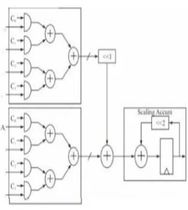
[image:3.595.314.506.282.498.2]Distributed architecture matrix contains only 0 and 1, which means the computation of Y can be obtained just by shifting and adding of the input vectors. The architecture of DA-DWT can save hardware to implement the computations.
Fig.4 Bit level arrangement for DA MAC
Fig. 5 Parallel implementation of DA technique
[image:3.595.316.501.523.729.2]Algorithm
BEGIN
Step 1: Loading of original signal
The signal which is taken from MIT-BIH arrhythmia data base
Step 2: Detect QRS complex in every heart beat
Step 3: Cut and align the signal beat by beat
prior to 2-D transformation
Step 4: Segmentation into NXN blocks (to construct 2-D array from 1-D array)
Step 5: Apply DWT to get the transformed information using by DA-DWT architecture
Step 6: Find the transformed coefficients
Step 7: Apply 2-D transform coding to achieve
the essential compression.
Step 8: Displaying the result
END
Algorithm 2
Step 1.The samples of input ECG signal are divided into even and odd in the first stage.
Step 2.Using shift register, upper four shift register store MSB bits and lower four shift register stores the LSB bits.
control logic configures the shift register as serial in parallel out, forming the address for the LUT.
Step 4.Partial products stored in the LUT are read simultaneously front all the four LUTs.
Step 5.Accumulated with previous values available across the shift register in the output stage.
Step 6.The output stage consisting of adders, accumulators and shift registers are used to
accumulate the LUT contents and compute the DWT output.
Distributed Arithmetic replaces multiplications by ROM look-up table so that DWT filter can achieve high computing speed with small area by 80% in hardware design.
5.
SIMULATION
AND
SYNTHESIS
METHODOLOGY
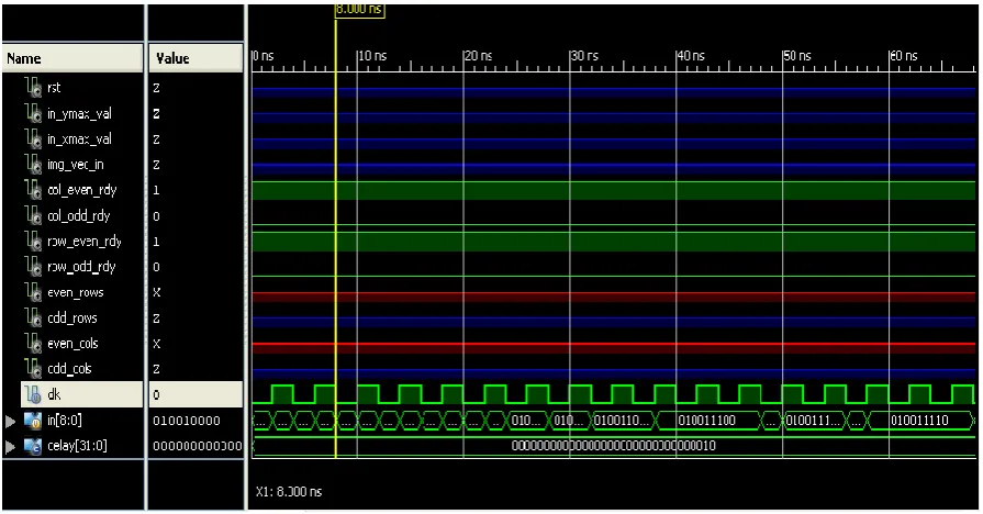
ECG data compression proposed here is tested for record 117 from MIT-BIH arrhythmia database using by MATLAB.
[image:4.595.74.522.454.688.2]DA-DWT was described in Verilog HDL. The architecture was divided into blocks level and blocks are divided into sub blocks. At sub block level, behavioral description was made and at block level, structural description connecting the sub blocks was constructed and validated using the Xilinx software.
Fig. 6 Snapshot of the simulation of DWT
Fig. 7 Snapshot of RTL view of DWT
[image:5.595.48.288.355.593.2]6.
EXPERIMENTAL RESULT
Table 1 Synthesis Results of DA-DWT
S.No Devices
No. of Devices
1
ROMs(2048x16-bit ROM) 42
Adder/Subtractors (9 bit Adder)26
3
Counters (3-bit and 9-bit up counter)2,2
4
Registers (1-bit reg.,16-bit,8-bit,9-bit regs.)32,28,4,2 (66)
5
Comparators 106
Multiplexers 47
Tristates 4Table 2 Performance Comparison
Scheme
Level-1
Efficiency
Filter based implementation
16x Multiplier N2
Lifting Based implementation
6x Multiplier N2
Proposed architecture
26x Adders N2/2
Above table 1 shows the performance of various architectures. The proposed architecture was obtained throughput by factor 2 than previous counterparts. The efficiency cycles were required to compute DWT of NxN image at same clock frequency.
7.
CONCLUSION
In this paper, design gives faster results using highly parallel Virtex field programmable gate array devices (FPGA).The proposed architecture has been correctly verified by the Verilog Hardware Descriptive Language (Verilog HDL). DA-DWT is better area optimization and increase the throughput. In this architecture achieve the clock frequency upto 310.207 MHZ and area utilization are reduced by 7% lesser then the normal approach. Simulation and synthesis results are obtained in figure 3 and figure 4 respectively.
8. REFERENCES
[1] Jalaleddine S.M.S, Hutchen C.G, Strattan R.D. and Coberly W.A ,April 1990, ECG data Compression Techniques- A unified approach, IEEE Trans. On biomed.Eng., 37(4):329-343.
[2] Cox J.R., Noile F.M., Fozzard H.A., and Olover G.C, 1968, AZTEC : preprocessing program for real time ECG rhythm analysis, IEEE Trans. Biomed.Eng., vol. 15, pp. 28-129.
[3] Ishijima M., Shin,S.B. Hostetter G.S., and Sklansky J., 1983, Scan-along polygonal approximation for data compression of electrocardiograms,IEEE Trans.Biomed. Eng., vol. 30, 11, pp. 723-729.
[6] Rajoub B.A., 2002,An efficient coding algorithm for the compression of ECG signal s using the wavelet transform, IEEE Trans. Biomed. Eng.,vol. 49, pp. 355-362.
[7] Hilton M.L, 1997, Wavelet and Wavelet packet compression of electrocardiograms, IEEE Trans Biomed. Eng., vol. 44, pp. 394-402.
[8] Moghaddam A.R.A and Nayebi K, 2001,A two dimensional wavelet packet approach for ECG compression, Signal processing and its applications,the 6th International, Symposium, pp.226-229.
[9] Wang B. and Yuan G, 1997,Compression of ECG data by vector quantization, IEEE Trans. Biomed. Eng., vol. 40,pp. 23-26.
[10] Nave G. and Cohen A, 1993,ECG compression using long-term prediction, IEEE Trans. Biomed. Eng., vol.40, pp. 877-885.
[11] Wei J.J, Chang C.J, Chou N.K, and Jan G.J, 2001ECG data compression using truncated singular value decomposition, IEEE Trans. Biomed. Eng., vol. 5, pp. 290-299.
[12] Morteza M.G, Taheri A, and Pooyan M, 2005,Efficient method for ECG compression using two dimensional Multiwavelet transform, An International journal of information technology, vol.2,4.
[13] Mohammadpour T.I and Mollaei M.R.K, 2009,ECG compression with thresholding of 2D wavelet transform oefficients and run length coding, European journal of scientific research, vol.27,no.2,pp.248-257.
Engineering, vol.52, no. 6,pp.999-1008.
[15] Sezgin M and Sankur B, 2004, Survey over image thresholding techniques and quantitative performance evaluation, Journal of Electronic Imaging, vol.13,no.1,pp.146-168.
[16] Bilgin A,Marcellin M.W and Altbach M.I,2003,Compression of electrocardiogram signals using JPEG2000, IEEE Transaction on consumer electronics, vol.49,no.4,pp.833-840.
[17] Zhang Qihui, Meng Nan, 2nd-5th Aug 2009 ,A low area pipelined 2D DCT architecture for JPEG encoder, MWSCAS’09,52nd IEEE international Midwest Symposium on Circuit and Systems, pp.747-750. Nagabushanam M., Raj P.C.P, Ramachandran,S.10-12 Feb.2011,Design and FPGA implementation of modified Distributed Arithmetic based DWT-IDWT processor for image compression,International conference on communications and signal processing (ICCSP), pp.1-4.
[18] Vander J, Sherman J.H , Luciano D.S, 1994,Human Physiology, McGraw-Hill, 6th edition, chap 14,pp393-472.
[19] Ruqaiya Khanam, Syed Naseem Ahmad, ,23-24 June 2012 , Peak Detection of ECG Signal Using Wavelet Transform” ,International Conference on Emerging Trends in Engineering & Management (ICETEM12-2012),pp.200-203.
[20] Mallat S, 1999, A Wavelet Tour of signal processing. Academia Press.
[21] MIT-BIH Arrhythmia Database,