International Journal of Scientific and Research Publications, Volume 9, Issue 9, September 2019 356 ISSN 2250-3153
Diagnostic Immunochemistry in Gynaecological
Neoplasia Guide to Diagnosis
S.S Pattnaik, J.Parija,S.K Giri, L.Sarangi, Niranjan Rout, S. Samantray, N. Panda,B.L Nayak, J.J Mohapatra, M.R Mohapatra, A.K Padhy
Presently Working As Senior Resident In Dept Of Gynaecology Oncology, At Ahrcc ,M.B.B.S , M.D (O&G)
DOI: 10.29322/IJSRP.9.09.2019.p9346
http://dx.doi.org/10.29322/IJSRP.9.09.2019.p9346
Abstract- AIM AND OBJECTIVE - This short review provides an updated overview of the essential immunochemical markers currently used in the diagnostics of gynaecological malignancies along with their molecular rationale. The new molecular markers has revolutionized the field of IHC
MATERIAL METHODS -We have reviewed the recent ihc markers according to literature revision and our experiencewe, have discussed the the use of ihc ,
CONCLUSION- The above facts will help reach at a diagnosis in morphologically equivocal cases of gynaecology oncology pathology, and guide us to use a specific the panel of ihc ,which will help us reach accurate diagnosis.
I. INTRODUCTION
HC combines microscopic morphology with accurate molecular identification and allows in situ visualisation of specific protein antigen . IHC has definite role in guiding cancer therapy.The role of pathologist is increasing beside tissue diagnoses , to perfoming IHC biomarker analyse ,assisting the development of novel markers. Ihc markers are being in used in new perspective , in guiding anticancer therapy.ihc represents a solid adjunct for the classification of gynaecological malignancies that improves intraobserver reproducibility and has potential of revealing unexpected features(1)
OVARIAN IHC:
• PAX -8 is the most specific marker, emerging to diagnose primary ovarian cancer,but it lacks sensibility as it is also expressed in metastasis from endocervix ,kidney and thyroid. Fig 1 table2(1)
FIG-1( A) Is the immunohistochemical algorithm for a PRIMARY AND METASTATIC CARCINOMA. The(A) algorithm addresses the markers which helps to distinguish morphologically equivocal primary ovarian cancers using five to nine markers. The (B) algorithm the step wise immunohistochemical approach of metastatic ovarian carcinoma, sx main immunospecific stains and tissue specific markers.CK-7 and CK-20are co expressed the are schematically represented as continuous vertical from the prevalent( CK-7) positivity at the upper end,to ck-20 positivity at the lower end.the frequency (upper end ) to prevalence CK-20 positve at lower end. The frequency of metastatic disease is correlated with font size. CC is clear cell carcinoma,ccRCC clear cell renal carcinoma,CK- cytokeratin,EAC- endo cervical adenocarcnoma,EMC- endometrod carcinoma,ER-estrogen geceptor,
FIG2
WT-1 – is most sensitive and specific marker of serous histiotype.(2)
• It can be used to discriminate serous from other histiotypes.
• Nuclear expression of WT-1,(muellerian marker)
• This marker is consistently nuclear expressed in normal tubal epithelium:thus coherently expressed in HGSC and LGOC
• Recently a practical approach to use ihc in classification of primary ovarian carcinoma using ihc algorithm.Basically the four markers WT-1,P53,Napsin a and progestrone receptors(PR).A modified ihc ALGORITHM that includes interchangeable markers,namely HNF1BETA and AMACR(racemase or p504s) for CCC.fig 1a and fig 1b (1)
• ER and vimentin for endometroid carcinomas
Endometroid versus HGSC
• HGSC withglandular and cribriform may closely resemble endometroid
• WT-1, 60- 70% OF THE HGSC. p53 is overexpressed,and diffusely and intensely nuclear staining of p53(3). 30% of high grade endometriod may have p53 +ve.
International Journal of Scientific and Research Publications, Volume 9, Issue 9, September 2019 360 ISSN 2250-3153
• HGSC - PTEN downregulation
• CCNE1 overexpression , absent in endometroid CLEAR CELL VERSUS HGSC-
• HGSC with morphology of cytoplasmic clearing from CCC or vice versa CCC with eosinophilic cytoplasm from HGSC
• WT-1 and ER chiefly expressed in HGSC, along with aberrant p53, as well as nuclear HNF1 beta
• Cytoplasmic napsin and AMACR POSITIVE IN CCC(4)
• ARIDIA can be negative in 57% cases of CCC
ADVANCED OVARIAN VERSUS UTERINE SEROUS CARCINOMA
AND SYNCHRONOUS PRIMARY OF ENDOMETRIUM AND OVARY
• Wt-1 – is diffusely positive in MAJORITY HGSC, whereas in uterine serous carcinoma upto one third of cases
• WT-1 negative in both points to primary endometrium
• If staining in different patterns at two different sites, suggest two synchronous primary.( 4-6)
Peritoneal serous carcinoma versus epetheloid mesothelioma
• The most reliable recent markerloss of BRCA –associated protein 1(BAP-1), deletion of P16 BY FISH for mesothelioma
• Mesothelioma - calretinin,keratin5/6,D2-40 positive
• Serous carcinoma-Pax-8, ER, claudin -4, MOC31 and Ber-EP4 positive in serous carcinoma(7-8)
MUCINOUS ADENOCARCINOMA- PRIMARY/METASTATIC
• There is a overlap of ihc between primary and metastatic gastrointestinal tumors
• CDX-2,CK20 and SATB2 are diffusely expressed and strongly positive in colorectal adenocarcinoma,
• Where as ,the above markers are less intense and weak, in comparision to CK-7 in Primary ovarian carcinomas, with exception of rare intestinal type mucinous ovarian tumors from teratomas
• Primary ovarian- positive for CA125, pax-8
• Ca125- BREAST, lung , pancreas,cervixand uterine carcinomas and mesothelioma , although ,CK20+/CK7-VE IS PROTO TYPICAL OF METASTATIC ADENOCARCINOMA HENCE SPECIC MARKERS pax-8 and sat-b2(9-10) both highly specific, butsensitivity is low.
• ER AND PR are negative in both intestinal type and metastatic carcinoma, cdx2 is a site unspecific
• marker of intestinal differentiation
• Smad4 (DPC4)- is lost in half pancreatic cancer
• Small cell carcinomas of hypercalcemia versus othe mimic(HGSC and adult granulosa)
• specific is loss of SMARCA4(BRG1) expression(11)
CCC VERSUS YOLK SAC TUMOR
Both share similar morphology- gycogen rich clear cells
CCC- is usually arises in a background of endometriosis, or clear cell adenofibroma and positive AMACR,CK-7,EMAand Napsin-a
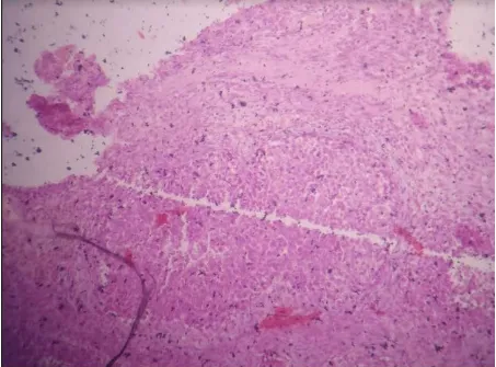
FIG-3 ihc staining image of staining of the metastaic and primary ovarian carcinoma
IHC OF NON-EPITHELIAL TUMORS-
ihc of primitive germs cell malignancies dysgerminoma- sall4+, OCT3/4+, SOX-2-VE YST- SALL4+, OCT3/5 -VE
EC- SALL4+ ,OCT3/4+VE,SOX2+VE
International Journal of Scientific and Research Publications, Volume 9, Issue 9, September 2019 362 ISSN 2250-3153
International Journal of Scientific and Research Publications, Volume 9, Issue 9, September 2019 364 ISSN 2250-3153
International Journal of Scientific and Research Publications, Volume 9, Issue 9, September 2019 366 ISSN 2250-3153
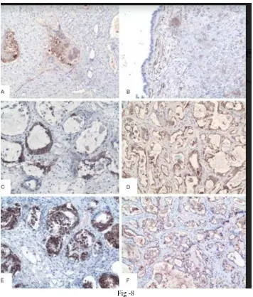
Fig -8
Hcg +ve in giant syncytotrophoblast(A) © cd-30+ve
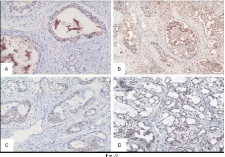
Fig -9
Afp found positive in yolk sac component(a), (b) AAT+VE(C)CD117+ve (d) glp3+ve
Ovarian small cell carcinoma of hypercalcemic type (OSCCHT) is a rare neoplasm with an aggressive behavior, broad differential diagnosis, and unknown histogenesis. d/d ofsmall round celltumors
Scst ,small call norphology, small round blue call tumor, neuroendocrine small cell,small cell carcinona of pulnomary type, this may be a componentof atypical ovarian surface epithelial – stromal tumor(14). In addition
International Journal of Scientific and Research Publications, Volume 9, Issue 9, September 2019 368 ISSN 2250-3153
Fig -4 ( round cell tumors)
Fig-10 SMALL ROUND CELL TUMOR OF UTEUS
• Cases were stained with AE1/3, EMA, BerEP4, CK5/6, calretinin, WT1, chromogranin, CD56, synaptophysin, CD99, NB84, desmin, S100, CD10, alpha inhibin, TTFI, and p53.
[image:13.612.80.534.55.546.2]Fig-11
• SLCT-FOXL2 –VE TUMORS , AT LEAST CALRETININ/OR ALPHA INHIBIN IS POSITIV
• RECNTLY DICER1 MUTATION
• SCCOHTs (small cell carcinoma of ovary hypercalcemic type)
• SMARC4 IHC SENSITIVE AND SPECIFIC IN DIAGNOSIS OF SCCOHT( THERE IS LOSS OF EXPRESSION OF
SMARC4(BRG1)
• POORLY DIFFERENTIATED TUMORS, RESEMBLING SMALL CELL OR ROUND CELL IN MORPHOLOGY
• D/D adult granulosa cell tumors , metastatic melanoma, ewings sarcoma,dysgerminoma and undifferentiated carcinoma.
International Journal of Scientific and Research Publications, Volume 9, Issue 9, September 2019 370 ISSN 2250-3153
•
•
Fig-12 ( ihc markers of small round cell tumors)
FALLOPIAN TUBE IHC
• Serous tubal intraepithelial carcinoma vs other mimics(HGSC)
• Stics – Marked cytological atypia with a high Ki-67>10%proliferation index and P53 ihc mutant form(16)
ENDOMETRIAL IHC
• the dualistic pathogenetic model has been proposed for endometrial cancer
• Type I- Endometroid variety
• Type II- USC,CCC,Malignant mixed muellerian tumor, undifferentiated
• the glandular cribriform USC, pappilary endometroid carcinoma and endometroid carcinomawith clear cells ,there distinction needs IHC
• Endometriod are ER and PR positive, whereas USC AND CCC and _ve
• P53 and mutant A POSITIVE IN CCC(17)
• Arias stella raection and some endometroid carcinomas exHNF1 beta
International Journal of Scientific and Research Publications, Volume 9, Issue 9, September 2019 372 ISSN 2250-3153
• UNDIFFERENTIATED –are negative or focal positive for CKS(AE1/AE3,8,18,8/18),vimentin,EMA,ER.PR,chromogranin,
synaptophysin,E-cadherin and CTNNB1,TP53,AND MMR in30%,30% and 50%(18)
• CD 34 +ve in 29%of undifferentiated, loss of expression of SMARCA4 AND SMARCA2(18)
• MMMT - high P53/wt-1 and low ER AND PR
• Muellerian adenosarcoma vs endometrial polyp
• Ki-67 +ve in adenosarcoma
• ENDOMETRIAL STROMAL SARCOMA
• LGESS- CD10 +VE,ER and PR+VE
• HGESS- CD-10, ER, PR –VE, WHERESAS STRONGLY NUCLEAR POSITIVE FOR CYCLIN D1 AND MEMBRANOUS/ CYTOPLASMIC REACTIVITY FOR C-KIT
• UNDIFFERENTIAED – VARIABLE EXPRESSION OF CD-10 ,ER ,PR mostly diagnosed by exclusion of leiomy
sarcoma,undifferentiatedcarcinoma,rhabdomyopsarcoma and diffuse B-LARGE LYMPHOMA.(19) IHC(REQUIRED FOR
UDEC)
• CK- CK-(ae1/ae3,8,18 )is frequently positive, CK- 18 more frequently positive(1)
• CAM5.2 (2)
• EMA- usually focally positive, very rarely diffusely positive
• special emphasis should be give to intensity of staining of keratin AND EMA than the percentage of staining(3)
IHC(REQUIRED FOR UDEC)
CK- CK-(ae1/ae3,8,18 )is frequently positive, CK- 18 more frequently positive(1) CAM5.2 (2)
EMA- usually focally positive, very rarely diffusely positive
special emphasis should be give to intensity of staining of keratin AND EMA than the percentage of staining(3)
ER/PR- CONFLICTING DATA(1)
• Relative frequency loss of SMARC4 and SMARC2
• Vimentin –ve or may be focally positive,Focally positive CD-10(2)
• Focal positivity for S-100, CD- 56
• Recently CD-34 expression,
UEC- is completely negative SMA, desmin ,HMB-45 OTHER IHC MARKERS FOR UEC
• Synaptophysin, chromogranin,CD56- usually –ve, may be focally positive(4,5)
• P-16-is diffusely / stronglypositive +/_ve
• P53 wt- weakly nuclear positivity seen
FIG 14
International Journal of Scientific and Research Publications, Volume 9, Issue 9, September 2019 374 ISSN 2250-3153
[image:19.612.118.500.363.534.2]FIG-16 UEC AND p-16 . p53 ihc
Fig-18
• ENDOMETRIAL HYPERPLASIA VS ENDOMETRIAL INTA EPITHE LIAL CARCINOMA VS ENDOMETROID CARCINOMA
• EIN- loss of PAX-2(20), NUCLESR BETA CATENIN, MLH1 OR PTEN LOSS
• ENDOMETROID – ADDITIONAL LOSS OF ARIDIA and incresed expression of Ki-67(21)
• Endometrial stromal vs uterine smooth muscle tumor
• cellular leiomyomas
• Smt are h- caldesmon desmin NOT CD 10
International Journal of Scientific and Research Publications, Volume 9, Issue 9, September 2019 376 ISSN 2250-3153
Fig-12
POORLY DIFFERENTIATED CARCINOMA OF ENDOMETRIUM OF SPINDLOID CELL MORPHOLGY FIG 20
SPINDLE CELL CARCINOMA IS A POORLY DIFFERENTIATED CARCINOMA. BASICALLY A DIAGNOSIS OF EXCLUSION-
GRADE 3 endometroid adenocarcinoma–
Spindle cell pattern firstly -the low grade endometroid carcinoma immature squamous differentiation
Solid pattern of serous cancer –
CARCINOSARCOMA
• Rhabdomyosarcoma
• Lymphoma
• Melanoma
International Journal of Scientific and Research Publications, Volume 9, Issue 9, September 2019 378 ISSN 2250-3153
uec/endometroid (7 )
Morphology UEC
ENDOMETROID
•
Growth Diffuse
solid and glandular
•
Glands absent
present in 1-4% of
UEC/SEROUS(8)
• Morphology - pappilary formation, psammo--bod ies
Solid pattern of serous cancer – ihc –p53 ,+,shows a diffuse ck positivity
Carcinosarcoma / uec(SPINDLE CELL MORPHOLOGY)(8)
• there is disinct compartmentalisation of the carcinoma and sarcomatous component.there are presence of heterologous elements.
• Morphological features is enough for diagnosis
• High +VE WT-1/P53 is helpful indifferentiating from spindle carcinoma( 9)
• RHABDOMYOSARCOMA / UEC (SPINDLE CELL MORHOLOGY)(10)
• Uniform spindle cell with a herring bone pattern
• Negative or else focal reactive FOR CK
• SMA -+VE
• DESMIN+VE, MYOGLOBIN+, CALDESMON
• +ve in spccBAF-47(INI-1 ) this protein is lost in rhabdoid tumor,
• nuclear expression of is maintained in UEC
IHC PANEL SPINDLE CELL
CARCINOMA-• Specific panel KERATIN(AE1/AE3) AND EMA
• This panel helps differentiating the SPCC from sarcoma
• Some cases of spcc show CEA and P63 positive(x3. neville etal elseveir pathology)
• These two above markers are additional epithelial markers may be done in few spcc, cytokeratin negative , with mesenchymal metaplasia and vimentin +.(5)
• Very rarely SPCC , may be SMA +VE THEN A KI-67. 60% IN SARCOMATOUS regions, 40% in ccarcinomatous regions(6)
CERVICAL CANCERS IHC
uec/endometroid (7 )
Morphology UEC
ENDOMETROID
•
Growth Diffuse
solid and glandular
•
Glands absent
International Journal of Scientific and Research Publications, Volume 9, Issue 9, September 2019 380 ISSN 2250-3153
• cervical squamous neoplasia vs bening mimic(reactive or metaplastic squamous changes, atrophy and cytologic atypia artefect)
• P 16- HPV INTEGRATION is STRONGLY diffusely positive both nuclear and cytoplasm , involving the basal third of epithelial cells(block staining’)
• P-16 block sensitivity posite for HSIL,AND ONE THIRD OF LSIL
• ALTHOUGH HSIL(cin3) is almost invariably positive for p16 the other extremity lsil(cin2) is negative
• P16 positive LSIL progresses whereas negative LSIL regresess
• Ki-67 – positive HSIL
• SCJ MARKERS(ck-7,ck17,mmp7)
• Ck-7 is widely used
• Positive lsil likely to be progressive (21)
D2-40 A(ALSO KNOWN AS -PODOPLANIN), is a mesothelial and lymphatic endothelial marker
• Besides gynaecological adenomatoid tumors, peritoneal mesothelioma, vascular tumors variable portion of ovarian carcinomas
• 10%-60% of serous carcinomas
• 0-16% mucinous carcinomas
• 0 -55%-CCC
• It is used to differentiate dysgerminoma from other germ cell malignancies,because invariably positive in dysgerminoma
• GATA-3 - IS COMMONLY USED IN BREAST AND UROTHELIAL DERIVATION
GAATA-3 IS FOCALLY POSITIVE IN ENDOCERVICAL, ENDOMETRIAL, OVARIAN ADENOCARCINOMAS IN UP TO-18%,23%,10%
Brenner diffusely GATA-3 POSITIVE
all GTN and totality of mesonephric carcinomas 95%express Gata-3 positive
International Journal of Scientific and Research Publications, Volume 9, Issue 9, September 2019 382 ISSN 2250-3153
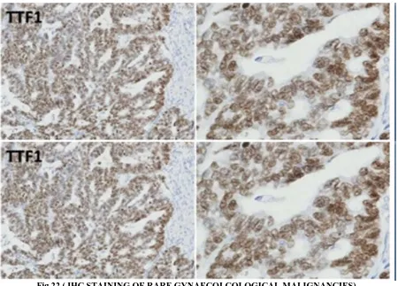
Fig 22 ( IHC STAINING OF RARE GYNAECOLCOLOGICAL MALIGNANCIES) ABBREVIATIONS – UEC- UNDIFFERENTIATED ENDOMETRIAL CANCER
EIN-ENDOMETRIAL INTRAEPITHELIAL NEOPLASIA SPCC – SPINDLE CELL CARCINOMA
CONCLUSION
Whenever the morphology or diagnostic dlilemma , we should consider frequency of primay and metastasis at the site.There should be a hiearchy in use of markers and then to specific marker pannel . The mmunochemistry also help in providing knowledge about the progression of lesio e.g P16 lsil , p53 in EIN
AFFILIATIONS-
S.SPATTNAIK – senior resident in dept gynaecology oncology at ahrcc
Co-responding author J.PARIJA – PROFHOD DEPT OF GYNAECOLOGY ONCOLOGY AHRCC S.KGIRI - PROF OF GYNAECOLOGY ONCOLOGY
L.SARNGI- DIRECTOR OF AHRCC N.ROUT- DEAN OF AHRCC
S.SAMANTRAY - PROF DEPT OF PATHOLOGY AHRCC N.PANDA –PROF HOD OF DEPT OF RADIOTHERAPY
[image:27.612.84.534.55.378.2][9] 9strckland S,ETAL INT IORNAL GYNAECOPATHOLOGY 2016 [10] 10.Vang. R, etal , Am J Surg pathology
[11] 11 KARNEIZan, Jpatho 2016
[12] 12 McCLOGGAGE WG, ET AL JOUR OF CLINICAL PATHOLOGY2012 [13] 13 RAMALINGAM, P,Malpic A,Silva ETAL AM JOURNALMPATHOLOGY [14] 14.Cao-d, guo s, allan rw ETAL AM JOURNAL PATHOLOGY
[15] 15. Kuhn E Kumari RJ ETA L INTERNATIONAL JOURNAL OF GYNAECOLOGY PATHOLOGY [16] 16. GUAN ET AL J.NAT CANCER
[17] 17 LIM .D ETAL AM J OF SURGIPATHO
[18] 18 KUHI HARA S ET ALAM JOURNAL OF SURG PAYHOLOGY [19] 19 JOINER ETAL
AUTHORS