Copyright © 1999, American Society for Microbiology. All Rights Reserved.
Quantitation of Ergosterol Content: Novel Method for Determination
of Fluconazole Susceptibility of
Candida albicans
BETH A. ARTHINGTON-SKAGGS, HODA JRADI, TEJAL DESAI,ANDCHRISTINE J. MORRISON*
Mycotic Diseases Branch, Division of Bacterial and Mycotic Diseases, National Center for Infectious Diseases, Centers for Disease Control and Prevention, Atlanta, Georgia 30333
Received 11 May 1999/Returned for modification 29 June 1999/Accepted 21 July 1999
MIC end points for the most commonly prescribed azole antifungal drug, fluconazole, can be difficult to determine because its fungistatic nature can lead to excessive “trailing” of growth during susceptibility testing by National Committee for Clinical Laboratory Standards broth macrodilution and microdilution methods. To overcome this ambiguity, and because fluconazole acts by inhibiting ergosterol biosynthesis, we developed a novel method to differentiate fluconazole-susceptible from fluconazole-resistant isolates by quantitating er-gosterol production in cells grown in 0, 1, 4, 16, or 64g of fluconazole per ml. Ergosterol was isolated from whole yeast cells by saponification, followed by extraction of nonsaponifiable lipids with heptane. Ergosterol was identified by its unique spectrophotometric absorbance profile between 240 and 300 nm. We used this sterol quantitation method (SQM) to test 38 isolates with broth microdilution end points of <8 g/ml
(susceptible), 16 to 32g/ml (susceptible dose-dependent [SDD]), or>64g/ml (resistant) and 10 isolates
with trailing end points by the broth microdilution method. No significant differences in mean ergosterol content were observed between any of the isolates grown in the absence of fluconazole. However, 18 susceptible isolates showed a mean reduction in ergosterol content of 72% after exposure to 1g of fluconazole/ml, an 84% reduction after exposure to 4g/ml, and 95 and 100% reductions after exposure to 16 and 64g of flucon-azole/ml, respectively. Ten SDD isolates showed mean ergosterol reductions of 38, 57, 73, and 99% after exposure to 1, 4, 16, and 64g of fluconazole/ml, respectively. In contrast, 10 resistant isolates showed mean reductions in ergosterol content of only 25, 38, 53, and 84% after exposure to the same concentrations of fluconazole. The MIC of fluconazole, by using the SQM, was defined as the lowest concentration of the drug which resulted in 80% or greater inhibition of overall mean ergosterol biosynthesis compared to that in the drug-free control. Of 38 isolates which gave clear end points by the broth microdilution method, the SQM MIC was within 2 dilutions of the broth microdilution MIC for 33 (87%). The SQM also discriminated between resistant and highly resistant isolates and was particularly useful for discerning the fluconazole susceptibilities of 10 additional isolates which gave equivocal end points by the broth microdilution method due to trailing growth. In contrast to the broth microdilution method, the SQM determined trailing isolates to be susceptible rather than resistant, indicating that the SQM may predict clinical outcome more accurately. The SQM may provide a means to enhance current methods of fluconazole susceptibility testing and may provide a better correlation of in vitro with in vivo results, particularly for isolates with trailing end points.
Rapid and reliable antifungal susceptibility testing has be-come particularly important in recent years because of the increased incidence of serious fungal infections and the con-comitant emergence of antifungal-drug resistance (20, 28). The National Committee for Clinical Laboratory Standards (NCCLS) recently published an approved broth macrodilution method (document M27-A) for in vitro testing of the suscep-tibilities ofCryptococcus neoformansand Candida species to amphotericin B, flucytosine, fluconazole, itraconazole, and ke-toconazole (14). This reference method, and simplified mi-crodilution adaptations of it, have significantly improved the inter- and intralaboratory reproducibility of antifungal suscep-tibility testing for most isolates. However, in tests with the most commonly used azole drug, fluconazole, some isolates do not give a clear-cut end point and exhibit a “trailing” growth effect, making interpretation of test results difficult (21). For isolates with trailing end points, MICs of less than 1g/ml at 24 h and of 64 g/ml or greater at 48 h are usually observed (21). Therefore, these isolates would be considered resistant by
NCCLS M27-A methodologies which recommend reading re-sults after 48 h of growth (14). Clinical outcomes for human immunodeficiency virus-infected patients with oropharyngeal candidiasis (21), as well as in vivo animal model data from our laboratory (4) and from others (23), have demonstrated that infections caused by organisms which produce trailing growth in vitro typically respond to low doses of fluconazole, suggest-ing that the lower MICs at 24 h better reflect host responsive-ness to therapy.
We therefore sought to improve the correlation of in vitro susceptibility testing results with in vivo therapeutic outcomes by developing a novel in vitro test which involves the quanti-tation of membrane sterols (the sterol quantiquanti-tation method [SQM]) to determine the MICs of fluconazole for clinical iso-lates ofCandida albicans. This test measures the sensitivity of ergosterol biosynthesis inC. albicansisolates to the effects of fluconazole by quantitation of steady-state amounts of ergos-terol following growth of the organism in various concentra-tions of fluconazole (2). The primary mechanism of action by which azole antifungal drugs inhibit yeast cell growth is through disruption of the normal sterol biosynthetic pathway, leading to a reduction in ergosterol biosynthesis (10). Ergos-terol is the major sErgos-terol component of the yeast cell membrane and is responsible for maintaining cell integrity and function
* Corresponding author. Mailing address: Centers for Disease Con-trol and Prevention, 1600 Clifton Rd., NE Mailstop G-11, Atlanta, GA 30333. Phone: (404) 639-3098. Fax: (404) 639-3546. E-mail: cjm3@cdc .gov.
3332
on May 15, 2020 by guest
http://jcm.asm.org/
(5, 24). Therefore, disruption of this pathway by azole drugs leads to fungistasis.
The SQM takes advantage of the unique spectral absorption pattern produced between 240 and 300 nm by extracted sterols, which is indicative of the ergosterol and 24(28)dehydroergos-terol [24(28)DHE, a late s24(28)dehydroergos-terol pathway intermediate] content. Both ergosterol and 24(28)DHE absorb at 281.5 nm, whereas only 24(28)DHE shows an intense spectral absorption band at 230 nm. Therefore, the amount of ergosterol can be deter-mined by calculating the total ergosterol-plus-24(28)DHE content and then subtracting from the total the amount of absorption due to 24(28)DHE only (6). Ergosterol content determined by the SQM is an absolute measurement, elimi-nating the need for subjective determination of growth inhibi-tion, as required for broth-based susceptibility testing methods. Therefore, in this regard, this method should be more objec-tive and reproducible than standard NCCLS methods.
Because decreased susceptibility to fluconazole is correlated with the ability ofC. albicans isolates to produce ergosterol even in the presence of azole drugs, we were able to determine fluconazole susceptibility by quantitating total intracellular er-gosterol production in cells grown in increasing concentrations of fluconazole and to assign unequivocal MIC end points to organisms which exhibit trailing growth during standard broth microdilution drug susceptibility testing. We compared MIC results obtained by the broth microdilution drug susceptibility method to those obtained by the SQM, using a panel of isolates determined by the broth microdilution method to be suscep-tible, susceptible dose-dependent (SDD), or resistant to flu-conazole, or classified as trailers.
MATERIALS AND METHODS
Isolates.A total of 48 oral or vaginalC. albicansisolates, 38 without trailing characteristics (Tables 1 through 3) and 10 with trailing characteristics (Table 4) were tested. Isolates were obtained from David A. Stevens (Stanford University, Palo Alto, and Santa Clara Valley Medical Center, San Jose, Calif.) and Dora Warren (Division of Reproductive Health, Centers for Disease Control and Prevention). Isolates were identified to the species level by the API 20C (Ana-lytab Products, Plainview, N.Y.) yeast identification system. Two reference
strains,Candida parapsilosisATCC 22019 andCandida kruseiATCC 6258, were included each day of broth microdilution testing to ensure quality control.
Isolates were retrieved from storage at⫺70°C and were subcultured twice on Sabouraud dextrose agar plates (BBL, Cockeysville, Md.) to ensure optimal growth. Prior to testing, subcultures on Sabouraud dextrose agar plates were incubated at 35°C for 24 h.
Broth microdilution method.Broth microdilution was performed according to the guidelines of NCCLS document M27-A (14). Analytical-grade powder of fluconazole was obtained as a gift from Pfizer (Groton, Conn.). A stock solution of fluconazole was prepared in sterile distilled water, diluted with RPMI-1640 medium (withL-glutamine but without bicarbonate) (Sigma Chemical Co., St.
Louis, Mo.), and buffered to pH 7.0 with 0.165 M morpholinopropanesulfonic acid (MOPS; Sigma). The final concentration range for fluconazole was 0.125 to 64g/ml.
Testing was performed in 96-well round-bottom microtitration plates. Cell suspensions were prepared in RPMI-1640 medium and were adjusted to give a final inoculum concentration of 0.5⫻103to 2.5⫻103cells/ml. The plates were
incubated at 35°C and were read after 48 h. The MIC of fluconazole was defined as the lowest concentration at which there was 80% inhibition of growth com-pared with that in a drug-free control.
SQM.Total intracellular sterols were extracted as reported by Breivik and Owades (6) with slight modifications. Briefly, a singleC. albicanscolony from an overnight Sabouraud dextrose agar plate culture was used to inoculate 50 ml of Sabouraud dextrose broth (Difco, Detroit, Mich.) containing 0, 1, 4, 16, or 64g of fluconazole per ml. The cultures were incubated for 16 h with shaking at 35°C. The stationary-phase cells were harvested by centrifugation at 2,700 rpm (model TJ-6 centrifuge; Beckman Instruments, Palo Alto, Calif.) for 5 min and washed once with sterile distilled water. The net wet weight of the cell pellet was determined. Three milliliters of 25% alcoholic potassium hydroxide solution (25 g of KOH and 35 ml of sterile distilled water, brought to 100 ml with 100% ethanol), was added to each pellet and vortex mixed for 1 min. Cell suspensions were transferred to 16- by 100-mm sterile borosilicate glass screw-cap tubes and were incubated in an 85°C water bath for 1 h. Following incubation, tubes were allowed to cool to room temperature. Sterols were then extracted by addition of a mixture of 1 ml of sterile distilled water and 3 ml ofn-heptane followed by vigorous vortex mixing for 3 min. The heptane layer was transferred to a clean borosilicate glass screw-cap tube and stored at⫺20°C for as long as 24 h. Prior to analysis, a 20-l aliquot of sterol extract was diluted fivefold in 100% ethanol and scanned spectrophotometrically between 240 and 300 nm with a Gilford Response Spectrophotometer (Ciba Corning Diagnostics Corp., Gilford Sys-tems, Oberlin, Ohio). The presence of ergosterol and the late sterol intermediate 24(28)DHE in the extracted sample resulted in a characteristic four-peaked curve (Fig. 1). The absence of detectable ergosterol in extracts was indicated by a flat line. A dose-dependent decrease in the height of the absorbance peaks was evident and corresponded to decreased ergosterol concentration.
Ergosterol content was calculated as a percentage of the wet weight of the cell by the following equations: % ergosterol⫹% 24(28)DHE⫽[(A281.5/
[image:2.612.55.546.73.283.2]290)⫻F]/pellet weight, % 24(28)DHE⫽[(A230/518)⫻F]/pellet weight, and
FIG. 1. UV spectrophotometric sterol profiles of representative fluconazole-susceptible (A), -SDD (B), and -resistant (C)C. albicansisolates. Isolates were grown for 16 h in Sabouraud dextrose broth containing 0 (curve A), 1 (curve B), 4 (curve C), 16 (curve D), or 64 (curve E)g of fluconazole per ml, sterols were extracted from cells, and spectral profiles between 240 and 300 nm were determined.
on May 15, 2020 by guest
http://jcm.asm.org/
% ergosterol⫽[% ergosterol⫹% 24(28)DHE]⫺% 24(28)DHE, whereFis the factor for dilution in ethanol and 290 and 518 are theEvalues (in percentages per centimeter) determined for crystalline ergosterol and 24(28)DHE, respec-tively. The wet weight of the cell pellet ranged from 1.09⫾0.14 g for organisms grown in 0g of fluconazole per ml to 0.97⫾0.12 g for organisms grown in 64 g of fluconazole per ml (n⫽48;P⬎0.05). The MIC of fluconazole was defined as the concentration of fluconazole which caused an 80% reduction in the total cellular ergosterol content compared to that in the drug-free control. MICs which fell between two fluconazole concentrations (i.e., less than 80% reduction at one concentration but more than 80% reduction at the next-higher concen-tration) were mathematically extrapolated based on the amount of reduction at the fluconazole concentration which gave results closest to an 80% reduction end point.
Analysis of results.Breakpoints for fluconazole susceptibility have been es-tablished for isolates ofCandidaspp. tested according to NCCLS guidelines. Organisms are classified as susceptible if the fluconazole MIC isⱕ8g/ml, as SDD if it is 16 to 32g/ml, and as resistant if it isⱖ64g/ml (14). Interpretive breakpoints for the SQM were based on those defined for the NCCLS method. MICs which fell between two susceptibility categories were assigned to the next-closest category (i.e., organisms with MICs of 14, 15, and 21g/ml were classified as SDD, and those with MICs of 52 and 61g/ml were classified as resistant; Table 3). The SQM MICs were compared with the microdilution MICs by using both on-scale and off-scale results. The high off-scale MICs were con-verted to the next-highest concentration, and the low off-scale MICs were left unchanged. SQM MICs were considered to be in agreement with the NCCLS microdilution MICs if they differed by no more than 2 drug dilutions.
Statistical analysis.Differences between means were analyzed by Student’st test, andPvalues of⬍0.05 were considered to represent statistically significant differences. Correlations between MICs and reductions in ergosterol levels were analyzed by Pearson’s correlation coefficient.
RESULTS
Correlation between susceptibilities determined by the broth microdilution method and the SQM. Table 1 summa-rizes the in vitro susceptibilities of 38 nontrailing isolates ofC. albicansto fluconazole as measured by the broth microdilution method and the SQM. The data are reported as MIC ranges and MICs required to inhibit 50 and 90% of the isolates (MIC50and MIC90, respectively). In each day of testing, MICs
of fluconazole for the two quality control strains were within the accepted limits defined by the NCCLS (14) (data not shown). The overall agreement between the results of the two methods was 87% for 38 isolates which gave unequivocal end points by the broth microdilution method (Table 1). Overall, agreement between methods was 100% for 18
fluconazole-susceptible isolates, 70% for 10 fluconazole-SDD isolates, and 80% for 10 fluconazole-resistant isolates.
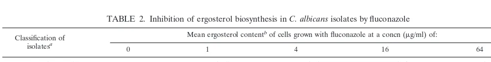
Table 2 summarizes the effect of fluconazole on ergosterol biosynthesis in fluconazole-susceptible, -SDD, and -resistantC. albicansisolates. The total ergosterol content was determined for each isolate grown in 0, 1, 4, 16, or 64g of fluconazole per ml. No significant differences in the mean amount of ergosterol produced by strains grown in the absence of fluconazole were observed regardless of the degree of fluconazole susceptibility (P⬎0.05). In contrast, a dose-dependent decrease in ergos-terol production was observed when isolates were grown in the presence of fluconazole (Table 2). The degree of sensitivity of the ergosterol biosynthetic pathway to the effects of flucon-azole decreased as the broth microdilution MIC of fluconflucon-azole increased (inverse correlation at 1 g of fluconazole/ml,r⫽
0.82; at 4g/ml,r⫽0.95; at 16g/ml,r⫽1.0; and at 64g/ml, r ⫽ 0.60). As shown in Table 2, the mean decrease in total cellular ergosterol content for susceptible isolates ranged from 72% for cells grown in 1g of fluconazole/ml to 100% for cells grown in 64g/ml. The mean decrease in total cellular ergos-terol content for SDD isolates ranged from 38% after exposure to 1g of fluconazole/ml to 99% after exposure to 64g/ml. In contrast, for resistant isolates, the mean decrease in total cel-lular ergosterol content ranged from 25% after exposure to 1
g of fluconazole/ml to 84% after exposure to 64g/ml. The less susceptible theC. albicansisolate was to fluconazole, the less sensitive ergosterol biosynthesis was to the inhibitory ef-fects of the drug. Incubation times from 16 to 24 h and inoc-ulum sizes from 105to 107cells/ml were also tested and did not
alter the SQM MICs (data not shown).
[image:3.612.53.552.85.161.2]The capacity of the SQM to discriminate more clearly the degrees of fluconazole resistance among nontrailing, resistant isolates relative to the broth microdilution method is presented in Table 3. For the 10 isolates tested, the broth microdilution MICs were ⱖ64 g/ml, while the SQM differentiated these isolates into three distinct groups. Specifically, three isolates (CA23, CA24, and CA28) classified as resistant by broth mi-crodilution (MICs of ⱖ64 g/ml) were SDD by the SQM (MICs of 14, 15, and 21g/ml), and two isolates (CA29 and CA30) for which the broth microdilution MICs were ⱖ64
TABLE 1. MICs of fluconazole forC. albicansisolates as determined by the NCCLS broth microdilution method and SQM
Classification of isolatesa
MIC (g/ml) as determined by:
% Agreement
Microdilution SQM
Range 50% 90% Range 50% 90%
Susceptible (n⫽18) 0.25–8 1 8 ⬍1–18 1 16 100
SDD (n⫽10) 16–32 16 32 3–29 17 52 70
Resistant (n⫽10) 64–⬎64 64 ⬎64 14–190 51 183 80
aBy the NCCLS broth microdilution assay.
TABLE 2. Inhibition of ergosterol biosynthesis inC. albicansisolates by fluconazole
Classification of isolatesa
Mean ergosterol contentbof cells grown with fluconazole at a concn (g/ml) of:
0 1 4 16 64
Susceptible (n⫽18) 1.4⫾0.07 0.4⫾0.1 (72)c 0.23⫾0.06 (84)c 0.07⫾0.03 (95)c 0⫾0 (100)c
SDD (n⫽10) 1.6⫾0.14 1.0⫾0.11 (38) 0.69⫾0.17 (57) 0.43⫾0.13 (73)c 0.005⫾0.003 (99)c
Resistant (n⫽10) 1.6⫾0.13 1.2⫾0.09 (25) 1.0⫾0.13 (38) 0.8⫾0.13 (53) 0.2⫾0.13 (84)c aBy the NCCLS broth microdilution assay.
bExpressed as a percentage of the wet weight of the cell⫾the standard error of the mean (followed in parentheses by the percent reduction in the mean cellular
ergosterol content compared with that of control cells grown without fluconazole).
cSignificant reduction compared with controls (P⬍0.05).
on May 15, 2020 by guest
http://jcm.asm.org/
[image:3.612.53.552.632.695.2]g/ml were strikingly more resistant to fluconazole (28 and 27% inhibition of ergosterol biosynthesis when these isolates were grown in 64g of fluconazole/ml, respectively) than the other isolates for which broth microdilution MICs wereⱖ64
g/ml (Table 3). The SQM further stratified isolates within the resistant category into “resistant” (MICs, 51 to 61g/ml) and “highly resistant” (MICs, 183 to 190 g/ml) subcategories, compared to the microdilution method, whereⱖ64g/ml is traditionally the highest MIC reported.
Using the SQM to differentiate fluconazole-resistant iso-lates from fluconazole-susceptible isoiso-lates which exhibit trail-ing by the broth microdilution method.TenC. albicansisolates which exhibited trailing growth in fluconazole, making end point determinations ambiguous by the broth microdilution method, and which were not included in the above analyses, were examined. Table 4 summarizes the in vitro susceptibilities of the isolates to fluconazole as measured by the broth mi-crodilution and SQM methods. There was no agreement be-tween broth microdilution MICs and SQM MICs for these isolates. By the NCCLS broth microdilution method, all were
susceptible (MIC ⱕ 1.0 g/ml) to fluconazole at 24 h and resistant (MICⱖ64g/ml) at 48 h. By the SQM, all 10 isolates were determined to be susceptible to fluconazole (MICsⱕ2
g/ml). Preliminary results using spectrophotometric MIC80
end point determination of the broth microdilution assay (13) did not improve agreement between the two methods (data not shown).
Interlaboratory reproducibility of MIC end point determi-nations for organisms which exhibit trailing.Table 5 summa-rizes the results of an interlaboratory comparison of flucon-azole susceptibility test results. Three of the 10 isolates exhibiting trailing by the broth microdilution method were retested in three other laboratories. The MICs of fluconazole for these isolates ranged from 0.25 to⬎64g/ml depending on the laboratory conducting the susceptibility testing (Table 5). Three of the four laboratories reported all of the isolates to be susceptible to fluconazole, and one laboratory reported all of the isolates to be resistant. SQM results revealed all three isolates to be susceptible to fluconazole, supporting the find-ings of all but one of the four laboratories conducting broth microdilution antifungal susceptibility testing.
DISCUSSION
The incidence of invasive fungal diseases and antifungal drug resistance has increased in recent years, making the de-velopment of reliable antifungal drug susceptibility tests more important (20). Substantial efforts have been made by the NCCLS first to standardize and then to simplify antifungal susceptibility testing, resulting in the publication of the M27-A guidelines and the acceptance of a standard broth macrodilu-tion format (14). Simplified broth microdilumacrodilu-tion adaptamacrodilu-tions of the M27-A method have been developed and have been shown to be useful (7, 8). However, problems with end point inter-pretation, particularly for isolates with trailing end points, re-main (21, 23).
Commercial companies have developed alternative antifun-gal susceptibility testing tools which offer simple and rapid approaches to antifungal susceptibility testing. Whereas the E test (AB Biodisk, Solna, Sweden) is an agar diffusion test, and the YeastOne (AccuMed International, Westlake, Ohio) and PASCO (Becton Dickinson, Pasco Division, Wheat Ridge, Colo.) tests are broth dilution systems, all of these methods rely on visual detection of growth inhibition as an indicator of drug susceptibility (1, 3, 16). Thus, they can be influenced by variables such as inoculum size, incubation time, cell culture medium, and subjective end point determination (17, 18).
Recently, efforts have been made to determine end points more objectively by reading broth microdilution plates with a spectrophotometer (13, 25). Unfortunately, this method still does not eliminate ambiguous end point determinations for trailing isolates. Adoption of a MIC50rather than a MIC80end
[image:4.612.52.295.92.236.2]point value may improve the correlation of in vitro suscepti-bility testing results with in vivo outcomes (4a). Alternatively,
TABLE 3. Stratification of fluconazole-resistantC. albicansisolates by the SQM versus the broth microdilution method
Isolate MIC determined by:
Ergosterol contentb(reductionc) of
cells grown with fluconazole at a concn (g/ml) of: Microdilutiona SQM 4 16 64
CA23 64 14d 0.7 (65) 0.2 (92) 0 (100)
CA28 ⱖ64 15d 0.5 (66) 0.2 (87) 0 (100)
CA24 64 21d 0.8 (66) 0.7 (62) 0 (100)
CA25 64 51 1.2 (0) 0.7 (42) 0 (100)
CA26 64 51 1.6 (16) 1.2 (37) 0 (100)
CA21 64 52 1.7 (26) 1.3 (44) 0.02 (99)
CA22 64 52 0.9 (40) 0.9 (40) 0.03 (98)
CA27 ⱖ64 61 0.7 (42) 0.3 (75) 0.2 (84) CA29 ⱖ64 183e 1.2 (33) 1.4 (23) 1.3 (28)
CA30 ⱖ64 190e 0.8 (27) 0.8 (27) 0.8 (27) aAfter 48 h of incubation.
bExpressed as a percentage of the wet weight of the cell.
cPercent reduction in ergosterol content compared with that of control cells
grown without fluconazole.
[image:4.612.53.294.553.704.2]dSDD by SQM. eHighly resistant by SQM.
TABLE 4. Ergosterol contents of 10C. albicansisolates with trailing MIC end points by the microdilution method
Isolate
Broth microdilution
MIC at:
Ergosterol contenta(reductionb)
of cells grown with fluconazole at
a concn (g/ml) of: SQM MIC 24 h 48 h 0 1 4, 16,or 64
CA2 0.2 64 1.0 0 (100) 0 (100) ⬍1.0
CA4 0.5 64 1.6 0 (100) 0 (100) ⬍1.0
CA5 0.5 64 2.1 0 (100) 0 (100) ⬍1.0
CA31 0.5 64 2.0 0.05 (98) 0 (100) ⬍1.0
CA32 0.5 64 1.3 0 (100) 0 (100) ⬍1.0
CA33 0.5 64 1.8 0 (100) 0 (100) ⬍1.0
CA35 1.0 64 1.6 0 (100) 0 (100) ⬍1.0
CA36 1.0 64 1.1 0 (100) 0 (100) ⬍1.0
CA37 1.0 ⱖ64 1.0 0.7 (33) 0 (100) 2.0
CA38 1.0 ⱖ64 1.5 0 (100) 0 (100) ⬍1.0
aExpressed as a percentage of the wet weight of the cell.
bPercent reduction in ergosterol content compared with that of control cells
grown without fluconazole.
TABLE 5. Interlaboratory variability in MIC end points for three C. albicansisolates which exhibit trailing growth when tested by the
broth microdilution method
Isolate Broth microdilution MIC (g/ml) SQMMIC Lab A Lab B Lab C Lab D
CA2 0.25 0.25 ⬎64 1.0–2.0 ⬍1.0
CA4 0.5 0.5 ⬎64 2.0–4.0 ⬍1.0
CA5 0.5 0.5 ⬎64 1.0–2.0 ⬍1.0
on May 15, 2020 by guest
http://jcm.asm.org/
adoption of a 24- rather than a 48-h end point reading for trailing isolates may achieve the same goal.
New methods using flow cytometric techniques for deter-mining the antifungal susceptibilities ofCandidaspecies have also been described (19) and have been shown to be rapid and sensitive alternatives to broth dilution methods. However, this approach requires costly equipment and the use of hazardous compounds, such as ethidium bromide (30).
Three general mechanisms of azole resistance have been described forCandidaspp. The first is alteration in the target enzyme, 14 alpha-demethylase, leading to its overexpression and/or reduced susceptibility to azole inhibition (12, 26, 31). Decreased drug accumulation, mediated by either diminished uptake or increased efflux of the drug, is the second mechanism (15, 27). The third is a deficiency in C5(6) sterol desaturase, which suppresses the accumulation of toxic sterol intermedi-ates, as a result of azole-mediated 14 alpha-demethylase inhi-bition (9, 11). The SQM is capable of detecting increased resistance due to any of the above mechanisms based on its ability to detect intracellular ergosterol following the exposure of the organisms to fluconazole.
The SQM provides definitive MIC end points in 18 h (16 h of incubation plus 2 h to complete the assay), uses common laboratory equipment (shaking incubator, tabletop centrifuge, water bath, and UV spectrophotometer), is simple to perform, and shows excellent agreement with the NCCLS broth mi-crodilution method for nontrailing isolates. Preliminary data collected by our laboratory have suggested that the SQM may be equally useful for the determination of the susceptibilities of otherCandidaspecies to fluconazole and other azoles (unpub-lished data). Ultimately, the best use of the SQM may be for the determination of the antifungal-drug susceptibilities of fil-amentous fungi, where determination of a visual or spectro-photometric end point may be problematic (8a).
In the design of the prototype SQM test, four concentrations of fluconazole were chosen to represent the different NCCLS-determined fluconazole susceptibility categories (susceptible, 1 and 4g/ml; SDD, 16g/ml; resistant, 64g/ml), thus simpli-fying a comparison of results with those of the broth microdi-lution method. Unlike a physical or chemical measurement, such as the determination of a drug level, a MIC determination by standardized broth dilution methodology is a function of the conditions selected by the tester (22). Variations in any con-dition can produce slight to dramatic variations in the mea-sured MIC (8, 29). The utility of the SQM as an index of antifungal drug susceptibility is that it is a physical measure-ment of total cellular ergosterol content. Stationary-phase cells are used so that steady-state levels of ergosterol are measured for all drug concentrations tested, making the assay far less sensitive to factors such as inoculum size and incubation time. Furthermore, determination of an exact numerical value elim-inates subjective interpretation of MIC end points when trail-ing growth occurs.
Trailing growth has been shown to be a major cause of interlaboratory variability in antifungal susceptibility testing (21, 23). This phenomenon complicates MIC end point deter-mination and often leads to misclassification of susceptible isolates (susceptibility based on animal models of candidiasis) as resistant (21, 23). Because the SQM is a direct measurement of total intracellular ergosterol content, MIC end point deter-mination is unequivocal. Although three of the four laborato-ries participating in our study determined that trailing isolates were susceptible by the broth microdilution method, one lab-oratory (25% of our sample) gave a significantly different end point interpretation (i.e., resistant). Because this laboratory is
one of the most experienced and widely used in the United States, this discrepancy is particularly significant.
For the isolates tested in this study, the intracellular ergos-terol contents of resistant and susceptible isolates grown in the absence of fluconazole were not significantly different. This observation suggests that increased resistance to fluconazole in these isolates is not due to a stable genetic change in an ergosterol biosynthetic gene leading to altered ergosterol con-tent but rather that these isolates are capable of reducing intracellular drug concentrations so that ergosterol biosynthe-sis is inhibited less by the presence of fluconazole.
Finally, the SQM offers the advantage of further differenti-ating isolates within a given susceptibility category based on their individual percentages of ergosterol inhibition. Thus, sub-tle changes leading to decreased fluconazole susceptibility of an isolate would be detected by the SQM even if the change was not large enough to shift the isolate to the next category of drug resistance. For example, isolates which were resistant to fluconazole by the broth microdilution method (MICⱖ 64
g/ml) demonstrated distinct degrees of resistance by the SQM. In addition, MIC end points which fall between the drug concentrations routinely tested by the broth microdilution method can be determined by the SQM without the need to test the organisms against additional fluconazole concentra-tions. This feature allows for additional stratification of de-grees of susceptibility within the NCCLS-established cate-gories of SDD and resistant. Adaptation of the SQM to a kit format would increase the usefulness of this test for clinical laboratory testing. Efforts are currently under way in our lab-oratory to accomplish this task.
In summary, the SQM demonstrated good agreement with the broth microdilution method forC. albicans isolates with unequivocal end points and gave clear MICs for isolates which trail in the broth microdilution test format. The SQM offered the additional advantage of enhanced discrimination of iso-lates within fluconazole-SDD and -resistant categories. The clinical impact of dividing isolates into “degrees” of suscepti-bility or resistance will require further analysis using animal models of candidiasis. Such studies will determine if the SQM offers increased clinical correlation and improved therapeutic decision making compared with standard antifungal suscepti-bility testing methods.
ACKNOWLEDGMENTS
We thank David A. Stevens and Dora Warren for isolates used in this study and Milwood Motley, Ana Espinel-Ingroff, and Michael Rinaldi for conducting blinded antifungal-drug susceptibility testing.
REFERENCES
1.Arikan, S., D. Gur, and M. Akova.1997. Comparison of E-test, microdilution and colorimetric dilution with reference broth macrodilution method for antifungal susceptibility testing of clinically significantCandidaspecies iso-lated from immunocompromised patients. Mycoses40:291–296.
2.Arthington-Skaggs, B. A., H. Jradi, T. Desai, E. Latimer, and C. J. Morrison.
1998. Determination of azole susceptibility by quantitation of ergosterol content, abstr. C-284, p. 178.InProgram and abstracts of the 98th General Meeting of the American Society for Microbiology. American Society for Microbiology, Washington, D.C.
3.Arthington-Skaggs, B. A., M. Motley, E. Reiss, and C. J. Morrison.1997. Evaluation of a commercially prepared microdilution panel for antifungal susceptibility testing, abstr. P509, p. 206.InProgram and abstracts of the 13th Congress of the International Society for Human and Animal Mycol-ogy.
4.Arthington-Skaggs, B. A., and C. J. Morrison.1999. Validation of the sterol quantitation method as a tool to predict in vivo response to fluconazole in experimental murine candidiasis.InProgram and abstracts of the 5th Can-dida and Candidiasis Conference. American Society for Microbiology, Washington, D.C.
4a.Barchiesi, F., M. D. Poeta, V. Morbiducci, F. Ancarani, and G. Scalise.1993. Turbidimetric and visual criteria for determining the in vitro activity of six
on May 15, 2020 by guest
http://jcm.asm.org/
antifungal agents againstCandidaspp. andCryptococcus neoformans. Myco-pathologia124:19–25.
5.Bard, M., N. D. Lees, L. S. Burrows, and F. W. Kleinhans.1978. Differences in crystal violet uptake and cation-induced death among yeast sterol mutants. J. Bacteriol.135:1146–1148.
6.Breivik, O. N., and J. L. Owades.1957. Spectrophotometric semi-microde-termination of ergosterol in yeast. Agric. Food Chem.5:360–363. 7.Davey, K. G., A. Szekely, E. M. Johnson, and D. W. Warnock.1998.
Com-parison of a new commercial colorimetric microdilution method with a standard method for in-vitro susceptibility testing ofCandidaspp. and Cryp-tococcus neoformans. J. Antimicrob. Chemother.42:439–444.
8.Espinel-Ingroff, A., J. L. Rodriguez-Tudela, and J. V. Martinez-Suarez.1995. Comparison of two alternative microdilution procedures with the National Committee for Clinical Laboratory Standards reference macrodilution method M27-P for in vitro testing of fluconazole-resistant and -susceptible isolates ofCandida albicans. J. Clin. Microbiol.33:3154–3158.
8a.Espinel-Ingroff, A., M. S. Barlett, R. Bowden, N. X. Chin, J. C. R. Cooper, A. Fothergill, M. R. McGinnis, P. Menezes, S. A. Messer, P. W. Nelson, F. C. Odds, L. Pasarell, J. Peter, M. A. Pfaller, J. H. Rex, M. G. Rinaldi, G. S. Shankland, T. J. Walsh, and I. Weitzman.1997. Multicenter evaluation of proposed standardized procedures for antifungal susceptibility testing of filamentous fungi. J. Clin. Microbiol.35:139–143.
9.Geber, A., C. A. Hitchcock, J. E. Swartz, F. S. Pullen, K. E. Marsden, K. J. Kwon-Chung, and J. E. Bennett.1995. Deletion of theCandida glabrata ERG3andERG11genes: effect on cell viability, cell growth, sterol compo-sition, and antifungal susceptibility. Antimicrob. Agents Chemother.39:
2708–2727.
10. Kelly, S. L., D. C. Lamb, A. J. Corran, B. C. Baldwin, and D. E. Kelly.1995. Mode of action and resistance to azole antifungals associated with the for-mation of 14␣-methylergosta-8,24(28)-dien-3,6␣-diol. Biochem. Biophys. Res. Commun.207:910–915.
11. Kelly, S. L., D. C. Lamb, D. E. Kelly, N. J. Manning, J. Loeffler, H. Hebart, U. Schunacher, and E. Einsele.1997. Resistance to fluconazole and cross-resistance to amphotericin B inCandida albicansfrom AIDS patients caused by defective sterol delta 5,6-desaturation. FEBS Lett.400:80–82. 12. Lamb, D. C., D. E. Kelly, W. H. Schunck, A. Z. Shyadehi, M. Akhtar, D. J.
Lowe, B. C. Baldwin, and S. L. Kelly.1997. The mutation T315A inCandida albicanssterol 14 alpha-demethylase causes reduced enzyme activity and fluconazole resistance. J. Biol. Chem.272:5682–5688.
13. Makimura, K., T. Sudo, M. Kudo, K. Uchida, and H. Yamaguchi.1998. Development of reference procedures for broth microdilution antifungal susceptibility testing of yeasts with standardized endpoint determination. Microbiol. Immunol.42:55–59.
14. National Committee for Clinical Laboratory Standards.1997. Reference method for broth dilution antifungal susceptibility testing of yeasts. Ap-proved standard. Document M27-A. National Committee for Clinical Lab-oratory Standards, Wayne, Pa.
15. Parkinson, T., D. J. Falconer, and C. A. Hitchcock.1995. Fluconazole resis-tance due to energy-dependent drug efflux inCandida glabrata. Antimicrob. Agents Chemother.39:1696–1699.
16. Pfaller, M. A., B. Buschelman, M. J. Bale, M. Lancaster, A. Espinel-Ingroff, J. H. Rex, and M. G. Rinaldi.1994. Multicenter comparison of a colorimetric microdilution broth method with the reference macrodilution method for in vitro susceptibility testing of yeast isolates. Mycology19:9–15.
17. Pfaller, M. A., and M. G. Rinaldi.1993. Antifungal susceptibility testing: current state of technology, limitations, and standardization. Infect. Dis. Clin. N. Am.7:435–444.
18. Pfaller, M. A., M. G. Rinaldi, J. N. Galgiani, M. S. Bartlett, B. A. Body, A.
Espinel-Ingroff, R. A. Fromtling, G. S. Hall, C. E. Hughes, F. C. Odds, and A. M. Sugar.1990. Collaborative investigation of variables in susceptibility testing of yeasts. Antimicrob. Agents Chemother.34:1648–1654. 19. Ramani, R., A. Ramani, and S. J. Wong.1997. Rapid flow cytometric
sus-ceptibility testing ofCandida albicans. J. Clin. Microbiol.35:2320–2324. 20. Rees, J. R., R. W. Pinner, R. A. Hajjeh, M. E. Brandt, and A. L. Reingold.
1998. The epidemiological features of invasive mycotic infections in the San Francisco Bay area, 1992–1993: results of population-based laboratory active surveillance. Clin. Infect. Dis.27:1138–1147.
21. Revankar, S. G., W. R. Kirkpatrick, R. K. McAtee, A. W. Fothergill, S. W. Redding, M. G. Rinaldi, and T. F. Patterson.1998. Interpretation of trailing endpoints in antifungal susceptibility testing by the National Committee for Clinical Laboratory Standards method. J. Clin. Microbiol.36:153–156. 22. Rex, J. H., M. A. Pfaller, J. N. Galgiani, M. S. Bartlett, A. Espinel-Ingroff,
M. A. Ghannoum, M. L. Lancaster, F. C. Odds, M. G. Rinaldi, T. J. Walsh, and A. L. Barry.1997. Development of interpretive breakpoints for antifun-gal susceptibility testing: conceptual framework and analysis of in vitro-in vivo correlation data for fluconazole, itraconazole, andCandidainfections. Clin. Infect. Dis.24:235–247.
23. Rex, J. H., P. W. Nelson, V. L. Paetznick, M. Lozano-Chiu, and A. Espinel-Ingroff.1998. Optimizing the correlation between results of testing in vitro and therapeutic outcome in vivo for fluconazole by testing clinical isolates in a murine model of invasive candidiasis. Antimicrob. Agents Chemother.
42:129–134.
24. Rodriguez, R. J., C. Low, C. D. K. Bottema, and L. W. Parks.1985. Multiple functions for sterols inSaccharomyces cerevisiae. Biochem. Biophys. Res. Commun.112:47–54.
25. Rodriguez-Tudela, J. L., J. Berenguer, J. V. Martinez-Suarez, and R. Sanchez.1996. Comparison of a spectrophotometric microdilution method with RPMI–2% glucose with the National Committee for Clinical Labora-tory Standards reference macrodilution method M27-P for in vitro suscep-tibility testing of amphotericin B, flucytosine, and fluconazole against Can-dida albicans. Antimicrob. Agents Chemother.40:1998–2003.
26. Sanglard, D., F. Ischer, L. Koymans, and J. Bille.1998. Amino acid substi-tutions in the cytochrome P-450 lanosterol 14 alpha-demethylase (CYP51A1) from azole-resistantCandida albicansisolates contribute to re-sistance to azole antifungal agents. Antimicrob. Agents Chemother.42:241– 253.
27. Sanglard, D., K. Kuchler, F. Ischer, J.-L. Pagani, M. Monod, and J. Bille.
1995. Mechanisms of resistance to azole antifungal agents inCandida albi-cansisolates from AIDS patients involve specific multidrug transporters. Antimicrob. Agents Chemother.39:2378–2386.
28. Vanden Bossche, H. V., F. Dromer, I. Improvisi, M. Lozano-Chiu, J. H. Rex, and D. Sanglard.1998. Antifungal drug resistance in pathogenic fungi. Med. Mycol.36(Suppl. 1):119–121.
29. Wanger, A., K. Mills, P. W. Nelson, and J. H. Rex.1995. Comparison of Etest and National Committee for Clinical Laboratory Standards broth macrodi-lution method for antifungal susceptibility testing: enhanced ability to detect amphotericin B-resistantCandidaisolates. Antimicrob. Agents Chemother.
39:2520–2522.
30. Wenisch, C., K. F. Linnau, B. Parschalk, K. Zedtwitz-Liebenstein, and A. Georgopoulos.1997. Rapid susceptibility testing of fungi by flow cytometry using vital staining. J. Clin. Microbiol.35:5–10.
31. White, T. C.1997. The presence of an R467K amino acid substitution and loss of allelic variation correlate with an azole-resistant lanosterol 14 alpha-demethylase inCandida albicans. Antimicrob. Agents Chemother.41:1488– 1494.