JOURNAL OFCLINICALMICROBIOLOGY, July 2003, p. 3181–3186 Vol. 41, No. 7 0095-1137/03/$08.00⫹0 DOI: 10.1128/JCM.41.7.3181–3186.2003
Copyright © 2003, American Society for Microbiology. All Rights Reserved.
Molecular Typing of Methicillin-Resistant
Staphylococcus aureus
:
Can PCR Replace Pulsed-Field Gel Electrophoresis?
A. Strande
´n,
1R. Frei,
2and A. F. Widmer
1*
Division of Hospital Epidemiology1and Bacteriology Laboratory,2University Hospital Basel,
CH-3031 Basel, Switzerland
Received 2 December 2002/Returned for modification 24 January 2003/Accepted 13 March 2003
Pulsed-field gel electrophoresis (PFGE) is considered the “gold standard” for molecular typing of methi-cillin-resistantStaphylococcus aureus(MRSA). However, the method is time-consuming and expensive, and its discriminatory power may not be necessary in outbreak situations. We used a rapid multiplex PCR-based method with published primers and compared the results with those obtained by PFGE. A total of 75 clinical isolates were typed: 59 strains originated from our prospectively collected clinical strains and were epidemi-ologically unrelated; 16 strains came from an outbreak that was epidemiepidemi-ologically well defined in time and space. A primer mix of thespagene, thecoagene, and the hypervariable region adjacent tomecA gene was used for multiplex PCR. Both PFGE and PCR clustered the 75 strains into 41 different genotypes. Concordance of the results was 100% for strains originating from the outbreak. Overall, both methods produced concordant results in 72% of cases. A total of 16% were clustered together by PFGE, but not by PCR and 12% were clustered together by PCR but not by PFGE, respectively. The turnaround time was only 8 h for PCR but 5 days for PFGE. This PCR-based method is excellent for rapid and inexpensive typing of MRSA in an outbreak setting, but the discriminatory power and reproducibility are still insufficient to replace PFGE in longitudinal studies in the endemic setting.
Nosocomial infections caused by methicillin-resistant strains ofStaphylococcus aureus(MRSA) belong to the most impor-tant multiresisimpor-tant pathogens worldwide (5). Monitoring and limiting the intra- and interhospital spread of MRSA strains requires the use of rapid and accurate epidemiologic typing systems. The emergence of recently documented vancomycin-resistantS. aureus(VRSA) amplifies the need for control of MRSA and VRSA (2). Some MRSA strains, epidemic MRSA, have been observed to disseminate quickly (7). Their prompt identification is crucial to control and eradicate an outbreak, while saving resources for unnecessary isolations of patients unrelated to an outbreak. A large number of molecular meth-ods have been developed for typing MRSA strains. DNA fin-gerprinting by pulsed-field gel electrophoresis (PFGE) is con-sidered to be one of the most reliable, discriminatory, and reproducible typing procedures to allow the detection of high degree DNA polymorphism (8, 10, 14, 21–23), but it is tech-nically demanding, time-consuming, and expensive. In an out-break setting a rapid typing tool should allow one to cluster epidemiologically linked cases and separate them from spo-radic cases.
The aim of the present study was to adapt a rapid PCR method to investigate MRSA strains in an outbreak setting. We compared PCR results from epidemiologically well-de-fined unrelated strains and outbreak strains with the results of PFGE.
MATERIALS AND METHODS
Bacterial isolates.A total of 75 MRSA isolates were investigated. Of these, 59 isolates originated from our prospectively collected epidemiologically unrelated clinical isolates; no association was found in terms of time and location. They were collected between 2000 and 2002. In February 2002, 16 isolates came from an outbreak on a single ward. Classification of the cases was done by our
prospective MRSA surveillance system (5). All strains were stored at⫺70°C.
Identification of MRSA.Colonies suspected to beS. aureuswere screened with the latex agglutination test Pastorex Staph Plus (Bio-Rad) and confirmed as such
with Gram stain, catalase, and aurease (Rapidec Staph; bioMe´rieux). Resistance
to oxacillin was determined according to the NCCLS guidelines by using an
oxacillin screen agar (MRSA screen agar; bioMe´rieux) and confirmed by
detec-tion of penicillin-binding protein 2a (MRSA-Screen; Denka Seiken). Any
dis-crepant results were resolved by PCR testing with primers targetingfemAand
mecAgene.
PFGE.The procedure was performed as described by Pfaller et al. (13). Briefly, a washed bacterial suspension was mixed with 2% SeaPlaque GTG agarose (FMC BioProducts) at 58°C and allowed to solidify into plug molds at room temperature. Chromosomal DNA was prepared in several incubating and washing steps by using EC buffer (6 mM Tris-HCl, 1 M NaCl, 0.1 M EDTA, 0.5% Brij-58, 0.2% deoxycholate, 0.5% Sarkosyl), lysostaphin, and proteinase K. After
digestion of the genomic DNA bySmaI (New England Biolabs), the restriction
fragments were separated by PFGE by using a temperature-controlled CHEF DR III system (Bio-Rad) with the following conditions: 6.0 V/cm for 24 h at switch times ramped from 5 to 60 s. After staining with ethidium bromide, the fragments were visualized by using a UV transilluminator and then documented by using a video gel documentation system (MWG-Biotech). For PFGE pattern analysis, the software GelCompar (Applied Maths) was applied. The dendro-grams were calculated by the unweighted pair group method by using arithmetic averages. Isolates were considered identical and closely related when there were
0 andⱕ2 banding differences, respectively. PFGE typing has routinely been
performed in our laboratory since 1992.
PCR.DNA was isolated as previously described (24). One loopful ofS. aureus
cells was harvested from agar plates. Cells were resuspended in 50l of
lyso-staphin (Sigma Chemical) at 100g/ml and incubated for 10 min at 37°C. Then,
50g of proteinase K solution/ml and 150l of 0.1 M Tris buffer (pH 7.5) were
added, followed by incubation for 10 min at 37°C and then 5 min at 95°C. DNA
was stored at⫺20°C until reuse.
A PCR was applied to simultaneously amplify a part of the hypervariable
region (HVR) adjacent to themecAgene, a part of thespagene coding for
* Corresponding author. Mailing address: Division of Hospital Ep-idemiology, University Hospital Basel, Petersgraben 4, CH-4031 Basel, Switzerland. Phone: 0041-61-265-38-50. Fax: 0041-61-265-38-54. E-mail: awidmer@uhbs.ch.
3181
on May 15, 2020 by guest
http://jcm.asm.org/
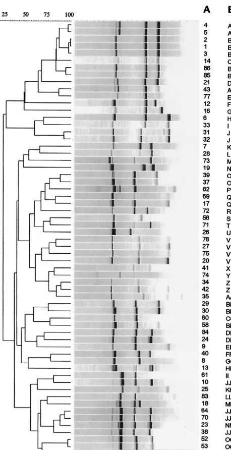
FIG. 1. Dendrogram of SmaI-PFGE of epidemiologically unrelated strains. Strain numbers are indicated in column A; PFGE types are indicated in column B.
3182
on May 15, 2020 by guest
protein A, and a part of thecoagene coding for coagulase, based on established
primers (25). The PCR was performed with 2l of the previously prepared cell
lysates, with HotStarTaq (Qiagen) in a final volume of 25l. The primer mix
contained 170 pmol of HVR1 and HVR2, 3.6 pmol of SPA1 and SPA2, and 6.0 pmol of COA1 and COA2. Cycling was performed in the GeneAmp PCR system 9600 (Perkin-Elmer). Samples were denaturated at 95°C for 15 min, followed by 35 cycles with the following parameters: denaturation at 94°C for 1 min, anneal-ing at 56°C for 1 min, and extension at 72°C for 3 min, with the final extension at 72°C for 5 min. With each run strain number 84 was amplified as an internal
control. Next, 8l of each amplicon was electrophoresed in a 2% agarose gel
(NuSieve3:1; BioWhittaker) with a subsequent ethidium bromide staining and visualized under UV light. A 100- to 1,500-bp ladder (Roche Applied Sciences) was used as a molecular weight marker. Images of gel lanes were normalized, and dendrograms prepared by using GelCompar software were printed. The inter-pretation criterion for identifying different strains was a single band difference (25). To evaluate the reproducibility, PCR was repeated with all of the strains.
Comparison and cost.Correlation of banding between PFGE and PCR was done independently by two investigators blinded to the results of the epidemi-ological investigations. The material costs for PCR and PFGE were estimated based on data from accounting, the real cost of the disposable, and the cost of labor. Costs for equipment and resources usually available in microbiology lab-oratories were not included.
RESULTS
Comparison of PCR with PFGE. PFGE grouped the 75 isolates into 41 types (Fig. 1), the first PCR run grouped the 75 isolates into 41 types (Fig. 2), and the second PCR run grouped the 75 isolates into 35 types. The concordance for the 16 isolates from the outbreak was 100% by PFGE versus PCR (first or second run) (Fig. 3).
[image:3.603.47.276.68.514.2]PFGE and the first run produced concordant results in 72% of the overall strains, PFGE and the second run produced concordant results in 76.7% of the overall strains (Table 1). In the first run 16% were clustered together by PFGE but not by PCR, and in the second one 8% were clustered together by
[image:3.603.310.537.307.693.2]FIG. 2. Dendrogram of PCR of epidemiologically unrelated strains. Strain numbers are indicated in column A; PCR types are indicated in column B.
FIG. 3. Dendrogram ofSmaI-PFGE (A) and PCR results (B) from epidemic strains. Strain numbers are indicated on the right side. Strain 84 is the unrelated control.
VOL. 41, 2003 MOLECULAR TYPING OF MRSA 3183
on May 15, 2020 by guest
http://jcm.asm.org/
PFGE but not by PCR. A total of 12% of the isolates were clustered together by PCR but not by PFGE in the first run versus a total of 15% clustered by the second run. In the first PCR run all isolates were typeable; in the second PCR run two strains (2.6%) were classified as not typeable, although only the band corresponding to thespagene could be detected.
[image:4.603.348.495.69.342.2]Reproducibility of the described PCR method.The concor-dance for the 16 isolates from the outbreak was 100% in both PCR runs. The two PCR runs produced concordant results in 72%, including both nonepidemic and epidemic strains. A total of 12% were clustered together by the first run but not by the second run, and 13.3% were clustered together by the second run but not by the first run. Figure 4 shows the PCR products for each primer. The first band, located between 1,200 and 1,500 bp, corresponded to thespagene and could be detected in all strains. The second band, located between 600 and 900 bp, corresponded to thecoagene and could be detected in all strains in the first run but was absent in two strains in the second run. The third band, located between 300 and 600 bp, corresponded to the HVR and was absent in 8 of 75 strains in the first run and in 18 strains in the second run. The intensity of HVR expression may vary between different runs. With each run, strain number 84 was amplified as an internal control and showed constant amplification fragment sizes in all reactions. Figure 5 shows the PCR products for primer mix for clinical unrelated and epidemic strains.
Cost and labor.By PCR DNA extraction, amplification and agarose gel electrophoresis were performed within 8 h, with a hands-on time of 2.5 h for a 17-sample batch. The material cost was estimated to be U.S. $2.5 per sample. PFGE was per-formed in 1 week, with a hands-on time of 9.5 h for a 17-sample batch. The material cost was estimated to be U.S. $6.0 per sample. In contrast to PCR, the hands-on time for PFGE is similar for 2 or 17 strains.
DISCUSSION
The PCR method with published primers described here proved to be reliable in an outbreak setting. It allows rapid separation of sporadic and epidemic cases. This method com-plements PFGE, which can be delayed until a dozen strains have been collected. Early detection of outbreaks is crucial to allocate resources in infection control (1). In the United States, MRSA has become endemic, and institutions frequently have
several MRSA patients on their wards. Expensive interven-tions are warranted if the isolated strains are identical, thus providing strong evidence for an outbreak. In contrast, re-sources, such as those used in the screening of health care workers, are wasted if there is only a coincidence of multiple MRSA patients.
PFGE is considered the gold standard technique for MRSA typing due to its high discriminatory power, its excellent repro-ducibility, and its good correlation with epidemiologically linked data (11, 13, 14, 17, 21–23). However, PFGE is a slow and time-consuming procedure that requires specifically trained personnel and sophisticated equipment. It also fails to provide results in a timely fashion. Faster molecular typing approaches have been published over the last decade; most of these are based on DNA amplification by PCR (3, 4, 6, 7, 10, 12, 18, 19). The PCR method used in the present study is quick, inexpensive, and reliable in an outbreak setting and uses es-tablished primers (25). In such situations, rapid and reliable results are more important than an excellent discriminatory power that allows longitudinal studies on relatedness of strains over long time periods. Both methods gave 41 different pat-terns. However, PCR grouped the same strains in 35 patterns in the second run, indicating limited reproducibility of the PCR method. We used the primer mix of thespagene, thecoagene, and the HVR adjacent to mecAgene for multiplex PCR. All three gene targets are known to be heterogeneous. The spa
gene encoding protein A and thecoagene encoding coagulase contain repeat units and are highly polymorphic with regard to the number and sequence of repeats (6, 16, 20). The DNA
FIG. 4. Agarose gel electrophoresis of PCR-amplified HVR adja-cent to themecA,spa, andcoagenes. Strain numbers are indicated on the top of the lanes. Lane M, size marker. Molecular sizes are indi-cated at the left in base pairs.
TABLE 1. Comparison of PCR versus PFGE typing resultsa
PCR round and result
No of strains tested by PFGE
Positive Negative
First
Positive 32 9
Negative 12 22
Second
Positive 35 11
Negative 6 21
a
Strains typed by both PFGE and PCR (⫹/⫹strains) were clustered together
by PFGE and PCR. Strains typed by neither method (⫺/⫺strains) were
unre-lated by PFGE and PCR. The⫹/⫺or⫺/⫹strains were clustered together by
PFGE but not by PCR or by PCR but not by PFGE as indicated. In the PCR second run, two strains were not typeable.
on May 15, 2020 by guest
http://jcm.asm.org/
[image:4.603.43.282.82.180.2]sequence between mecA and IS431 consists of direct repeat units and is hypervariable due to its length polymorphism (12, 15). The variability in the spa gene has been considered as good marker for short-term epidemiological studies during nosocomial outbreaks, being able to distinguish between the epidemic and sporadic strains (7). The slower rate of change associated with coa gene is more appropriate for answering questions of global epidemiology (19). The reproducibility of thespagene was 100%, that of thecoagene was 97%, and that of themecAHVR varied between 76 and 89% in the present study. In the nonepidemic setting, the differences in concor-dance of the two typing systems are not surprising: the muta-tion rate (including point mutamuta-tions), genetic rearrangements, and horizontal transfers of mobile DNA elements strongly influence the stability of typing patterns in longitudinal studies. Therefore, PCR is more prone to variable results over long time periods. PFGE is more appropriate in this setting. De-plano et al. (3) reported inter-IS256PCR typing that achieved intracenter reproducibility and discrimination similar to those of PFGE. However, we could not confirm these good results for this PCR typing method (data not shown). Moreover, De-plano et al. also noted limited discriminatory power for strains
originating from the United States. At this time, the method appears not yet suitable for a clinical microbiology laboratory. A few limitations of this typing technique deserve consider-ation. (i) The reproducibility of 72% is less than optimal, although it is in a range concordant with previously published data (3, 4). (ii) Multiplex PCR is more prone to variable PCR products and may explain the unstable band corresponding to HVR. In fact, all but one banding pattern difference in the second run was due to changes in HVR. (iii) The study here was performed in a single laboratory, which facilitates repro-ducibility of the results.
This PCR method turned out to be significantly less expen-sive than PFGE, as also reported in a study by Montesinos et al. (9).
[image:5.603.121.469.67.425.2]In conclusion, the PCR-based method described here is ex-cellent for rapid and inexpensive typing of MRSA in an out-break setting. In addition, it provides reasonable results for epidemiologically unrelated strains. PFGE still provides more information due to its high discriminatory power, established interpretation criteria, and intra- and interlaboratory repro-ducibility (23). However, the PCR-based method presented here can assist in an outbreak investigation and help to guide
FIG. 5. Agarose gel electrophoresis of PCR products of clinical unrelated isolates (A) and epidemic strains (B), with strain 84 as a positive amplification control. K, negative control. Strain numbers are indicated at the top of the lanes. Lane M, molecular size marker.
VOL. 41, 2003 MOLECULAR TYPING OF MRSA 3185
on May 15, 2020 by guest
http://jcm.asm.org/
infection control interventions long before PFGE results be-come available. These results provide additional evidence that PCR-based methods can complement PFGE but cannot yet replace PFGE for MRSA typing in longitudinal studies.
ACKNOWLEDGMENTS
We are grateful for the technical support of E. Schultheiss and for critical reading of the manuscript by J. Pettypool.
REFERENCES
1. Boyce, J. M.1995. Strategies for controlling methicillin-resistant Staphylo-coccus aureusin hospitals. J. Chemother.7(Suppl. 3):81–85.
2. Centers for Disease Control.2002.Staphylococcus aureusresistant to
van-comycin—United States. JAMA288:824–825.
3. Deplano, A., A. Schuermans, J. Van Eldere, W. Witte, H. Meugnier, J. Etienne, H. Grundmann, D. Jonas, G. T. Noordhoek, J. Dijkstra, A. van Belkum, W. van Leeuwen, P. T. Tassios, N. J. Legakis, A. van der Zee, A. Bergmans, D. S. Blanc, F. C. Tenover, B. C. Cookson, G. O’Neil, M. J. Struelens, et al.2000. Multicenter evaluation of epidemiological typing of
methicillin-resistantStaphylococcus aureusstrains by repetitive-element PCR
analysis. J. Clin. Microbiol.38:3527–3533.
4. Deplano, A., M. Vaneechoutte, G. Verschraegen, and M. J. Struelens.1997.
Typingof Staphylococcus aureusandStaphylococcus epidermidisstrains by
PCR analysis of inter-IS256spacer length polymorphisms. J. Clin. Microbiol.
35:2580–2587.
5. Fluckiger, U., and A. F. Widmer.1999. Epidemiology of methicillin-resistant
Staphylococcus aureus. Chemotherapy45:121–134.
6. Goh, S. H., S. K. Byrne, J. L. Zhang, and A. W. Chow.1992. Molecular typing ofStaphylococcus aureuson the basis of coagulase gene polymorphisms.
J. Clin. Microbiol.30:1642–1645.
7. Hoefnagels-Schuermans, A., W. E. Peetermans, M. J. Struelens, S. Van Lierde, and J. Van Eldere.1997. Clonal analysis and identification of
epi-demic strains of methicillin-resistantStaphylococcus aureusby antibiotyping
and determination of protein A gene and coagulase gene polymorphisms.
J. Clin. Microbiol.35:2514–2520.
8. Maslow, J. N., M. E. Mulligan, and R. D. Arbeit.1993. Molecular epidemi-ology: application of contemporary techniques to the typing of
microorgan-isms. Clin. Infect. Dis.17:153–162.
9. Montesinos, I., E. Salido, T. Delgado, M. Cuervo, and A. Sierra.2002.
Epidemiologic genotyping of methicillin-resistantStaphylococcus aureusby
pulsed-field gel electrophoresis at a university hospital and comparison with antibiotyping and protein A and coagulase gene polymorphisms. J. Clin.
Microbiol.40:2119–2125.
10. Nada, T., S. Ichiyama, Y. Osada, M. Ohta, K. Shimokata, N. Kato, and N. Nakashima.1996. Comparison of DNA fingerprinting by PFGE and PCR-RFLP of the coagulase gene to distinguish MRSA isolates. J. Hosp. Infect.
32:305–317.
11. Na’was, T., A. Hawwari, E. Hendrix, J. Hebden, R. Edelman, M. Martin, W. Campbell, R. Naso, R. Schwalbe, and A. I. Fattom.1998. Phenotypic and
genotypic characterization of nosocomial Staphylococcus aureus isolates
from trauma patients. J. Clin. Microbiol.36:414–420.
12. Nishi, J., H. Miyanohara, T. Nakajima, I. Kitajima, M. Yoshinaga, I.
Ma-ruyama, and K. Miyata.1995. Molecular typing of the methicillin resistance
determinant (mec) of clinical strains ofStaphylococcusbased onmec
hyper-variable region length polymorphisms. J. Lab. Clin. Med.126:29–35.
13. Pfaller, M. A., R. J. Hollis, and H. S. Sader.1992. Chromosomal restriction fragment analysis by pulsed-field gel electrophoresis, p. 10.5.c.1.–10.5.c.12.
InH. D. Isenberg et al. (ed.), Clinical microbiology procedures handbook.
American Society for Microbiology, Washington, D.C.
14. Prevost, G., B. Jaulhac, and Y. Piemont.1992. DNA fingerprinting by pulsed-field gel electrophoresis is more effective than ribotyping in
distin-guishing among methicillin-resistantStaphylococcus aureusisolates. J. Clin.
Microbiol.30:967–973.
15. Ryffel, C., R. Bucher, F. H. Kayser, and B. Berger-Bachi.1991. The Staph-ylococcus aureus mecdeterminant comprises an unusual cluster of direct
repeats and codes for a gene product similar to theEscherichia coli sn
-glycerophosphoryl diester phosphodiesterase. J. Bacteriol.173:7416–7422.
16. Sakurada, J., Z. Li, K. Seki, M. Murai, A. Usui, M. Morihigashi, H. Jit-sukawa, H. K. Seong, M. Mutou, and S. Masuda.1994. Biochemical and genetic heterogeneity of staphylococcal protein A. FEMS Microbiol. Lett.
119:59–63.
17. Schlichting, C., C. Branger, J. M. Fournier, W. Witte, A. Boutonnier, C. Wolz, P. Goullet, and G. Doring.1993. Typing ofStaphylococcus aureusby pulsed-field gel electrophoresis, zymotyping, capsular typing, and phage
typ-ing: resolution of clonal relationships. J. Clin. Microbiol.31:227–232.
18. Senna, J.-P. M., C. A. Pinto, L.-P. S. Carvalho, and D. S. Santos.2002. Comparison of pulsed-field gel electrophoresis and PCR analysis of
poly-morphisms on themechypervariable region for typing methicillin-resistant
Staphylococcus aureus. J. Clin. Microbiol.40:2254–2256.
19. Shopsin, B., M. Gomez, M. Waddington, M. Riehman, and B. N. Kreiswirth.
2000. Use of coagulase gene (coa) repeat region nucleotide sequences for
typing of methicillin-resistantStaphylococcus aureusstrains. J. Clin.
Micro-biol.38:3453–3456.
20. Shuttleworth, H. L., C. J. Duggleby, S. A. Jones, T. Atkinson, and N. P. Minton.1987. Nucleotide sequence analysis of the gene for protein A from
Staphylococcus aureusCowan 1 (NCTC8530) and its enhanced expression in
Escherichia coli. Gene58:283–295.
21. Struelens, M. J., R. Bax, A. Deplano, W. G. Quint, and A. van Belkum.1993.
Concordant clonal delineation of methicillin-resistantStaphylococcus aureus
by macrorestriction analysis and polymerase chain reaction genome
finger-printing. J. Clin. Microbiol.31:1064–1070.
22. Struelens, M. J., A. Deplano, C. Godard, N. Maes, and E. Serruys.1992. Epidemiologic typing and delineation of genetic relatedness of
methicillin-resistant Staphylococcus aureus by macrorestriction analysis of genomic
DNA by using pulsed-field gel electrophoresis. J. Clin. Microbiol.30:2599–
2605.
23. Tenover, F. C., R. Arbeit, G. Archer, J. Biddle, S. Byrne, R. Goering, G. Hancock, G. A. Hebert, B. Hill, R. Hollis, et al.1994. Comparison of
tradi-tional and molecular methods of typing isolates ofStaphylococcus aureus.
J. Clin. Microbiol.32:407–415.
24. Unal, S., J. Hoskins, J. E. Flokowitsch, C. Y. Wu, D. A. Preston, and P. L. Skatrud.1992. Detection of methicillin-resistant staphylococci by using the
polymerase chain reaction. J. Clin. Microbiol.30:1685–1691.
25. Wichelhaus, T. A., K. P. Hunfeld, B. Boddinghaus, P. Kraiczy, V. Schafer, and V. Brade.2001. Rapid molecular typing of methicillin-resistant Staphy-lococcus aureusby PCR-RFLP. Infect. Control Hosp. Epidemiol.22:294– 298.