0095-1137/05/$08.00⫹0 doi:10.1128/JCM.43.10.5136–5142.2005
Copyright © 2005, American Society for Microbiology. All Rights Reserved.
Transmission of
Burkholderia cepacia
Complex: Evidence for New
Epidemic Clones Infecting Cystic Fibrosis Patients in Italy
S. Campana,
1* G. Taccetti,
1N. Ravenni,
1F. Favari,
4L. Cariani,
4A. Sciacca,
6D. Savoia,
7A. Collura,
8E. Fiscarelli,
9G. De Intinis,
10M. Busetti,
11A. Cipolloni,
12A. d’Aprile,
13E. Provenzano,
14I. Collebrusco,
15P. Frontini,
16G. Stassi,
17M. Trancassini,
18D. Tovagliari,
19A. Lavitola,
20C. J. Doherty,
3T. Coenye,
2J. R. W. Govan,
3and P. Vandamme
2Department of Paediatrics, Cystic Fibrosis Center, University of Florence, Florence, Italy1; Laboratorium voor Microbiologie, Ghent University, Ghent, Belgium2; Medical Microbiology, Centre for Infectious Diseases, University of Edinburgh, Edinburgh, United Kingdom3; and Ospedale Civile Maggiore, Verona, Italy4; Istituti Clinici di Perfezionamento, Milan, Italy5; Universita` di Catania, Catania, Italy6; Universita` di Torino, Turin, Italy7; Ospedale dei Bambini G. de Cristina, Palermo, Italy8
;
Ospedale Bambin Gesu`, Rome, Italy9; Azienda Ospedaliera OIRM S. Anna, Turin, Italy10; Istituto di Igiene, Trieste, Italy11; Ospedale M. Bufalini, Cesena, Italy12; Ospedale G. Tatarella, Cerignola, Italy13; Divisione Pediatrica, Ospedale di Soverato, Soverato, Italy14; Ospedale Calai,Gualdo Tadino, Italy15; Ospedale G. Salesi, Ancona, Italy16; Azienda Ospedaliera Universitaria di Messina, Messina, Italy17; Universita` la Sapienza, Rome, Italy18; Universita` di Parma, Parma, Italy19; Universita` di Napoli, Naples, Italy20
Received 3 March 2005/Returned for modification 30 April 2005/Accepted 17 July 2005
To analyze national prevalence, genomovar distribution, and epidemiology of theBurkholderia cepaciacomplex in Italy, 225 putativeB. cepaciacomplex isolates were obtained from 225 cystic fibrosis (CF) patients attending 18 CF centers. The genomovar status of these isolates was determined by a polyphasic approach, which included whole-cell protein electrophoresis andrecArestriction fragment length polymorphism (RFLP) anal-ysis. Two approaches were used to genotypeB. cepaciacomplex isolates: BOX-PCR fingerprinting and pulsed-field gel electrophoresis (PFGE) of genomic macrorestriction fragments. A total of 208 (92%) of 225 isolates belonged to theB. cepaciacomplex, withBurkholderia cenocepaciaas the most prevalent species (61.1%). Clones delineated by PFGE were predominantly linked to a single center; in contrast, BOX-PCR clones were composed of isolates collected either from the same center or from different CF centers and comprised multi-ple PFGE clusters. Three BOX-PCR clones appeared of special interest. One clone was composed of 17
B. cenocepacia isolates belonging to recARFLP type H. These isolates were collected from six centers and
represented three PFGE clusters. The presence of insertion sequence IS1363in all isolates and the comparison with PHDC reference isolates identified this clone as PHDC, an epidemic clone prominent in North American CF patients. The second clone included 22 isolates from eight centers and belonged torecARFLP type AT. The genomovar status of strains with the latter RFLP type is not known. Most of these isolates belonged to four different PFGE clusters. Finally, a third clone comprised nineB. pyrrociniaisolates belonging torecARFLP type Se13. They represented three PFGE clusters and were collected in three CF centers.
In the late 1970s and 1980s, reports on the recovery of
Burkholderia cepaciafrom cystic fibrosis (CF) specimens be-gan to appear (29), and the emergence of this pathogen was subsequently reviewed (25, 35, 36). Polyphasic taxonomic stud-ies identified bacteria tentatively classified asB. cepaciaas a complex of at least nine closely related species (genomovars). ThisB. cepacia complex consists of B. cepacia, Burkholderia multivorans, Burkholderia cenocepacia, Burkholderia stabilis,
Burkholderia vietnamiensis, Burkholderia dolosa, Burkholderia ambifaria, Burkholderia anthina, and Burkholderia pyrrocinia
(representing genomovars I through IX, respectively) (10, 11, 64–67).
From the first description of the occurrence of different genomovars in CF patients (64), it was evident that all
B. cepaciacomplex species were potentially capable of human infection. Studies of the distribution of B. cepacia complex
species in CF patients revealed a highly disproportionate distribution, with most isolates belonging to eitherB. multi-voransorB. cenocepacia; in contrast,B. stabilis,B. ambifaria, andB. anthinawere rarely isolated (1, 2, 37, 53, 58). Compel-ling evidence thatB. cepaciacomplex strains can spread be-tween CF patients either by social contacts or via hospital admission was provided by several studies (25, 33, 34, 48).
B. cepaciacomplex infections have a considerable impact on clinical outcome, due to the intrinsic antimicrobial resistance of these organisms and a lack of effective antibiotics. Chronic pulmonaryB. cepacia complex infections are associated with increased rates of morbidity and mortality and in some patients are responsible for a dramatic and fatal deterioration of lung function known as cepacia syndrome (29, 33). Poor outcome in lung transplant recipients has also led some clinicians to con-sider these infections as a contraindication to transplantation (7, 19). Some CF patients may be transiently colonized, and others may be chronically or asymptomatically colonized, sug-gesting differences in host response or pathogenic potential amongB. cepaciacomplex bacteria. Prevention of pulmonary infection is vital for CF patients; hence, the importance of * Corresponding author. Mailing address: Cystic Fibrosis Center,
Department of Pediatrics, Anna Meyer Children’s Hospital, University of Florence, 50132 Florence, Italy. Phone: 39 055 5662509. Fax: 39 055 5662474. E-mail: s.campana@meyer.it.
5136
on May 15, 2020 by guest
http://jcm.asm.org/
stringent infection control policies, including patient segrega-tion, has been strongly emphasized (24, 36, 49, 54, 63). How-ever, it is also recognized that segregation places considerable social and personal burdens on CF patients and their families. To clarify how broadly such protocols should be applied, a more complete knowledge of the distribution, transmissibility, and clinical impact ofB. cepaciacomplex members is urgently needed. The degree to which national and international guide-lines are followed varies among microbiology laboratories (57), and misidentification ofB. cepacia complex is common (45). Furthermore, due to the taxonomic complexity of these bacte-ria and the emergence of phenotypically similar species, the identification of individual genomovars is often a difficult chal-lenge (10, 11, 43, 56, 64–66).
A better understanding of the epidemiology and pathogenic potential ofB. cepaciacomplex species is vital to ensure opti-mal prevention and management of CF lung disease. The goal of the present study was to evaluate the epidemiology of
B. cepacia complex bacteria infecting CF patients in Italy by using BOX-PCR analysis and pulsed-field gel electrophoresis (PFGE) of genomic macrorestriction fragments. The latter approach has a higher index of discrimination and is opti-mal for studying local B. cepaciacomplex epidemiology (3), whereas BOX-PCR fingerprinting is considered to be more appropriate for long-termB. cepaciacomplex epidemiology (3, 12, 15). BOX-PCR fingerprinting is a repetitive element se-quence-based typing method that differentiates microorgan-isms by using primers complementary to interspersed repeti-tive consensus sequences that amplify diverse-sized DNA fragments between the repetitive BOX elements (47). While originally detected in Streptococcus pneumoniae (47), BOX elements with BOX-A1R as the single primer in the PCR assay were subsequently used as target for PCR-based fingerprinting in a range of different bacteria, includingB. cepaciacomplex bacteria (3, 12, 15, 32).
MATERIALS AND METHODS
Bacterial strains.To analyze the prevalence, genomovar status, and epidemi-ology of theB. cepaciacomplex in Italy, all patients presumptively colonized by theB. cepaciacomplex attending 18 of 20 Italian CF centers from 1997 to 2003 were included in the study. A total of 225 isolates were obtained from sputum samples of 225 CF patients. All bacterial strains were isolated, following inter-national guidelines, and were tentatively identified as members of theB. cepacia complex by commercial tests, including API20NE (Biomerieux, Rome, Italy) and BBL Cristal (Becton Dickinson, Milan, Italy).
All isolates were stored at⫺80°C.B. cepaciacomplex genomovar status was determined by a polyphasic approach, including whole-cell protein electrophore-sis andrecArestriction fragment length polymorphism (RFLP) analysis by means of the restriction enzymes HaeIII and MnlI.
SDS-PAGE of whole-cell proteins.Whole-cell protein extracts were prepared, and sodium dodecyl sulfate-polyacrylamide gel electrophoresis (SDS-PAGE) was performed as previously described (51). The densitometric analysis, normal-ization, and interpolation of the protein profiles and numerical analyses were performed with the GelCompar software package (Applied Maths). The Pearson product moment correlation coefficient was used to calculate similarity levels between the patterns.
recARFLP analysis.RFLP analyses were performed as described previously (43). Electrophoretic separation of the restriction fragments was, however, per-formed by SDS-PAGE. Briefly, 10 ml of a 40% (wt/vol), 29:1 acrylamide:bisac-rylamide monomer solution (National Diagnostics) was mixed with 5 ml 10⫻
Tris-borate-EDTA (TBE) buffer and 34.5 ml sterile MilliQ Ultrapure water to obtain an 8% polyacrylamide gel. PhiX174 DNA/HinfI (Promega) comprising 20 DNA fragments ranging from 24 to 726 bp was used as a size marker. Patterns
were stored and analyzed with the BioNumerics 3.5 software package (Applied Maths).
Pulsed-field gel electrophoresis analysis.Pulsed-field gel electrophoresis was performed as described by Butler et al. (5) with the following modifications. A suspension of bacteria was made in 75 mM NaCl–25 mM EDTA (pH 7.5) (SE buffer), standardized, and mixed with an equal volume of molten 1.2% low-melt agarose (Bio-Rad, Hemel Hempstead, United Kingdom). Bacteria were lysed using lysis buffer containing Triton X-100 (5l/ml). DNA, restricted with XbaI and SpeI (Gibco BRL, Paisley, United Kingdom) was separated by PFGE by using the CHEF DRII system (Bio-Rad) in 0.5⫻TBE Buffer with pulse times of 2.9 to 35.4 s at 200 V for 20 h at 14°C. Clonality was identified based on the recommendations of Tenover et al. (61).
BOX-PCR.DNA from each isolate was prepared by heating one colony at 95°C for 15 min in 20l of lysis buffer containing 0.25% (wt/vol) SDS and 0.05 M NaOH. Following lysis, 180l of distilled water was added, and the DNA solutions were stored at 4°C. Repetitive sequence-based PCR typing with a BOX-A1R primer (5⬘-CTACGGCAAGGCGACGCTGACG-3⬘) was carried as described previously (52). Briefly, 1l of DNA solution was mixed with 2 U of TaqDNA polymerase (Red Goldstar; Eurogentec), 1.25l of 25 mM (each) of deoxynucleoside triphosphates (Pharmacia), 2.5l of dimethyl sulfoxide, 0.2l of bovine serum albumin (10 mg/ml) (Boehringer), 5l of 5⫻Gitschier buffer (52), and 1l of primer (0.3g/ml) in a final volume of 25l. Amplification was carried out as follows: after initial denaturation for 7 min at 95°C, 30 amplifica-tion cycles were completed, each consisting of 1 min at 94°C, 1 min at 52°C, and 8 min at 65°C. A final extension of 16 min at 65°C was applied. PCR products were separated on a 1.5% agarose gel in 0.5⫻TBE buffer (55 V for 960 min at 4°C). A reference marker comprising a mixture of 100-bp and 500-bp molecular rulers (Bio-Rad) was used multiple times on each gel to allow normalization. Following staining with ethidium bromide and visualization by UV illumination, gels were analyzed with the software package BioNumerics 3.5 (Applied Maths).
Insertion sequence IS1363 PCR.The assay for the detection of insertion sequence IS1363 (specific forB. cenocepacia strains ET12 and PHDC) was performed as previously described (40). Briefly, template DNA was prepared as described above. IS1363-specific PCR was performed with oligonucleotide prim-ers P1 (5⬘-GCTTAATAGGATGGTCAG-3⬘) and P2 (5⬘-TCCATGACCACC GTACAACTC-3⬘) that target sequences within IS1363. PCR was performed in 25-l reaction mixtures containing 2l of bacterial cell lysate, 1 U ofTaq polymerase (QIAGEN), 0.25 mM deoxynucleoside triphosphates, 0.2M (each) primers P1 and P2, and 2.5l 10⫻PCR buffer (QIAGEN). Amplification was carried out by using a PTC-100 programmable thermal cycler (MJ Research) under the following conditions: 95°C for 2 min; 30 cycles, each consisting of 95°C for 45 s, 55°C for 45 s, and 72°C for 45 s; and 72°C for 10 min. The resulting 235-bp amplicon was visualized after gel electrophoresis and staining with ethidium bromide. DNA fromB. cenocepaciaAU1054 was used as a positive control.
RESULTS
Species identification. Of 225 isolates, 208 isolates were confirmed asB. cepaciacomplex bacteria, representing 73.5% of Italian CF patients colonized byB. cepaciacomplex (22, 44). In 2002, the Italian Register of Cystic Fibrosis reported 3,791 living patients (52a); therefore, the overall prevalence of B. cepaciacomplex is 7.5%. To identify non-B. cepaciacomplex isolates, whole-cell protein electrophoretic profiles of all iso-lates were prepared and compared with a database comprising a large number of gram-negative nonfermenting bacteria that are regularly misidentified as members of theB. cepacia com-plex (10, 11, 13). Of the isolates, 10 were identified as Burk-holderia gladioli, two asStenotrophomonas maltophilia, one as
Achromobacter xylosoxidans, one as Herbaspirillum huttiense, one asInquilinus limosus, and two asPseudomonassp. (data not shown). The misidentified organisms represented 8% of the received isolates (17 of 225), showing the good perfor-mance of the Italian laboratories. Among the 208 isolates con-firmed asB. cepaciacomplex bacteria, 13 isolates were identi-fied asB. multivoransand 2 were identified asB. vietnamiensis
by whole-cell protein electrophoresis (data not shown). As the
on May 15, 2020 by guest
http://jcm.asm.org/
latter technique is often not sufficiently discriminatory to dif-ferentiate isolates belonging to the remainingB. cepacia com-plex genomovars (10, 11), allB. cepaciacomplex isolates were further identified by means ofrecARFLP analysis using HaeIII and MnlI as restriction enzymes (43). Table 1 presents the distribution of B. cepacia complex species among Italian centers. Sixteen isolates (7.7%) were identified asB. cepacia
(genomovar I): 11 isolates belonged to HaeIIIrecARFLP type E, 3 isolates belonged to HaeIII-recA RFLP type K, and 2 belonged to a novel HaeIII-recARFLP type designated Se20. The 13 B. multivorans isolates (6.3%) belonged to
HaeIII-recA RFLP types F (11 isolates) and R (2 isolates). The
B. cenocepaciaisolates represented the major fraction among the Italian isolates (61.1%) and belonged to HaeIII-recA
RFLP types G (50 isolates), H (21 isolates), I (38 isolates), and U (18 isolates). Thirteen isolates (6.3%) belonged toB. stabilis
HaeIII-recARFLP type J. One isolate each (1%) belonged to
B. vietnamiensis HaeIII-recA RFLP types A and B and to
B. ambifariaHaeIII-recARFLP type L. NineB. pyrrocinia iso-lates (4.3%) belonged to HaeIII-recARFLP type Se13. For 27 isolates (13%), the genomovar status remained undetermined, as they represented RFLP types for which the taxonomic status has not yet been clarified; 22 isolates belonged to HaeIII-recA
RFLP type AT (10.6%), 2 (1%) belonged to HaeIII-recA
RFLP type W, and 1 belonged to HaeIII-recARFLP type S. Two isolates had well-established HaeIII-recARFLP profiles
(H and J) but upon digestion of the amplicon by MnlI proved to be aberrant from the typical type H and J isolates.
Strain typing.To investigate the epidemiology ofB. cepacia
complex in Italy over a 7-year period, BOX-PCR fingerprints were generated for all isolates (3, 12, 15). The reproducibility of the BOX-PCR fingerprints for isolates representing various
B. cepacia complex species was in the range of 80 to 85%, which corresponds with results from previous studies (3, 12). In contrast with other PCR-based typing methods, the discrimi-natory power of BOX-PCR fingerprinting was not as high as for PFGE analysis, for instance; however, BOX-PCR profiles have been found useful for strain typing, as well as for the identification of infraspecific clusters of strains (23, 41).
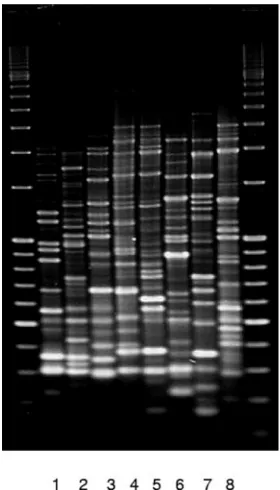
[image:3.585.44.546.80.207.2]For this reason, the similarity cutoff level to identify clonally related isolates was set as stringently as possible (at 80%) for all B. cepacia complex species (referred to below as BOX
FIG. 1. BOX-PCR profiles of isolates belonging to the majorrecA
[image:3.585.351.491.459.704.2]RFLP types AT, E, G, H, I, J, Se13, and U (lanes 1 to 8). TABLE 1. Distribution ofB. cepaciacomplex species in 18 Italian centers
B. cepaciacomplex species
No. of strains in centera:
Total no. (%) of strains
a b c d e f g h i l m n o p q r s t
B. cepaciab 1 3 3 4 1 3 1 16 (7.7)
B. multivorans 1 4 3 2 2 1 13 (6.3)
B. cenocepacia 7 2 30 2 4 1 18 13 1 5 3 1 7 7 20 6 127 (61.1)
B. stabilis 1 4 1 1 1 2 1 2 13 (6.3)
B. vietnamiensis 1 1 2 (1)
B. ambifaria 1 1 (0.5)
B. pyrrocinia 3 5 1 9 (4.3)
Undetermined 8 3 3 1 6 2 1 2 1 27 (13)
Total 8 3 49 2 5 1 21 24 12 11 11 1 3 2 10 14 23 8 208
a
One strain per patient has been isolated.
b
[image:3.585.44.283.587.707.2]B. cepaciaconstitutes genomovar I.
TABLE 2. MajorB. cepaciacomplex clones identified by BOX-PCR
BOX cluster
No. of isolatesa
HaeIII-recA RFLP type
B. cepacia complex species
No. of participating centers (designation) 1 22 AT Undetermined 8 (n, h, o, g, l, r, i, c)
2 7 E B. cepaciab 4 (m, h, r, c)
3 12 G B. cenocepacia 5 (l, t, o, e, d)
4 31 G B. cenocepacia 7 (h, c, r, g, t, i, q)
5 17 H B. cenocepacia 6 (c, g, q, h, s, m)
6 13 I B. cenocepacia 4 (s, h, c, m)
7 14 I B. cenocepacia 3 (c, h, g)
8 9 J B. stabilis 6 (h, c, i, s, m, q)
9 9 Se13 B. pyrrocinia 3 (i, m, c)
10 17 U B. cenocepacia 6 (a, r, q, g, h, b)
aOne isolate per patient has been isolated. bB. cepaciaconstitutes genomovar I.
on May 15, 2020 by guest
http://jcm.asm.org/
clusters). Ten major BOX clusters, encompassing the majority (72.6%) of the isolates examined, were identified. The BOX profiles of remaining isolates were unique or represented small clusters comprising two to three isolates (data not shown). Table 2 gives an overview of the number of isolates belonging to each of the major BOX clusters, their HaeIII-recARFLP type, genomovar, and CF centers involved. Figure 1 illustrates the BOX-PCR profiles of isolates belonging to the majorrecA
RFLP types (AT, E, G, H, I, J, Se13, and U), while the
HaeIII-recARFLP profiles of the same types are shown in Fig. 2. All 208 isolates were subsequently typed by PFGE.
The criteria used to define clonality for PFGE were as de-scribed by Tenover et al. (61): profiles showing differences of up to three bands were regarded as closely related, while those showing a four- to six-band difference were considered to be possibly related, provided there was an epidemiological link between the isolates.
BOX-PCR cluster 1 (HaeIII-recARFLP type AT) included 22 isolates, which were distributed among four PFGE clusters as follows. Two PFGE clusters were composed of isolates from the same center (seven and five isolates, respectively), another PFGE cluster included six isolates from four different centers, and the last PFGE cluster was composed of three isolates collected from three centers. The remaining isolate showed a unique PFGE pattern. Figure 3 shows the four different PFGE profiles produced by these isolates. The BOX-PCR clone 5 (HaeIII-recARFLP type H) included 17 isolates representing three different PFGE clusters composed of 2, 3, and 5 isolates, respectively. Isolates within each of the PFGE clusters were from a single center. The remaining seven isolates all had unique PFGE profiles (data not shown). In addition, we also investigated all BOX cluster 5 isolates for the presence of
IS1363using specific IS1363PCR. All BOX cluster 5 isolates contained this genetic marker (data not shown).
All but two (seven isolates) of BOX-PCR clone 9
(HaeIII-recARFLP type Se13) isolates belonged to a single clone and were from two different centers. The remaining two isolates in this group failed to produce any PFGE profile (data not shown).
Among the remaining isolates examined, several additional center-specific PFGE clusters were identified, corroborating results obtained by BOX-PCR analysis (data not shown).
DISCUSSION
The present study reports on the epidemiology ofB. cepacia
complex bacteria infecting CF patients in Italy. Strains from 208 out of 283 (73.5%) Italian CF patients colonized byB. cepacia
complex were collected (22, 44; data are from reference 44 and the present study). In 2002, the Italian Register of Cystic Fi-brosis reported 3,791 living CF patients (52a); therefore, the overall prevalence ofB. cepaciacomplex is 7.5%. Similar re-sults have been reported from previous studies of patients in Italy (22, 60), confirming the efficacy of the segregation policies actually adopted by all Italian CF centers (22). Higher preva-lences (about 20%) have been observed in some Italian centers and were associated with local spread ofB. cenocepacia(1, 44). Of 208 isolates collected from 208 patients attending 18 Italian CF centers, the majority (61.1%) of isolates belonged to
B. cenocepacia, whereas 4.3 to 7.7% of isolates belonged to
B. cepacia, B. multivorans, B. stabilis, and B. pyrrocinia. The other genomovars accounted for 1% or fewer of the isolates received (B. vietnamiensis and B. ambifaria) or were absent (B. dolosa and B. anthina). These data confirm and extend results from previous studies ofB. cepacia complex bacteria infecting Italian CF patients (1, 44, 49). About 13% of the
[image:4.585.81.246.71.313.2]B. cenocepaciaisolates belonged to HaeIII-recARFLP type U, which was reported as a major problem in the Genoa CF center (44). The HaeIII-recA RFLP type U isolates of the FIG. 2. HaeIII-recARFLP profiles of types AT, E, G, H, I, J, Se13,
and U (lanes 1 to 8).
FIG. 3. PFGE profiles representing the four different clusters within the 22 isolates of BOX-PCR cluster 1 (HaeIII-recARFLP type AT).
on May 15, 2020 by guest
http://jcm.asm.org/
present study were obtained from six CF centers throughout Italy.
This predominance ofB. cenocepaciain CF patients in Italy resembles the situation reported in Canada (80% prevalence) (58), the United States (50%) (53), the United Kingdom (76%) (9), and the Prague and Lisbon CF centers in Portugal, with prevalences of 90% and 52%, respectively (17, 21). Some national reports showed that other genomovars reach signifi-cant percentages, such asB. cepacia(genomovar I) in Australia (29%) (31);B. multivoransin France (51%) (4), the United States (38%) (37), the United Kingdom (39%) (62), and Belgium (18); orB. stabilis(54%) in the Slovak Republic (21). In the present study, the genomovar status of 13% of the strains was undetermined (Table 1). Isolates belonging to HaeIII-recARFLP type AT accounted for most of these iso-lates. Isolates with the same RFLP type were recovered as contaminants from dialysis equipment in Brazil (42). BOX-PCR was chosen as the initial genotyping tool because it was previously shown that it is a useful tool to study the large-scale epidemiology ofB. cepaciacomplex bacteria (3, 12). With the exception of small clusters comprising two to three isolates, 10 major BOX clusters were delineated. An analysis of the iso-lates by PFGE revealed that each of these BOX clusters com-prised isolates that were considered unrelated by conventional criteria (61). This difference in typing resolution forB. cepacia
complex bacteria confirmed previous reports (12). Three BOX clusters appeared of special interest. We report the first case of epidemic spread of HaeIII-recA RFLP type AT among CF patients (BOX cluster 1). The latter clone was isolated from 22 patients in eight CF centers (Table 2) and comprised multiple clusters of isolates, which were also considered clonal following interpretation of PFGE patterns.
The 17 BOX cluster 5 isolates were derived from patients in six CF centers (Table 2). Their BOX fingerprints were identi-cal to those of strain PHDC (16), a well-characterized epi-demicB. cenocepaciastrain recovered from numerous CF pa-tients and environmental samples in the United States (8, 39). The presence of IS1363, an insertion sequence specific for the highly epidemic strains B. cenocepacia ET12 (HaeIII-recA
RFLP type G) and PHDC (HaeIII-recARFLP type H) was confirmed in all BOX cluster 5 isolates. Identification as PHDC was further confirmed by comparison of random am-plified polymorphic DNA profiles, as reported previously (16). Most remarkably, this epidemic clone was recognized in CF specimens from patients in several other European countries (16). When examined by PFGE, seven of these isolates repre-sented a single PFGE cluster and several isolates with unique profiles.
Finally, BOX cluster 9 comprised nine isolates from three centers, which were identified asB. pyrrocinia, an organism not previously associated with spread in CF patients. When exam-ined by PFGE, these isolates represented two PFGE clusters (specific for a single center each).
In general, the comparison between the PFGE and BOX-PCR genotyping data demonstrated that BOX clusters com-prised isolates from multiple centers representing multiple PFGE clusters. In contrast, most PFGE clusters comprised isolates specific for a single center. While the latter is most likely indicative of intracenter spread through patient-to-pa-tient contact (1, 2, 6, 44), the former is more difficult to explain.
Although not common, isolates with the same PFGE profile have been observed in different Italian centers in the present study (e.g., in one of the HaeIII-recARFLP type AT clones) and in a previous study in Italy (1). We consider isolates with the same BOX-PCR profile but with different PFGE profiles as examples of recent divergence from a common ancestral strain (14). Patients may become infected by such isolates through patient-to-patient transfer or through acquisition from a com-mon source. The latter implies that such strains would be widely distributed in the environment and would have the potential to infect CF patients.
This latter hypothesis is supported by a number of observa-tions. Govan et al. (27) provided genomic and phenotypic evidence of a clonal relationship between theB. cepaciatype strain, isolated by W. Burkholder in the 1940s from a decaying onion, and an isolate recovered over 50 years later from the respiratory secretions of a patient with CF. In addition, LiPuma et al. (38) reported the isolation of the PHDC clone from numerous patients and agricultural soils of several years (14, 15, 38). These observations confirm that genotypically divergingB. cepaciacomplex clones can prevail in environmen-tal sources and infect CF patients on multiple occasions. Al-though to our knowledge the highly transmissibleB. cenocepa-cia lineage ET12 has not been isolated from environmental sources, it has been recovered from CF specimens from pa-tients in Europe, North America, Australia, and New Zealand (1, 14, 15, 25, 26, 30, 50, 59) with minor differences in genotype and phenotypic properties (50). It seems unlikely that this global distribution can be explained through patient-to-patient contacts alone. Interestingly, similar evidence is emerging for other CF pathogens as well. Genotypically diverse isolates of
P. aeruginosaclone C have been recovered from CF, non-CF, and environmental specimens throughout Europe (20); since the early 1990s, European-wide clones ofStaphylococcus au-reushave been isolated from CF and non-CF sources without epidemiological relationships (55).
The present study provides an overview of the prevalence and distribution ofB. cepaciacomplex species and strains in Italy and a comparison with other countries. It confirms that the epidemiology of the B. cepaciacomplex bacteria in Italy is mainly characterized by the acquisition and spread of
B. cenocepaciastrains and presents the first evidence of spread ofB. pyrrociniaand of a strain belonging to HaeIII-recARFLP type AT. Furthermore, our study confirms the presence of PHDC, an epidemic lineage ofB. cenocepacia, in patients from multiple CF centers in Italy. These findings substantiate pre-vious reports of the spread of non-B. cenocepacia B. cepacia
complex species including B. cepacia (genomovar I) (3),
B. multivorans(28, 46, 68), andB. dolosa(3). Finally, the oc-currence of isolates with distinct PFGE profiles but with the same BOX-PCR profiles suggests the nationwide environmen-tal distribution of genetically diverging clones.
REFERENCES
1.Agodi, A., E. Mahenthiralingam, M. Barchitta, V. Giannino, A. Sciacca, and S. Stefani.2001.Burkholderia cepaciacomplex infection in Italian patients with cystic fibrosis: prevalence, epidemiology, and genomovar status. J. Clin. Microbiol.39:2891–2896.
2.Bevivino, A., C. Dalmastri, S. Tabacchioni, L. Chiarini, M. L. Belli, S. Piana, A. Materazzo, P. Vandamme, and G. Manno.2002.Burkholderia cepacia complex bacteria from clinical and environmental sources in Italy:
on May 15, 2020 by guest
http://jcm.asm.org/
var status and distribution of traits related to virulence and transmissibility. J. Clin. Microbiol.40:846–851.
3.Biddick, R., T. Spilker, A. Martin, and J. J. LiPuma.2003. Evidence of transmission ofBurkholderia cepacia,Burkholderia multivoransand Burk-holderia dolosaamong persons with cystic fibrosis. FEMS Microbiol Lett. s228:57–62.
4.Brisse, S., C. Cordevant, P. Vandamme, P. Bidet, C. Loukil, G. Chabanon, M. Lange, and E. Bingen.2004. Species distribution and ribotype diversity of Burkholderia cepaciacomplex isolates from French patients with cystic fibro-sis. J. Clin. Microbiol.42:4824–4827.
5.Butler, S., C. J. Doherty, J. E. Hughes, J. W. Nelson, and J. R. W. Govan.
1995. Burkholderia cepacia and cystic fibrosis: do natural environments present a potential hazard? J. Clin. Microbiol.33:1001–1004.
6.Campana, S., G. Taccetti, N. Ravenni, F. Favari, L. Cariani, A. Sciacca, O. Bianco, T. Pensabene, E. Fiscarelli, G. De Intinis, M. Busetti, A. Cipolloni, A. d’Aprile, F. Gioffre`, I. Collebrusco, P. Frontini, G. Stassi, A. Magni, D. Tovagliari, A. Lavitola, C. J. Doherty, J. R. W. Govan, and P. Vandamme.
2003. Transmission ofBurkholderia pyrrocinia:a report of two outbreaks between Italian cystic fibrosis patients. Pediatr. Pulmonol.25(Suppl.):299. 7.Chaparro, C., J. Maurer, M. Gutierrez, M. Krajden, T. Chan, S. Winston, S.
Keshavjee, M. Scavuzzo, E. Tullis, M. Hutcheon, and S. Kesten.2001. Infection withBurkholderia cepaciain cystic fibrosis. Outcome following lung transplantation. Am. J. Respir. Care Med.163:43–48.
8.Chen, J. S., K. Witzmann, T. Spilker, R. Fink, and J. J. LiPuma.2001. Endemicity and inter-city spread ofBurkholderia cepaciagenomovar III in cystic fibrosis. J. Pediatr.139:643–649.
9.Clode, F. E., M. E. Kaufmann, H. Malnik, and T. L. Pitt.2000. Distribution of genes encoding putative transmissibility factors among epidemic and non-epidemic strains ofBurkholderia cepaciafrom cystic fibrosis patients in the United Kingdom. J. Clin. Microbiol.38:1763–1766.
10.Coenye, T., E. Mahenthiralingam, D. Henry, J. J. LiPuma, S. Laevens, M. Gillis, D. P. Speert, and P. Vandamme.2001.Burkholderia ambifariasp. nov., a novel member of theBurkholderia cepaciacomplex comprising biocontrol and cystic-fibrosis related isolates. Int. J. Syst. Evol. Microbiol.51:1481–1490. 11.Coenye, T., P. Vandamme, J. R. W. Govan, and J. J. LiPuma.2001. Taxon-omy and identification of theBurkholderia cepaciacomplex. J. Clin. Micro-biol.39:3427–3436.
12.Coenye, T., T. Spilker, A. Martin, and J. J. LiPuma.2002. Comparative assessment of genotyping methods for epidemiologic study ofBurkholderia cepaciagenomovar III. J. Clin. Microbiol.40:3300–3307.
13.Coenye, T., J. Goris, T. Spilker, P. Vandamme, and J. J. LiPuma.2002. Characterization of unusual bacteria isolated from respiratory secretions of cystic fibrosis patients and description ofInquilinus limosusgen. nov., sp. nov. J. Clin. Microbiol.40:2052–2069.
14.Coenye, T., and J. J. LiPuma.2003. Population structure analysis of Burk-holderia cepaciagenomovar III: varying degrees of genetic recombination characterize major clonal complexes. Microbiology149:77–88.
15.Coenye, T., and J. J. LiPuma.2003. Molecular epidemiology ofBurkholderia species. Front. Biosci.8:e55–e67.
16.Coenye, T., T. Spilker, A. Van Schoor, J. J. LiPuma, and P. Vandamme.
2004. Recovery ofBurkholderia cenocepaciastrain PHDC from cystic fibrosis patients in Europe. Thorax59:952–954.
17.Cunha, M. V., J. H. Leitao, E. Mahenthiralingam, P. Vandamme, L. Lito, C. Barreto, M. J. Salgado, and I. Sa-Correia. 2003. Molecular analysis of Burkholderia cepaciacomplex isolates from a Portuguese cystic fibrosis cen-ter: a 7-year study. J. Clin. Microbiol.41:4113–4120.
18.De Boeck, K., A. Malfroot, L. Van Schil, P. Lebecque, C. Knoop, J. R. Govan, C. Doherty, S. Laevens, P. Vandamme, et al.2004. Epidemiology of Burk-holderia cepaciacomplex colonisation in cystic fibrosis patients. Eur. Respir. J.23:851–856.
19.De Soyza, A., K. Morris, A. McDowell, C. Doherty, L. Archer, J. Perry, J. R. W. Govan, P. A. Corris, and K. Gould.2004. Prevalence and clonality in UK patients with cystic fibrosis referred for lung transplantation. Thorax
59:526–528.
20.Dinesh, S. D., H. Grundmann, T. L. Pitt, and U. Ro¨mling.2003. European-wide distribution ofPseudomonas aeruginosaclone C. Clin. Microbiol. Infect.
9:1228–1233.
21.Drevinek, P., O. Cinek, J. Melter, L. Langsadl, Y. Navesnakova, and V. Vavrova.2003. Genomovar distribution of theBurkholderia cepaciacomplex differs significantly between Czech and Slovak patients with cystic fibrosis. J. Med. Microbiol.52:603–604.
22.Festini, F., S. Ballarin, C. Loganes, T. Codamo, R. Doro, A. Adamo, R. Adorni, M. Cucci, F. Di Marco, R. Lovallo, S. Omenetti, R. Panebianco, G. Pisano, A. Russo, M. C. Sciabacucchi, and M. L. Zunino.2004. Prevention and control of respiratory tract infections in the network of Italian Centers for Cystic Fibrosis. Assist. Inferm. Ric.23:14–20. (In Italian.)
23.Goris, J., W. Dejonghe, E. Falsen, E. De Clerck, B. Geeraerts, A. Willems, E. M. Top, P. Vandamme, and P. De Vos.2002. Diversity of transconjugants that acquired plasmid pJP4 and pEMT1 after inoculation of a donor strain in the A- and B-horizon of an agricultural soil and description of Burkhold-eria hospitasp. nov. andBurkholderia terricolasp. nov. Syst. Appl. Microbiol.
25:340–352.
24.Govan, J. R. W., and V. Deretic.1996. Microbial pathogenesis in cystic fibrosis: mucoidPseudomonas aeruginosaandBurkholderia cepacia. Micro-biol. Rev.60:539–574.
25.Govan, J. R. W., P. H. Brown, J. Maddison, C. Doherty, C. J. Nelson, M. Dodd, A. P. Greening, and A. K. Webb.1993. Evidence for transmission of Pseudomonas cepaciaby social contact in cystic fibrosis patients. Lancet
342:15–19.
26.Govan, J. R. W., J. E. Hughes, and P. Vandamme.1996.Burkholderia cepa-cia: medical, taxonomic and ecological issues. J. Med. Microbiol.45:395–407. 27.Govan, J. R. W., J. Balandreau, and P. Vandamme.2000.Burkholderia
cepacia—friend and foe. ASM News66:124–125.
28.Heath, D. G., K. Hohneker, C. Carriker, K. Smith, J. Routh, J. J. LiPuma, R. M. Aris, D. Wber, and P. H. Gilligan.2002. Six-year molecular analysis of Burkholderia complexisolates among cystic fibrosis patients at a referral center for lung transplantation. J. Clin. Microbiol.40:1188–1193. 29.Isles, A., M. Maclusky, M. Corey, R. Gold, C. Prober, P. Fleming, and H.
Levinson.1984.Pseudomonas cepaciainfection in cystic fibrosis: an emerging problem. J. Pediatr.104:206–210.
30.Johnson, W. M., S. D. Tyler, and K. R. Rozee. 1994. Linkage analysis of geographic and clinical clusters inPseudomonas cepaciainfections by multilocus enzyme electrophoresis and ribotyping. J. Clin. Microbiol.32:
924–930.
31.Kidd, T. J., S. C. Bell, and C. Coulter.2003. Genomovar diversity amongst Burkholderia cepaciacomplex isolates from an Australian adult cystic fibrosis unit. Eur. J. Clin. Microbiol. Infect. Dis.22:434–437.
32.Koeuth, T., J. Versalovic, and J. R. Lupski.1995. Differential subsequence conservation of interspersed repetitive Streptococcus pneumoniae BOX el-ements in diverse bacteria. Genome Res.5:408–418.
33.Liou, T. G., F. R. Adler, S. C. FitzSimmons, B. C. Cahill, J. R. Hibbs, and B. C. Marshall.2001. Predictive 5-years survivorship model of cystic fibrosis. Am. J. Epidemiol.153:345–352.
34.LiPuma, J. J., S. E. Dasen, D. W. Nielson, R. C. Stern, and T. L. Stull.1990. Person-to-person transmission ofPseudomonas cepacia between patients with cystic fibrosis. Lancet336:1094–1096.
35.LiPuma, J. J., A. Marks-Austin, D. S. Holsclaw, B. Winnie, P. H. Gilligan, and T. S. Stull.1994. Inapparent transmission ofPseudomonas(Burkholderia) cepa-ciaamong patients with cystic fibrosis. Pediatr. Infect. Dis.13:716–719. 36.LiPuma, J. J.1998.Burkholderia cepacia: management issues and new
in-sights. Clin. Chest Med.19:473–486.
37.LiPuma, J. J.1998.Burkholderia cepaciaepidemiology and pathogenesis: implications for infection control. Curr. Opin. Pulm. Med.4:337–441. 38.LiPuma, J. J., T. Spilker, L. H. Gill, P. W. Campbell III, L. Liu, and E.
Mahenthiralingam.2001. Disproportionate distribution ofBurkholderia ce-paciacomplex species and transmissibility markers in cystic fibrosis. Am. J. Respir. Crit. Care Med.164:92–96.
39.LiPuma, J. J., T. Spilker, T. Coenye, and C. F. Gonzales.2002. An epidemic Burkholderia cepaciacomplex strain identified in soil. Lancet359:2002–2003. 40.Liu, L., T. Spilker, T. Coenye, and J. J. LiPuma.2003. Identification by subtractive hybridization of a novel insertion element specific for two wide-spreadBurkholderia cepaciagenomovar III strains. J. Clin. Microbiol.41:
2471–2476.
41.Louws, F. J., D. W. Fulbright, C. T. Stephens, and F. J. de Bruijn.1994. Specific genomic fingerprints of phytopathogenicXanthomonasand Pseudo-monaspathovars and strains generated with repetitive sequences and PCR. Appl. Environ. Microbiol.60:2286–2295.
42.Magalha˜es, M., C. Doherty, J. R. W. Govan, and P. Vandamme.2003. Polyclonal outbreak ofBurkholderia cepaciacomplex bacteremia in haemo-dialysis patients. J. Hosp. Infect.54:120–123.
43.Mahenthiralingam, E., J. Bischof, S. K. Byrne, C. Radomski, J. E. Davies, Y. Av-Gay, and P. Vandamme.2000. DNA-based approaches for identification ofBurkholderia cepaciacomplex,Burkholderia vietnamiensis,Burkholderia multivorans,Burkholderia stabilis, andBurkholderia cepacia genomovars I and III. J. Clin. Microbiol.38:3165–3173.
44.Manno, G., C. Dalmastri, S. Tabacchioni, P. Vandamme, R. Lorini, L. Minicucci, L. Romano, A. Giannattasio, L. Chiarini, and A. Bevivino.2004. Epidemiology and clinical course ofBurkholderia cepaciacomplex infections, particularly those caused by different Burkholderia cenocepacia strains, among patients attending an Italian cystic fibrosis center. J. Clin. Microbiol.
42:1491–1497.
45.McMenamin, J. D., T. M. Zaccone, T. Coenye, P. Vandamme, and J. J. LiPuma.2000. Misidentification ofBurkholderia cepaciain U.S. cystic fibro-sis treatment centers: an analyfibro-sis of 1051 recent sputum isolates. Chest
117:1661–1665.
46.Millar-Jones, L., H. C. Ryley, A. Paull, and M. C. Goodchild.1998. Trans-mission and prevalence ofBurkholderia cepaciain Welsh cystic fibrosis pa-tients. Respir. Med.92:178–183.
47.Olive, D. M., and P. Bean.1999. Principles and applications of methods for DNA-based typing of microbial organisms. J. Clin. Microbiol.37:1661–1669. 48.Pegues, D. A., L. A. Carson, O. C. Tablan, S. C. FitzSimmons, S. B. Roman, J. M. Miller, and W. R. Jarvis.1994. Acquisition ofPseudomonas cepaciaat summer camps for patients with cystic fibrosis. J. Pediatr.124:694–702. 49.Petrucca, A., P. Cipriani, P. Valenti, D. Santapaola, C. Cimmino, G. L.
on May 15, 2020 by guest
http://jcm.asm.org/
Scoarughi, L. Santino, S. Stefani, R. Sessa, and M. Nicoletti.2003. Molec-ular characterization ofBurkholderia cepaciaisolates from cystic fibrosis (CF) patients in an Italian CF center. Res. Microbiol.154:491–498. 50.Pitt, T. L., M. E. Kaufmann, P. S. Patel, L. C. Gaskin, and D. M. Livermore.
1996. Type characterization and antibiotic susceptibility of Burkholderia (Pseudomonas)cepaciaisolates from patients with cystic fibrosis in the United Kingdom and the Republic of Ireland. J. Med. Microbiol.44:203–210. 51.Pot, B., P. Vandamme, and K. Kersters.1994. Analysis of electrophoretic
whole-organism protein fingerprints, p. 493–521. In M. Goodfellow and A. G. O’Donnel (ed.), Modern microbial methods. Chemical methods in prokaryotics systematics. J. Wiley and Sons, Ltd., Chichester, United Kingdom.
52.Rademaker, J. L. W., F. J. Louws, and F. J. de Bruijn.1998. Characterization of the diversity of ecologically important microbes by rep-PCR fingerprint-ing, p. 1–26.InA. D. L. Akkermans, J. D. van Elsas, and F. J. de Bruijn (ed.), Molecular microbial ecology manual, supplement 3. Kluwer Academic Pub-lishers, Dordrecht, The Netherlands.
52a.Registro Italiano Fibrosi Cistica.2002. Annual Report. Registro Italiano Fibrosi Cistica, Milan, Italy. (In Italian.)
53.Reik, R., T. Spilker, J. J. LiPuma.2005. Distribution ofBurkholderia cepacia complex species among isolates recovered from persons with or without cystic fibrosis. J. Clin. Microbiol.43:2926–2928.
54.Saiman, L., and J. Siegel.2004. Infection control in cystic fibrosis. Clin. Microbiol. Rev.17:57–71.
55.Schlichting, C., C. Branger, J. M. Fournier, W. Witte, A. Boutonnier, C. Wolz, P. Goullet, and G. Do¨ring.1993. Typing ofStaphylococcus aureusby pulsed-field gel electrophoresis, zymotyping, capsular typing, and phage typ-ing: resolution of clonal relationships. J. Clin. Microbiol.31:227–232. 56.Segonds, C., T. Heulin, N. Marty, and G. Chabanon.1999. Differentiation of
Burkholderiaspecies by PCR-restriction fragment length polymorphism anal-ysis of the 16S rRNA gene and application to cystic fibrosis isolates. J. Clin. Microbiol.37:2201–2208.
57.Shreve, M. R., S. Butler, H. J. Kaplowitz, H. R. Rabin, D. Stokes, M. Light, and W. E. Regelmann for North American Scientific Advisory group and Investigators for the Epidemiologic Study of Cystic Fibrosis.1999. Impact of microbiology practice on cumulative prevalence of respiratory tract bacteria in patients with cystic fibrosis. J. Clin. Microbiol.37:753–757.
58.Speert, D. P., D. Henry, P. Vandamme, M. Corey, and E. Mahenthiralingam.
2002. Epidemiology ofBurkholderia cepaciacomplex in patients with cystic
fibrosis in Canada: geographical distribution and clustering of strains. Emerg. Infect. Dis.8:181–187.
59.Sun, L., R. Z. Jiang, S. Steinbach, A. Holmes, C. Campanelli, J. Forstner, U. Sajjan, Y. Tan, M. Riley, and R. Goldstein.1995. The emergence of a highly transmissible lineage of cbl⫹Pseudomonas(Burkholderia)cepaciacausing CF centre epidemics in North America and Britain. Nat. Med.1:661–666. 60.Taccetti, G., and S. Campana.1997. Microbiologic data overview of Italian
cystic fibrosis patients. Eur. J. Epidemiol.13:323–327.
61.Tenover, F. C., R. D. Arbeit, R. V. Goering, P. A. Mickelsen, B. E. Murray, D. A. Persing, and B. Swaminathan.1995. Interpreting chromosomal DNA restriction patterns produced by pulsed-field gel electrophoresis: criteria for bacterial strain typing. J. Clin. Microbiol.33:2233–2239.
62.Turton, J. F., M. E. Kaufmann, N. Mustafa, S. Kawa, F. E. Clode, and T. L. Pitt. 2003. Molecular comparison of isolates ofBurkholderia multivorans from patients with cystic fibrosis in the United Kingdom. J. Clin. Microbiol.
41:5750–5754.
63.UK Cystic Fibrosis Trust Infection Control Group.2004. TheBurkholderia cepaciacomplex. Suggestions for prevention and infection control. Cystic Fibrosis Trust, Bromley, Kent, United Kingdom.
64.Vandamme, P., B. Holmes, M. Vancanneyt, T. Coenye, B. Hoste, R. Coopman, H. Revets, S. Lauwers, M. Gillis, K. Kersters, and J. R. Govan.1997. Oc-currence of multiple genomovars ofBurkholderia cepaciain cystic fibrosis patients and proposal ofBurkholderia multivoranssp. nov. Int. J. Syst. Bac-teriol.47:1188–1200.
65.Vandamme, P., E. Mahenthiralingam, B. Holmes, T. Coenye, B. Hoste, P. De Vos, D. Henry, and D. P. Speert.2000. Identification and population structure ofBurkholderia stabilissp. nov. (formerly Burkholderia cepacia genomovar IV). J. Clin. Microbiol.38:1042–1047.
66.Vandamme, P., B. Holmes, T. Coenye, J. Goris, E. Mahenthiralingam, J. J. LiPuma, and J. R. W. Govan.2003.Burkholderia cenocepaciasp. nov.—a new twist to an old story. Res. Microbiol.154:91–96.
67.Vermis, K., T. Coenye, J. J. LiPuma, E. Mahenthiralingam, H. J. Nelis, and P. Vandamme.2004. Proposal to accommodateBurkholderia cepacia geno-movar VI asBurkholderia dolosasp. nov. Int. J. Syst. Evol. Microbiol.54:
689–691.
68.Whiteford, M. L., J. D. Wilkinson, J. H. McColl, F. M. Conlon, J. R. Michie, T. J. Evans, and J. Y. Paton.1995. Outcome ofBurkholderia(Pseudomonas) cepaciacolonisation in children with cystic fibrosis following a hospital out-break. Thorax50:1194–1208.