and Review
Sophie Edouard, Cecile Nabet, Hubert Lepidi, Pierre-Edouard Fournier, Didier Raoult
Aix Marseille Université, URMITE, UM63, CNRS 7278, IRD198, Inserm 1095, Marseille, France
Bartonellaspp. are fastidious bacteria that cause blood culture-negative endocarditis and have been increasingly reported. In this study, we included all patients retrospectively and prospectively diagnosed withBartonellaendocarditis in our French refer-ence center between 2005 and 2013. Our diagnosis was based on the modified Duke criteria and microbiological findings, includ-ing serological and PCR results. To review the published literature, we searched all humanBartonellaendocarditis cases pub-lished in the PubMed database between January 2005 and October 2013. We report here a large series of 106 cases, which include 59 cases that had not previously been reported or mentioned. Indirect immunofluorescence assays, Western blotting, and real-time PCR from total blood, serum, and valve tissue exhibited sensitivities of 58%, 100%, 33%, 36%, and 91%, respectively. The number of cases reported in the literature between 2005 and 2013 increased to reach a cumulative number of 196 cases. The number of cases reported in the literature by other centers is increasing more rapidly than that reported by our French reference center (P<10ⴚ2). Currently, there is a lack of criteria for the diagnosis ofBartonellaendocarditis. We suggest that a positive PCR result from a cardiac valve or blood specimen, an IgG titer of>800 using an immunofluorescence assay, or a positive West-ern blot assay be considered major Duke criteria forBartonellaendocarditis. There is no real increase in the incidence of these infections but rather a better understanding and interest in the disease resulting from the improvement of diagnostic tools.
B
artonellaspecies are small Gram-negative hemotropic bacilliclassified within the classAlphaproteobacteria. These
faculta-tive intracellular bacteria, which are generally transmitted by
ar-thropod vectors (1), cause various clinical syndromes in
immu-nocompetent and immunocompromised patients. To date, 23
Bartonellaspecies have been described, 11 of which are proven or
likely human pathogens (2).Bartonellaspecies were first
recog-nized as endocarditis agents in 1993 when 3 such cases were
re-ported (3–5). After the seminal work of Drancourt et al. (6), 120
cases were reported in the literature until 2006 (7). Most reported
cases were attributed toBartonella quintana, the agent of trench
fever that may also cause endocarditis, chronic bacteremia, and
bacillary angiomatosis, and Bartonella henselae, which mainly
causes cat scratch disease, endocarditis, bacillary angiomatosis,
peliosis hepatis, and osteomyelitis (8).B. quintanaprimarily
in-fects homeless people infested with body lice, whereasB. henselae
is associated with patients with a previous valvulopathy and
con-tact with cats (2,7,8). Single cases of endocarditis caused by other
Bartonellaspecies, includingBartonella elizabethae(4),Bartonella vinsoniisubsp.berkhoffii(9,10),B. vinsoniisubsp.arupensis(11),
Bartonella koehlerae(12),Bartonella alsatica(13,14), and “ Candi-datusBartonella mayotimonensis” (15), have occasionally been reported in the literature. Epidemiological data suggest a
Europe-an-African gradient distribution in the prevalence ofBartonella
endocarditis, the highest prevalence being reported in southern
countries (7). In Europe, the prevalence ofBartonellaendocarditis
among those with infective endocarditis is 0% in Sweden, 1.1% in
the United Kingdom, and 3% in France and Germany (7), and it
reaches 15.6% and 9.8% in Algeria (16) and Tunisia (17),
respec-tively.
Blood culture-negative endocarditis (BCNE) represents 2.5%
to 31% of cases of endocarditis, depending on the series (18). In
France, approximately 20% to 30% of all documented cases of
BCNE areBartonellaendocarditis, which represents the second
most common cause of endocarditis followingCoxiella burnetii
(19,20). Over the past few years, in our national reference center
for Q fever andBartonellainfection, we have developed diagnostic
tools for the diagnosis of BCNE, includingBartonellaendocarditis
(19). BecauseBartonellaspp. are fastidious, serological analysis
using an indirect immunofluorescence assay (IFA) remains the most common method for diagnosing endocarditis caused by these bacteria. However, these infections can also be successfully diagnosed by Western blotting or specific real-time PCR
(RT-PCR), allowing for the rapid detection ofBartonellaDNA in blood
or resected valve samples. The bacteria can also be isolated by
inoculation onto agar plates or in tissue culture (2) or visualized in
valvular tissues using Warthin-Starry staining and
immunohisto-chemical examination (2).
From this study, we report a series of 106 patients with
Barto-nellaendocarditis who were diagnosed in our laboratory during a 9-year period, and we compare our data to the increasing number of diagnosed cases reported in the literature during the same pe-riod.
Received1 October 2014Returned for modification25 October 2014 Accepted19 December 2014
Accepted manuscript posted online24 December 2014
CitationEdouard S, Nabet C, Lepidi H, Fournier P-E, Raoult D. 2015.Bartonella, a common cause of endocarditis: a report on 106 cases and review. J Clin Microbiol 53:824 – 829.doi:10.1128/JCM.02827-14.
Editor:R. Patel
Address correspondence to Didier Raoult, [email protected].
S.E. and C.N. contributed equally to this article.
Supplemental material for this article may be found athttp://dx.doi.org/10.1128 /JCM.02827-14.
Copyright © 2015, American Society for Microbiology. All Rights Reserved.
doi:10.1128/JCM.02827-14
on May 16, 2020 by guest
http://jcm.asm.org/
MATERIALS AND METHODS
Patient recruitment.The French Reference Center for Rickettsial Dis-eases is located in Marseille, southern France. We received samples from patients from all parts of France and other countries forBartonella diag-nosis. Between January 2005 and October 2013, we systematically searched forBartonellaspecies as the causative agents of BCNE using serology and/or RT-PCR, depending on the available samples. We tested 54,401 serum samples for antibodies toBartonellaspp., including 4,381 serum samples from patients suspected to have BCNE, and we received 750 cardiac valve samples that were tested by RT-PCR from patients with a BCNE diagnosis.
Serological assays.The IFA was performed as previously described to detect the titer of antibodies againstB. henselaeandB. quintana(21). An IFA was considered positive when the IgG antibody titer wasⱖ1:100, but for the diagnosis of endocarditis, we use a titer of 1:800 as the cutoff (22). To confirm positive serological results or when serology was negative in the context of BCNE, we performed Western blotting as previously de-scribed (23). This technique shows a very specific profile forBartonella endocarditis (23). Six protein bands (Bq83, Bq65, Bq45, Bq30, Bq20, Bq10) react more frequently with antibodies from serum samples of pa-tients presenting with B. quintanaendocarditis and 4 protein bands (Bh65, Bh45, Bh35, Bh23) react more frequently with antibodies from serum samples of patients presenting withB. henselaeendocarditis (23). Due to cross-reactions, current serological assays may not reliably distin-guish antibody responses against different species ofBartonella. In such cases, Western blotting with cross-adsorption enables identification of the Bartonellaspecies that generated the antibody response. We usually per-form Western blotting using antigens from 5Bartonellaspecies, including B. quintana,B. henselae,B. vinsoniisubsp.berkhoffii,B. elizabethae, andB. alsatica, which are proven endocarditis agents, and the serum of a patient diluted to 1:200 in Tris-buffered saline (TBS)–3% nonfat milk. Each se-rum sample was tested before and after cross-adsorption withB. quintana andB. henselae. When cross-adsorption is performed with the species involved in the disease, homologous and heterologous antibodies disap-pear, whereas only heterologous antibodies are removed when cross-ad-sorption is performed with a species that is not involved in the disease (Fig. 1).
PCR assays.Extraction of bacterial DNA was performed from surgi-cally excised valves or EDTA blood samples using a QIAamp tissue kit (Qiagen, Hilden, Germany) according to the manufacturer’s recommen-dations. RT-PCR amplification was performed using a LightCycler ther-mal cycler (Roche Diagnostics, Meylan, France) between 2005 and 2010 and a CFX thermal cycler (Bio-Rad, Marne la Coquette, France) between 2011 and 2013. All the samples were screened using primers and TaqMan probes targeting a portion of theBartonella16S-23S RNA intergenic re-gion, as previously described (19). All positive reactions were confirmed using primers and TaqMan probes targeting thepap31gene forB. henselae and theyopPgene forB. quintana, as previously described (2,24). A case was considered definite when 2 positive PCR results in assays targeting 2 differentBartonellasp. genes with a threshold cycle (CT) value ofⱕ35
were obtained. Identification of otherBartonellaspecies was obtained by sequencing the amplified 16S-23S RNA intergenic region (25). DNAs fromB. henselaeandB. quintanastrains were used as positive controls, and sterile water was used as a negative control. We verified the quality of the extracted DNA by detecting the human actin gene by RT-PCR.
All the valvular specimens were also tested using a broad-range 16S rRNA PCR assay, followed by sequencing when positive, as previously described (26).
Immunohistochemical analysis and Warthin-Starry staining. War-thin-Starry staining and immunohistochemical analysis were performed on paraffin-embedded valve sections. Anti-B. henselaeor anti-B. quintana polyclonal rabbit antibodies were used for immunohistochemistry as pre-viously described (27).
Case definition.Between January 2005 and October 2013,Bartonella endocarditis was diagnosed according to the modified Duke criteria, which includes positive blood culture, positive histologic examination of valve tissue, and vegetations on echocardiography as major criteria (28). We consideredBartonellato be the etiological agent of endocarditis and a major diagnosis criterion when we found either an elevated serological titer of anti-Bartonellaantibodies with an IgG titer ofⱖ1:800 using IFA (22), a Western blot analysis with a typical endocarditis profile (23), a positive PCR from valve or blood specimens using two different target genes (25), or a positive immunohistochemical analysis (26). The caus-ativeBartonellaspecies was identified by PCR and sequencing (25) or FIG 1(A) Western immunoblotting of serum from a patient withB. quintanaendocarditis presenting with negative serology and a positive PCR for the cardiac valve. The utilized antigens includedB. quintana(lanes 1),B. henselaestrain Houston (lanes 2),B. elizabethae(lanes 3),B. vinsoniisubsp.berkhoffii(lanes 4), and B. alsatica(lanes 5). (B)B. quintanaadsorbed sera. (C)B. henselaeadsorbed sera. This Western blot presents a typical profile observed forBartonella-induced endocarditis cases. Molecular masses (in kilodaltons) are given on the left.
on May 16, 2020 by guest
http://jcm.asm.org/
[image:2.585.100.488.67.297.2]Western blotting following cross-adsorption (23). A standardized ques-tionnaire, including criteria used in the diagnostic score of the modified Duke criteria, was completed for each patient with documented Barto-nellaendocarditis.
Literature review.To review the published literature, we searched the PubMed database (http://www.ncbi.nlm.nih.gov/pubmed/) using the keywords “Bartonella” and “endocarditis” and included all human Barto-nellaendocarditis case reports published between January 2005 and Oc-tober 2013. We compared the number of published and unpublished cases diagnosed in our laboratory between 2005 and 2013 to those reported in the literature during the same period. Redundant cases were excluded. All references used for this analysis are listed in the supplemental material. To establish a cumulative frequency curve, we used the year of publication for the published cases or the year of diagnosis for the unpublished cases.
Statistical analysis.The statistical analysis was performed using Epi Info 3.5.1 (Centers for Disease Control and Prevention, Atlanta, GA, USA). Differences were considered statistically significant when theP value was⬍0.05.
RESULTS
During the study period, we diagnosed 106 cases ofBartonella
endocarditis, 47 of which have been mentioned or reported (11,
13–15, 19). Serum, EDTA blood, and valvular specimens were available for 102, 60, and 52 of these patients, respectively. The IFA was positive in 93 of the 102 tested patients (91%) and negative in 9 cases, 8 of which had a positive Western blot result. Western blotting was not performed for 1 patient. Among the 93 patients
with positive IFA titers, 34 (37%) had an IgG titer of⬍1:800, and
59 (63%) patients had an IgG titer ofⱖ1:800. In the 34 patients
with an IgG titer of⬍1:800, the diagnosis ofBartonella
endocar-ditis was confirmed by Western blotting for 25 patients, by RT-PCR from valve samples of 7 patients, and by RT-RT-PCR from the blood of 2 patients. Among the 59 patients with an IgG titer of
ⱖ1:800, Western blotting was performed and positive for 40
pa-tients (Table 1).
RT-PCR was performed on 182 samples from 103 patients and was positive for 48/52 (92%) valvular specimens, 20/60 (33%) blood samples, and 25/70 (36%) tested serum samples. Sera and blood were tested by PCR for 40. The PCR was negative for the sera and blood from 16 patients and positive on the 2 samples from 8 patients. However, a dissonance was observed between the 2 samples for 15 patients; PCR was positive for serum and negative for blood from 9 patients and positive for blood and negative for sera from 6 patients. In addition, 16S RNA gene amplification was positive for 21/35 (60%) cardiac specimens and 0/15 blood sam-ples from the tested patients. 16S rRNA PCR on valvular biopsy
specimens was significantly less sensitive than RT-PCR (P ⫽
0.0003 according to the chi-square test). Immunohistochemistry and Warthin-Starry staining were performed on 28 paraffin-em-bedded valve sections and were positive for 8 (29%) and 13 (46%) of them, respectively.
Overall, the IFA and Western blotting exhibited sensitivities of 58% (59/102) and 100% (72/72), respectively, and PCR from val-vular, blood, and serum samples exhibited sensitivities of 92% (48/52), 33% (20/60), and 36% (25/70), respectively. As 33% of
our cases presentedBartonellaendocarditis with low antibody
ti-ters (IgG⬍1:800), we reevaluated the positive predictive value
(PPV) of theBartonellaIgG titer for an endocarditis diagnosis. We
included 461 patients with positive serology forBartonellaspp.
from our laboratory between 2005 and 2013, and we defined cases of endocarditis according to serological or PCR findings, as de-scribed above. The PPVs for the diagnosis of endocarditis accord-ing to the IgG titers were 20%, 32%, 54%, 94%, 91%, and 100% for IgG titers ranging between 1:100 and 1:6,400, 1:200 and 1:6,400, 1:400 and 1:6,400, 1:800 and 1:6,400, 1:1600 and 1:6,400,
and 1:3200 and 1:6,400 (Table 2), respectively.
The species causing endocarditis was identified in 91 patients (Table 3). Four Bartonella species were identified as causative agents of endocarditis in our study. The two main etiologic agents wereB. quintana, which was detected in 48 cases (53%), andB. henselae, which was detected in 39 cases (43%). We also diagnosed
3 cases (3%) ofB. alsaticaand 1 case (1%) ofB. vinsoniisubsp.
berkhoffiiinfections.
The mean age (⫾the standard deviation [SD]) of the 48
pa-tients withB. quintanaendocarditis was 54⫾14 years, and 43
[image:3.585.40.286.87.321.2](90%) were male. Epidemiologic information was not available for all 48 patients, but we identified 18 homeless individuals, 8
TABLE 1Microbiologic diagnosis of 106 patients withBartonella endocarditis
Test type and criteria
No. of positive samples/no. of samples tested (%)
IFA
IFA with IgG titerⱖ100 93/102 (91)
IFA with IgG titerⱖ800 59/93 (63)
IFA with IgG titer from 1:100 to 1:800 34/93 (37)
Negative IFA 9/102 (9)
Western blotting
Total 73/73 (100)
Patient with IgG titerⱖ1:800 40/40 (100)
Patient with IgG titer from 1:100 to 1:800 25/25 (100)
Patient with negative IFA 8/8 (100)
Specific RT-PCR forBartonellaspp.
Cardiac valves 48/52 (92)
Blood 20/60 (33)
Serum 25/70 (36)
16S RNA amplification
Cardiac valves 21/35 (60)
Blood 0/15 (0)
TABLE 2Positive predictive value (PPV) ofBartonellaIgG titers for patients with endocarditis, France, 2005–2013
Patients (n⫽461)
Result for patients with IgG titer range of:
1:100–1:6,400 1:200–1:6,400 1:400–1:6,400 1:800–1:6,400 1:1,600–1:6,400 1:3,200–1:6,400
No. of patients with definite endocarditis 93 87 72 59 32 9
Total no. of patients 461 267 133 63 35 9
PPV, definite endocarditis (%) 20 32 54 94 91 100
on May 16, 2020 by guest
http://jcm.asm.org/
[image:3.585.38.546.659.724.2]alcoholic men, 4 intravenous drug users, and 3 patients who had contact with body lice. Seventeen (35%) patients originated from Marseille, 24 (50%) from other parts of France, and 7 (15%) from other countries (2 cases from Ireland and 1 case each from Bel-gium, Chile, Great Britain, Italy, and the United States). IFA titers
ofⱖ1:800 were observed in 28/47 (60%)B. quintanacases, and
Western blotting was positive in 100% of the cases. PCR of valve samples was positive in 26/27 (96%) tested patients. PCR of the blood samples was positive in 13/28 (46%) cases, and PCR of the serum samples was positive in 13/32 (41%) cases.
The mean age (⫾SD) of the 39 patients withB. henselae
endo-carditis was 49⫾21 years, and 26 patients (67%) were male.
Twenty-two patients (56%) reported contact with cats. Six pa-tients (15%) originated from Marseille, 30 (77%) from other cities in France, and 3 (8%) from other countries (Belgium, Thailand,
and the United States). IFA titers ofⱖ800 were obtained in 19/36
(53%)B. henselae cases, PCR of valve samples was positive in
19/20 (95%) cases, PCR of blood was positive in 6/24 (25%) tested
patients, and PCR of serum samples was positive in 12/24 (50%) cases.
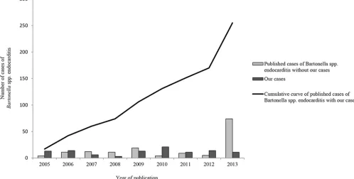
The annual number of cases diagnosed in our laboratory did not increase during the 9-year study period. We diagnosed a mean
(⫾SD) of 12⫾6Bartonellaendocarditis cases per year, with a
minimum of 3 in 2008 and a maximum of 21 in 2010. Following a literature review from January 2005 to October 2013 (see supple-mental material), we observed that the number of cases reported in the literature doubled every 1.5 years, reaching a cumulative
number of 196 cases in 2013 (Fig. 2). In this study, we added 59
unpublished cases. Between 2005 and 2006, we reported more than half of the published cases; however, since 2007, we observed a slight increase in the number of cases reported by other centers, with a more important accentuation in 2013. Using a chi-square test to examine the trend, we found that the number of cases reported in the literature by other centers (excluding ours) is in-creasing significantly more rapidly than the number of cases
[image:4.585.41.539.438.690.2]diag-nosed at our French reference center (P⬍10⫺2) (Fig. 2).
TABLE 3Epidemiologic features and biological data of the 91 patients with endocarditis induced byBartonellaspp. identified to the species level
Species
No. of cases
Mean age (yr)
Sex ratio
(no. male/no. female)
No. of samples positive in the indicated test/total no. of samples tested
IFA with IgG
ⱖ1:800
Western blotting
PCR on valve sample
PCR on blood sample
PCR on serum sample
B. quintana 48 54 43/5 28/47 35/35 26/27 13/28 13/32 B. henselae 39 49 26/13 19/36 28/28 19/20 6/24 12/24
B. alsatica 3 64 2/1 0/3 3/3 1/1 0/3 0/1
B. vinsonii 1 19 1/0 1/1 1/1 ND 1/1 0/1
FIG 2Number of cases of endocarditis induced byBartonellaspp. between January 2005 and October 2013 diagnosed in our center and number of cases reported in the published literature by other centers. A cumulative frequency curve of the total cases of endocarditis induced byBartonellaspp. was generated. Chi-square test for trends:P⬍10⫺2.
on May 16, 2020 by guest
http://jcm.asm.org/
DISCUSSION
Here, we report a large series of 106 patients diagnosed in our
laboratory withBartonellaendocarditis. We employed a strategy
of performing strict, validated protocols with specific RT-PCR, serology by IFA or Western blot analysis, and immunohistochem-istry to avoid false-positive and false-negative results and to in-crease the sensitivity of diagnosis. The combination of these dif-ferent techniques provided a specificity of 100%. In our study, the
IFA contributed to the diagnosis of endocarditis induced by
Bar-tonellaspp. in 91% of the tested samples but was confirmatory
with IgG titers ofⱖ800 in only 58% of cases. Western blotting
provided the diagnosis in 100% of the tested samples, and RT-PCR and 16S rRNA RT-PCR of valvular specimens were positive in 92% and 60% of the tested samples, respectively.
Serological testing is the easiest and most frequently used tool
for the laboratory diagnosis ofBartonellaendocarditis. IFA is the
reference method, despite the cross-reactivity amongBartonella
spp. and withChlamydiaspp. andC. burnetii(22, 29). All the
samples in our study were also tested by serology and PCR forC.
burnetii. Only 2 patients presented a cross-reaction withC. bur-netiiby IFA, butBartonellaendocarditis was determined through a positive Western blot analysis and a positive PCR on sera and cardiac valves, respectively. In our center, we have estimated that
an IgG titer ofⱖ1:800 has a positive predictive value of 95% in
patients with infective endocarditis (22). However, in this study,
we observed that such a titer was found in fewer than 60% of the
cases. Therefore, an IgG titer of⬍800 does not exclude the
diag-nosis ofBartonella endocarditis in patients with valvulopathy.
Moreover, in our study, we found 8 cases of endocarditis caused byBartonellaspp. with a negative IFA for which the serological diagnosis was confirmed only by Western blotting. Western blot-ting exhibited a sensitivity of 100%, as it was positive for all
pa-tients from whom direct evidence ofBartonellaendocarditis was
obtained by PCR. In a previous study, we demonstrated the high
PPV of Western blotting for Bartonellaendocarditis (23). Our
study suggests that any patient with aBartonellaIgG titer of⬍800
and a medical history evocative of endocarditis should be tested by Western blotting and RT-PCR following cardiac valve removal.
RT-PCR is a useful, sensitive, specific, and rapid tool for the
diagnosis ofBartonellaendocarditis. We observed a high
sensitiv-ity of RT-PCR for valvular biopsy specimens (92%), whereas this technique exhibited lower sensitivities when applied to blood and serum samples (33% and 36%, respectively). However, RT-PCR on blood may enable the diagnosis before cardiac surgery. Our results confirmed those from a previous study in which a positive PCR result was found in 97.8% of the available valvular biopsy specimens, despite the fact that 62.2% of the specimens were
ob-tained from patients receiving antibiotic therapy (8). Similar
re-sults demonstrating the high value of RT-PCR on valves in the
diagnosis ofBartonella endocarditis were recently reported by
Chaloner et al. (30), with positive results in 13 of 14 patients.
Furthermore, as previously reported (19,31), RT-PCR is
signifi-cantly more sensitive than 16S rRNA gene amplification for the
diagnosis ofBartonellaendocarditis in valvular tissues and blood.
Immunohistochemistry and Warthin-Starry staining were the less sensitive techniques. In our series, Warthin-Starry staining was the most sensitive of these 2 methods. Five samples were pos-itive for Warthin-Starry staining and negative in the immunohis-tochemical analysis; however, this histologic stain is not specific
forBartonellaspp. Because the infective process may be confined, a negative immunohistochemistry result does not definitively
ex-clude the diagnosis ofBartonellaendocarditis (27). Development
of a technique based on fluorescencein situhybridization would
increase the sensitivity of detection of bacteria and in assessing their viability by bacterial RNA detection.
Bartonellaendocarditis occurs in people with preexisting val-vular abnormalities that promote the development of infective endocarditis and generates significant destruction of the valve; therefore, valvular surgery is required in more than 90% of cases, which is higher than that required for patients with endocarditis caused by other pathogens. In our study, we found a rate of valve replacement inferior to those described earlier, but information on this criterion is missing in our study, and this number is prob-ably underestimated. However, we did not observe a significant difference in the rates of valvular surgery depending on the species ofBartonella(56% forB. quintana, 51% forB. henselae, and 33% forB. alsatica).
The number of reported cases ofBartonellaendocarditis
ap-pears to have increased between 2005 and 2013 (Fig. 2), and in this
study, we added 59 cases to the 196 previously reported to reach a cumulative number of 255 cases. The emergence of an infectious disease can usually be linked to one or more of the following factors: an increase in clinician interest in the infection, an im-provement in diagnostic techniques, or a real increase in incidence
(32). If we consider the distribution of sera tested forBartonella
spp. and the numbers of cases ofBartonellaendocarditis in our
laboratory from 2005 to 2013, an increase in the diagnosed cases was not observed. The observation of an increasing number of cases in the literature by our group and other centers can be
ex-plained by the fact that clinicians request moreBartonellatests for
patients with endocarditis. Therefore, we believe that there is no real increase in the incidence of these infections but rather a better understanding of and interest in the disease resulting from the improvement of diagnostic tools.
The strategy for diagnosing Bartonella endocarditis is not
clearly established, and specific diagnostic tools were available in only a few laboratories. We showed recently that expert centers play a key role in improving the diagnosis, management, and
pre-vention of Q fever endocarditis (33, 34). In 2012, a new score
based on evidence of endocardial involvement and microbiologi-cal results was proposed specifimicrobiologi-cally for the diagnosis of Q fever
endocarditis (35). Currently, there is a lack of criteria for the
di-agnosis ofBartonellaendocarditis, and we think that the
establish-ment of a new score-based diagnosis would be a valuable tool for
clinicians in diagnosingBartonellaendocarditis. We have
demon-strated that RT-PCR from valve samples can be a major element in the diagnosis of these infections, and we suggest that a positive PCR result from a valvular biopsy specimen can be considered a
definite criterion (28). In addition, we suggest that a positive PCR
from blood, an IgG titer ofⱖ800 using an IFA, and a positive
Western blot analysis may be considered major criteria in patients with BCNE.
ACKNOWLEDGMENT
We declare no competing interests.
REFERENCES
1.Chomel BB, Kasten RW, Williams C, Wey AC, Henn JB, Maggi R, Carrasco S, Mazet J, Boulouis HJ, Maillard R, Breitschwerdt EB.2009.
on May 16, 2020 by guest
http://jcm.asm.org/
Bartonellaendocarditis: a pathology shared by animal reservoirs and pa-tients. Ann N Y Acad Sci1166:120 –126.http://dx.doi.org/10.1111/j.1749 -6632.2009.04523.x.
2.Edouard S, Raoult D.2010.Bartonella henselae, an ubiquitous agent of proteiform zoonotic disease. Med Mal Infect40:319 –330.http://dx.doi .org/10.1016/j.medmal.2009.11.004.
3.Spach DH, Callis KP, Paauw DS, Houze YB, Schoenknecht FD, Welch DF, Rosen H, Brenner DJ.1993. Endocarditis caused byRochalimaea quintanain a patient infected with human immunodeficiency virus. J Clin Microbiol31:692– 694.
4.Daly JS, Worthington MG, Brenner DJ, Moss CW, Hollis DG, Weyant RS, Steigerwalt AG, Weaver RE, Daneshvar MI, O’Connor SP.1993. Rochalimaea elizabethaesp. nov. isolated from a patient with endocarditis. J Clin Microbiol31:872– 881.
5.Hadfield TL, Warren R, Kass M, Brun E, Levy C.1993. Endocarditis caused byRochalimaea henselae. Hum Pathol24:1140 –1141.http://dx.doi .org/10.1016/0046-8177(93)90196-N.
6.Drancourt M, Mainardi JL, Brouqui P, Vandenesch F, Carta A, Lehnert F, Etienne J, Goldstein F, Acar J, Raoult D.1995.Bartonella( Rochali-maea)quintanaendocarditis in three homeless men. N Engl J Med332: 419 – 423.http://dx.doi.org/10.1056/NEJM199502163320702.
7.Brouqui P, Raoult D.2006. New insight into the diagnosis of fastidious bacterial endocarditis. FEMS Immunol Med Microbiol47:1–13.http://dx .doi.org/10.1111/j.1574-695X.2006.00054.x.
8.Fournier PE, Lelievre H, Eykyn SJ, Mainardi JL, Marrie TJ, Bruneel F, Roure C, Nash J, Clave D, James E, Benoit-Lemercier C, Deforges L, Tissot-Dupont H, Raoult D.2001. Epidemiologic and clinical character-istics ofBartonella quintanaandBartonella henselaeendocarditis: a study of 48 patients. Medicine (Baltimore)80:245–251.http://dx.doi.org/10 .1097/00005792-200107000-00003.
9.Roux V, Eykyn SJ, Wyllie S, Raoult D.2000.Bartonella vinsoniisubsp. berkhoffiias an agent of afebrile blood culture-negative endocarditis in a human. J Clin Microbiol38:1698 –1700.
10. Olarte L, Ampofo K, Thorell EA, Sanderson S, Doby E, Pavia AT, Rosado H, Raoult D, Socolovschi C, Hersh AL.2012.Bartonella vinsonii endocarditis in an adolescent with congenital heart disease. Pediatr Infect Dis J31:531–534.http://dx.doi.org/10.1097/INF.0b013e31824ba95a. 11. Fenollar F, Sire S, Raoult D.2005.Bartonella vinsoniisubsp.arupensisas
an agent of blood culture-negative endocarditis in a human. J Clin Micro-biol43:945–947.http://dx.doi.org/10.1128/JCM.43.2.945-947.2005. 12. Avidor B, Graidy M, Efrat G, Leibowitz C, Shapira G, Schattner A,
Zimhony O, Giladi M.2004.Bartonella koehlerae, a new cat-associated agent of culture-negative human endocarditis. J Clin Microbiol42:3462– 3468.http://dx.doi.org/10.1128/JCM.42.8.3462-3468.2004.
13. Raoult D, Roblot F, Rolain JM, Besnier JM, Loulergue J, Bastides F, Choutet P.2006. First isolation ofBartonella alsaticafrom a valve of a patient with endocarditis. J Clin Microbiol44:278 –279.http://dx.doi.org /10.1128/JCM.44.1.278-279.2006.
14. Jeanclaude D, Godmer P, Leveiller D, Pouedras P, Fournier PE, Raoult D, Rolain JM.2009.Bartonella alsaticaendocarditis in a French patient in close contact with rabbits. Clin Microbiol Infect15(Suppl):S110 –S111.
http://dx.doi.org/10.1111/j.1469-0691.2008.02187.x.
15. Lin EY, Tsigrelis C, Baddour LM, Lepidi H, Rolain JM, Patel R, Raoult D.2010. CandidatusBartonella mayotimonensisand endocarditis. Emerg Infect Dis16:500 –503.http://dx.doi.org/10.3201/eid1603.081673. 16. Benslimani A, Fenollar F, Lepidi H, Raoult D.2005. Bacterial zoonoses
and infective endocarditis, Algeria. Emerg Infect Dis11:216 –224.http: //dx.doi.org/10.3201/eid1102.040668.
17. Znazen A, Rolain JM, Hammami N, Kammoun S, Hammami A, Raoult D.2005. High prevalence ofBartonella quintanaendocarditis in Sfax, Tunisia. Am J Trop Med Hyg72:503–507.
18. Brouqui P, Raoult D.2001. Endocarditis due to rare and fastidious
bac-teria. Clin Microbiol Rev14:177–207.http://dx.doi.org/10.1128/CMR.14 .1.177-207.2001.
19. Fournier PE, Thuny F, Richet H, Lepidi H, Casalta JP, Arzouni JP, Maurin M, Célard M, Mainardi JL, Caus T, Collart F, Habib G, Raoult D.2010. Comprehensive diagnostic strategy for blood culture-negative endocarditis: a prospective study of 819 new cases. Clin Infect Dis51:131– 140.http://dx.doi.org/10.1086/653675.
20. Houpikian P, Raoult D.2005. Blood culture-negative endocarditis in a reference center: etiologic diagnosis of 348 cases. Medicine (Baltimore) 84:162–173.http://dx.doi.org/10.1097/01.md.0000165658.82869.17. 21. Maurin M, Rolain JM, Raoult D.2002. Comparison of in-house and
commercial slides for detection by immunofluorescence of immunoglob-ulins G and M againstBartonella henselaeandBartonella quintana. Clin Diagn Lab Immunol 9:1004 –1009.http://dx.doi.org/10.1128/CDLI.9.5 .1004-1009.2002.
22. Fournier PE, Mainardi JL, Raoult D.2002. Value of microimmunoflu-orescence for diagnosis and follow-up ofBartonellaendocarditis. Clin Diagn Lab Immunol9:795– 801.http://dx.doi.org/10.1128/CDLI.9.4.795 -801.2002.
23. Houpikian P, Raoult D.2003. Western immunoblotting forBartonella endocarditis. Clin Diagn Lab Immunol 10:95–102.http://dx.doi.org/10 .1128/CDLI.10.1.95-102.2003
24. Angelakis E, Rolain JM, Raoult D, Brouqui P.2011.Bartonella quintana in head louse nits. FEMS Immunol Med Microbiol62:244 –246.http://dx .doi.org/10.1111/j.1574-695X.2011.00804.x.
25. Roux V, Raoult D.1995. Inter- and intraspecies identification of Barto-nella(Rochalimaea) species. J Clin Microbiol33:1573–1579.
26. Drancourt M, Bollet C, Carlioz A, Martelin R, Gayral JP, Raoult D. 2000. 16S ribosomal DNA sequence analysis of a large collection of envi-ronmental and clinical unidentifiable bacterial isolates. J Clin Microbiol 38:3623–3630.
27. Lepidi H, Fournier PE, Raoult D.2000. Quantitative analysis of valvular lesions duringBartonellaendocarditis. Am J Clin Pathol114:880 – 889.
http://dx.doi.org/10.1309/R0KQ-823A-BTC7-MUUJ.
28. Li JS, Sexton DJ, Mick N, Nettles R, Fowler VG Jr, Ryan T, Bashore T, Corey GR.2000. Proposed modifications to the Duke criteria for the diagnosis of infective endocarditis. Clin Infect Dis30:633– 638.http://dx .doi.org/10.1086/313753.
29. La Scola B, Raoult D.1996. Serological cross-reactions between Barto-nella quintana,Bartonella henselae, andCoxiella burnetii. J Clin Microbiol 34:2270 –2274.
30. Chaloner GL, Harrison TG, Birtles RJ.2013.Bartonellaspecies as a cause of infective endocarditis in the UK. Epidemiol Infect141:841– 846.http: //dx.doi.org/10.1017/S0950268812001185.
31. Edouard S, Million M, Lepidi H, Rolain JM, Fournier PE, La Scola B, Grisoli D, Raoult D.2013. Persistence of DNA in cured patient and positive culture in cases with low antibody levels questioned the diagnosis of Q fever endocarditis. J Clin Microbiol51:3012–3017.http://dx.doi.org /10.1128/JCM.00812-13.
32. Raoult D.2009. Reemergence of Q fever after 11 September 2001. Clin Infect Dis48:558 –559.http://dx.doi.org/10.1086/596706.
33. Edouard S, Million M, Royer G, Giorgi R, Grisoli D, Raoult D.2014. Reduction in incidence of Q fever endocarditis: 27 years of experience of a national reference center. J Infect68:141–148.http://dx.doi.org/10.1016/j .jinf.2013.10.010.
34. Million M, Walter G, Thuny F, Habib G, Raoult D.2013. Evolution from acute Q fever to endocarditis is associated with underlying valvu-lopathy and age and can be prevented by prolonged antibiotic treatment. Clin Infect Dis57:836 – 844.http://dx.doi.org/10.1093/cid/cit419. 35. Raoult D.2012. Chronic Q fever: expert opinion versus literature analysis
and consensus. J Infect65:102–108.http://dx.doi.org/10.1016/j.jinf.2012 .04.006.