0095-1137/07/$08.00⫹0 doi:10.1128/JCM.02482-06
Analysis of Multiple Differing Copies of the 16S rRNA Gene in Five
Clinical Isolates and Three Type Strains of
Nocardia
Species
and Implications for Species Assignment
䌤
Patricia S. Conville* and Frank G. Witebsky
Microbiology Service, Department of Laboratory Medicine, Warren G. Magnuson Clinical Center, National Institutes of Health, U.S. Department of Health and Human Services, 10 Center Drive, MSC 1508, Bethesda, Maryland 20882-1508
Received 12 December 2006/Returned for modification 31 January 2007/Accepted 6 February 2007
Five clinical isolates ofNocardiathat showed ambiguous bases within the variable region of the 16S rRNA
gene sequence were evaluated for the presence of multiple copies of this gene. The type strains of threeNocardia
species,Nocardia concava,Nocardia ignorata, andNocardia yamanashiensis, which also showed ambiguous bases
in the variable region, were also examined. Cloning experiments using an amplified region of the 16S rRNA that contains the variable region showed that each isolate possessed 16S rRNA genes with at least two different sequences. In addition, hybridization studies using a 16S rRNA gene-specific probe and extracted genomic
DNA of the patient isolates and of the type strain ofN.ignoratashowed that each isolate possessed at least three
copies of the gene. These multiple differing copies of the 16S rRNA gene and the results of DNA-DNA hybridization studies indicate problems of species definition and identification for such isolates. A broader species concept than that currently in vogue may be required to accommodate such organisms.
The presence of multiple different copies of the 16S rRNA gene has been demonstrated for numerous genera and species of bacteria (3, 14, 16, 20); an analysis of the genomic sequence of aNocardia farcinicaclinical strain revealed the presence of three copies of the 16S rRNA operon (10). Recently, selected
isolates of Nocardia nova have also been shown to contain
multiple copies of this gene (8). The presence of multiple differing 16S rRNA gene copies is suggested by the observation of overlapping peaks at specific base loci on the sequence chromatogram, resulting in ambiguous base designations. In
Nocardia spp., these multiple peaks most frequently occur within the variable region of the 16S rRNA gene
(correspond-ing to bases 150 through 169 of the sequence of Nocardia
asteroidesATCC 19247T; GenBank accession number X84850).
These base ambiguities cannot be resolved on repeat sequence analysis of this region.
An analysis of five patient isolates which were determined to be nonidentifiable to the species level using multiple molecular
approaches and threeNocardiatype strains revealed base
am-biguities within the variable regions of their 16S rRNA gene sequences; these patient isolates and type strains were evalu-ated for the presence of multiple differing 16S rRNA genes. Species level identification and definition problems for such isolates are discussed.
MATERIALS AND METHODS
Organisms.The German Collection of Microorganisms and Cell Cultures type strains ofNocardia concava(DSM 44804),Nocardia ignorata(DSM 44496), and
Nocardia yamanashiensis(DSM 44669) and five isolates from four patients being
treated at the Clinical Center of the National Institutes of Health were examined. Single isolates from three patients, isolates 1, 4, and 5, were recovered from tissue biopsy, lung biopsy, and bronchoalveolar lavage samples, respectively; two isolates (isolates 2 and 3), one from sputum and one from lung biopsy, were recovered from a single patient on consecutive days. Each patient isolate was considered to be a significant pathogen for those patients. All organisms were grown on Sabouraud dextrose agar (Emmons modification; Hardy Diagnostics, Santa Monica, CA), were modified acid fast positive, and exhibited aerial hy-phae. Molecular studies on all isolates were performed on subcultures derived from a single colony. DNA for sequencing studies was extracted from all isolates as previously described (7).
Direct 16S rRNA gene sequencing.The 16S rRNA gene sequences (minimum of 1,386 bases) of each organism were determined as previously described (6, 7). The sequences of both the forward and the reverse strands of all isolates were determined.
HSP gene sequence.A 441-bp region of the heat shock protein (HSP) gene of the five patient isolates was amplified using primers previously described by Telenti et al. (19) with tails containing M-13 binding sites attached. The sequences of the primers were as follows: 5⬘-GTA-AAA-CGA-CGG-CCA-G AC-CAA-CGA-TGG-TGT-GTC-CAT-3⬘ and 5⬘-CAG-GAA-ACA-GCT-ATG-AC C-TTG-TCG-AAC-CGC-ATA-CCC-T-3⬘(sequences of the tail are indicated in bold type). Amplification was performed according to the method of Steingrube et al. (18). Amplification products were electrophoresed on 2% SeaKem agarose gels in Tris-borate-EDTA buffer, the resultant bands were excised, and the DNA was purified using the GFX PCR DNA and gel band purification kit (GE Healthcare, Fairfield, CT). Cycle sequencing was performed using M13⫺20 forward (5⬘-G TA-AAA-CGA-CGG-CCA-G-3⬘) and M13 reverse (5⬘ -CAG-GAA-ACA-GCT-ATG-AC-3⬘) primers and procedures as previously described (6).
secA1gene sequence.A 520-bp region of the five patient isolates was amplified and sequenced using primers and procedures as previously described (9).
Sequence analysis.Sequences for all genes and clones were assembled using the Lasergene SeqMan II software (DNAStar, Inc., Madison, WI). Chromato-grams were carefully analyzed to detect the presence of overlapping peaks within the variable region. Sequences were aligned using the Clustal W algorithm with the Lasergene MegAlign software (DNAStar, Inc.). Amino acid sequences were deduced from the HSP andsecA1gene sequences using the MegAlign software. Direct sequences and sequences of clones were subjected to BLAST analysis and were compared to the sequences of type strains in the NIH database. In all gene sequence comparisons, ambiguous bases were considered mismatches. Percent similarity was determined by counting the number of base differences and relat-ing the number of these differences to sequence length.
Cloning.A 531-bp region of the 16S rRNA gene closest to the 5⬘terminus of the gene (bases 2 to 532 ofN. asteroidesX84850) was amplified for each patient
* Corresponding author. Mailing address: Microbiology Service, De-partment of Laboratory Medicine, Warren G. Magnuson Clinical Cen-ter, National Institutes of Health, 10 Center Drive, MSC 1508, Be-thesda, MD 20892-1508. Phone: (301) 496-4433. Fax: (301) 402-1886. E-mail: [email protected].
䌤Published ahead of print on 14 February 2007.
1146
on May 16, 2020 by guest
http://jcm.asm.org/
isolate and type strain; the amplified product was cloned for sequence analysis as previously described (8). Briefly, amplified DNA was ligated into pCR2.1 (In-vitrogen Corporation, Carlsbad, CA) using the TA cloning kit (In(In-vitrogen Cor-poration) and plasmids were transformed into One Shot INV␣F⬘ chemically competentEscherichia coli(Invitrogen Corporation). Transformants were plated on Luria-Bertani medium with ampicillin, X-Gal (5-bromo-4-chloro-3-indolyl- -D-galactopyranoside), and IPTG (isopropyl--D-thiogalactopyranoside) (K-D Medical, Columbia, MD), and colonies showing inserts were subcultured in Lennox L broth (Quality Biologicals, Gaithersburg, MD). Plasmids were recov-ered from between 17 and 22 colonies for each organism studied. Plasmids were checked for insertion of the appropriately sized insert using EcoRI digestion (New England Biolabs, Beverly, MA). The sequences of the clones were deter-mined using the M13⫺20 forward and M13 reverse primers as previously described (8); both the forward and the reverse strands were sequenced, and only clones with unambiguous sequences within the variable region were used in the analysis.
Preparation of genomic DNA.Genomic DNA was extracted from the patient isolates and from the type strains ofN. ignorataandN. concavausing a procedure based on that of Loeffelholz and Scholl (13). Briefly, isolates were incubated at 28°C for 48 h in Middlebrook 7H9 broth (Remel, Lenexa, KS), transferred to Mueller-Hinton broth (BBL, Sparks, MD) with glycine (American Bioanalytical, Natick, MA) and glycerol (Sigma-Aldrich, Inc., St. Louis, MO), and incubated for another 48 h. Suspensions were concentrated, incubated overnight at 35°C in 150 mg lysozyme (Sigma-Aldrich) and 25% sucrose (ICN Biomedicals, Inc., Aurora, OH), and then incubated at 60°C in 25% sodium dodecyl sulfate (Phoenix Biotechnologies, Huntsville, AL), 50g/ml proteinase K (Sigma-Al-drich), and 1 M Tris (pH 9.5) (Molecular Biologicals, Columbia, MD) for 30 min. After centrifugation, the supernatant was washed once in phenol-chloroform-isoamyl alcohol (25:24:1) (Sigma-Aldrich), with two additional washes in chlo-roform-isoamyl alcohol (24:1) (Sigma-Aldrich). After concentration and drying, the pellet was resuspended in 0.5 M Tris-EDTA (pH 8.0) (Quality Biologicals) and incubated in 25 mg/ml RNase (Sigma-Aldrich), followed by incubation with 20 mg/ml proteinase K, both at 35°C for 30 min each. The DNA suspension was washed three times with chloroform-isoamyl alcohol and precipitated with 3 M
sodium acetate (Quality Biologicals) and 95% ethanol (Warner-Graham Co., Cockeysville, MD). After concentration and drying, the pellet was washed with 70% ethanol, dried, and reconstituted with molecular-grade water. Extracted DNA was held at 4°C.
Southern blot analysis.Genomic DNA from the patient isolates and from the type strain ofN. ignoratawas digested with SphI (isolates were determined to have no SphI recognition sites within their 16S rRNA sequence) and electro-phoresed on an agarose gel. Resulting fragments were transferred to a nylon membrane, and hybridization was performed using an 89-bp probe (8) specific to a conserved region of the 16S rRNA gene. Chemiluminescence detection was performed (8).
DNA-DNA hybridization.Genomic DNA of the type strain ofN. concavawas labeled with [32
P]dCTP. The labeledN. concavawas hybridized with the genomic DNA of patient isolate 2 as previously described (1, 2). Subsequently, the genomic DNA of patient isolate 2 was similarly labeled and hybridized with the unlabeled genomic DNA of the type strain ofN. concava. Positive controls (labeled DNA and unlabeled DNA of the same species) and negative controls (labeled DNA only) were run with each analysis and gave acceptable results.
Nucleotide sequence accession numbers.The 16S rRNA gene sequences of theNocardia type strains have been deposited in GenBank under accession numbers EF177464 (N. concava), DQ659907 (N. ignorata), and DQ659920 (N. yamanashiensis).
RESULTS
Analysis of the direct 16S rRNA gene sequences obtained for the five patient isolates and the three type strains showed between 6 and 13 ambiguous bases within the 20-base variable region (Table 1). BLAST analysis of the direct 16S rRNA gene sequences of the patient isolates resulted in no definitive iden-tification for any of the isolates but confirmed their
[image:2.585.43.546.88.345.2]identifica-tion asNocardiaspecies, as onlyNocardiaspecies were listed as
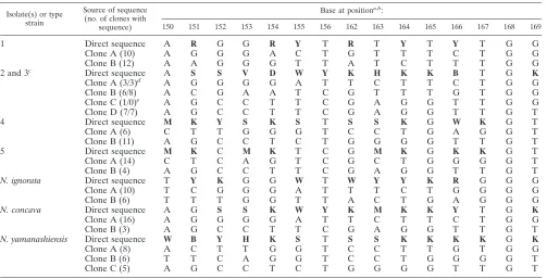
TABLE 1. Direct sequences of the variable region of the 16S rRNA genes, and sequences of the variable region of clones of patient isolates and the type strains ofN. ignorata,N. concava, andN. yamanashiensis
Isolate(s) or type strain
Source of sequence (no. of clones with
sequence)
Base at positiona,b:
150 151 152 153 154 155 156 162 163 164 165 166 167 168 169
1 Direct sequence A R G G R Y T R T Y T Y T G G
Clone A (10) A G G G A C T G T T T C T G G
Clone B (12) A A G G G T T A T C T T T G G
2 and 3c Direct sequence A S S V D W Y K H K K B T G K
Clone A (3/3)d A G G G G A T T C T T C T G G
Clone B (6/8) A C G A A T C G T T T G T G G
Clone C (1/0)e A G C C T T C G A G G T T G G
Clone D (7/7) A G C C T T C G A G G T T G T
4 Direct sequence M K Y S K S T S S K G W K G T
Clone A (6) C T T G G G T C C T G A G G T
Clone B (11) A G C C T C T G G G G T T G T
5 Direct sequence M K C M K T C G M K G K K G T
Clone A (14) C T C A G T C G C T G G G G T
Clone B (4) A G C C T T C G A G G T T G T
N. ignorata Direct sequence T Y K G G W T W Y Y K R G G G
Clone A (10) T C G G G A T T T C T G G G G
Clone B (6) T T T G G T T A C T G A G G G
N. concava Direct sequence A G S S K W Y K M K K Y T G K
Clone A (16) A G G G G A T T C T T C T G G
Clone B (3) A G C C T T C G A G G T T G T
N. yamanashiensis Direct sequence W B Y H K S T S S K K K K G K
Clone A (8) A C T T G G T C C T T G T G G
Clone B (6) T T C A G G T C C T G G G G T
Clone C (5) A G C C T C T G G G G T T G T
a
Compared toN. asteroidesATCC 19247T
(GenBank accession no. X84850), bases at positions 157 through 161 are identical for all isolates. b
Bold letters indicate ambiguous bases. Abbreviations: R represents A or G; Y represents C or T; S represents C or G; V represents A, C or G; D represents A, G, or T; W represents A or T; K represents G or T; H represents A, C, or T; B represents C, G, or T; M represents A or C.
c
Isolated from the same patient. d
Indicates number of clones sequenced from isolates 2 and 3, respectively. e
This clone not found in isolate 3.
on May 16, 2020 by guest
http://jcm.asm.org/
the most likely matches (Table 2). By the interpretive
stan-dards for identification ofNocardiaisolates by 16S rRNA gene
sequence currently in use in our institution, all patient isolates
would be reported asNocardiaspp., as a comparison to related
species showed⬍99.8% similarity to a type strain of any
spe-cies (5).
Three of the five patient isolates (isolates 1, 4, and 5) showed various degrees of similarity to multiple different species when direct sequences of the three genes (16S rRNA, HSP, and
secA1) were analyzed (Table 2), resulting in inconclusive iden-tifications for these isolates. In no case were gene sequences most similar to a single species by all genes for these three isolates.
Isolates 2 and 3 (identical isolates recovered from the same patient on consecutive days) showed the most sequence
simi-larity to N. concava by all three genes examined (Table 2),
suggesting that these isolates may be clinical isolates of N.
concava. In addition, the sequence of one of the 16S rRNA gene clones examined from patient isolates 2 and 3 (clone A) was identical to that of a clone derived from the type strain of
N. concava(N. concavaclone A) (Table 1). Repeated
DNA-DNA hybridization experiments using isolate 2 and the N.
concava type strain gave inconclusive results. Three experi-ments in which the patient isolate was labeled showed the
isolate to be nonrelated to the N. concava type strain, with
relative binding ratios ofⱕ58.6 and divergence values ofⱕ2.9.
Three experiments in which the type strain ofN. concavawas
labeled showed the isolates to be related to theN. concavatype
strain, with relative binding ratios of 89.6, 86.6, and 85.9 and divergence values of 4.7, 3.5, and 3.4, respectively.
Isolate 4 showed a high degree of HSP gene similarity to
Nocardia seriolae(99.4%); however, sequence comparison of
the 16S rRNA andsecA1genes toN. seriolaewas inconclusive,
showing 97.0% similarity to theN. seriolaetype strain for both
genes (data not shown).
Except for isolates 2 and 3, the deduced HSP amino acid sequence was not discriminatory enough to assign a species identification to the patient isolates on the basis of that se-quence. The deduced HSP amino acid sequences for isolates 2
and 3 were identical to that of theN. concavatype strain. For
all patient isolates, the deducedSecA1 amino acid sequence
was not discriminatory enough to assign a species identification (data not shown).
Sequence analysis of clones derived from an amplified re-gion of the 16S rRNA gene showed that clones with between two and four different sequence patterns within the variable region were obtained for each of the five patient isolates and the three type strains (Table 1). These sequence patterns were determined for multiple clones from each isolate (with be-tween 3 and 14 separate clones showing identical sequences), except for one sequence pattern found in a single clone derived from isolate 2. For each isolate, the sequences of all the clones corresponded to the ambiguous bases seen on analysis of the direct sequence of this region of the 16S rRNA gene (Table 1). Except for the one clone sequence (clone A from patient isolates 2 and 3) that showed significant similarity (99.6%) to
the type strain of N. concava, BLAST analysis of all other
cloned 16S rRNA genes showedⱕ98.8% sequence similarity
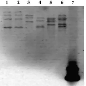
to the sequence of the closest type strain (data not shown). Hybridization of the digested genomic DNA with a 16S rRNA gene-specific probe verified the presence of multiple copies of the 16S rRNA gene in each of the patient isolates and
in the type strain ofN. ignorata. Patient isolates 1 and 5 showed
at least three copies, isolates 2, 3, and 4 showed at least four
copies, and the N. ignorata type strain showed at least five
copies (Fig. 1).
DISCUSSION
The use of the 16S rRNA gene sequence has become the
“gold standard” for the identification ofNocardiaspecies due
to the inadequacy of traditional biochemicals for the discrim-ination of an increasing number of clinically significant species in this genus. The presence of ambiguous bases in the direct
16S rRNA gene sequence of someNocardiaisolates has been
noted (8, 15) and has been shown to be due to the presence of multiple copies of the 16S rRNA gene, at least for some
iso-lates of N. nova (8). Data presented here show that some
strains ofNocardiamay possess up to five copies of the 16S
rRNA gene, and cloning studies show that significant base pair substitutions exist within the variable regions of these copies. A determination of the transcriptional activity of these genes was not addressed in this study.
[image:3.585.43.283.91.198.2]The variable region examined is near the 5⬘terminus of the
[image:3.585.351.496.540.685.2]FIG. 1. DNA hybridization of SphI digests of genomic DNA with a probe complementary to an 89-bp region of the 16S rRNA gene. Lane 1, isolate 2; lane 2, isolate 3; lane 3, isolate 5; lane 4, isolate 1; lane 5, isolate 4; lane 6,N. ignorataDSM 44496T; lane 7, positive control.
TABLE 2. Most closely related type strain to patient isolates by gene sequence of direct 16S rRNA, HSP, andsecA1
Patient isolate(s)
Most closely related type strain (% similarity) by sequence of:
Direct 16S rRNAa
HSPb
secA1c
1 Nocardia africana
(97.8)
Nocardia niigatensis
(96.6)
N. niigatensis
(95.9) 2 and 3 N. concava(99.2) N. concava(97.5) N. concava
(98.9) 4 N. yamanashiensis
(97.8)
N. seriolae(99.4) N. concava
(98.2) 5 N. seriolae(97.8) N. seriolae(97.2) N. niigatensis
(97.3)
aA total of 1,299 bases were analyzed.
bA total of 356 bases were analyzed.
cA total of 440 bases were analyzed.
on May 16, 2020 by guest
http://jcm.asm.org/
16S rRNA gene (corresponding to bases 150 to 169 of the
sequence ofN. asteroidesATCC 19247T; GenBank accession
number X84850) and has been shown to be specific for
numer-ous species ofNocardia(7). Alignment of this region of the 16S
rRNA gene sequence of the type or reference strains of 39 species or taxa shows unique sequences within this region for 24 species or taxa (data not shown). It is in part the variability of this region that allows identification of some species by partial sequencing of the 16S rRNA gene using the MicroSeq 500 system (4, 15).
Hybridization studies with a 16S rRNA gene-specific probe verified the presence of multiple 16S rRNA genes in all of the
patient isolates and in the type strain ofN. ignorataand, in
most cases, demonstrated the presence of more 16S rRNA gene copies than the number of different sequences derived from clones suggested. It is possible that two or more gene copies share the same sequence or that an insufficient number of clones was evaluated to detect all sequence variations present.
We are confident that the data presented here represent multiple 16S rRNA gene copies, as all molecular work was performed using subcultures from a single colony. In addition, for seven of the eight isolates, multiple clones with identical sequences were obtained for all sequence patterns. Cloning experiments using isolates 2 and 3 showed multiple clones with three different patterns and a fourth pattern that was seen in only one clone from isolate 2. Except for the single clone found in isolate 2, the presence of multiple clones with the same sequence indicates that the sequence differences noted in the clones are not merely the result of transcription errors that occurred during PCR.
We do not think that any of the patient isolates examined in this study can be assigned to any currently recognized species because, in most cases, none of the multiple 16S rRNA gene sequences obtained from the clones is sufficiently similar to the sequences of any currently described species. In addition, for patient isolates 1, 4, and 5, an analysis of both the HSP gene
and thesecA1gene sequences gave inconclusive identifications
or gave identifications which were not in agreement with each other or with the identification obtained for the 16S rRNA gene sequence (Table 2). Although all genes examined for isolates 2 and 3 indicated a high degree of similarity to the type
strain ofN. concavaand one 16S rRNA gene clone was
iden-tical to a clone from N. concava, DNA-DNA hybridization
studies failed to show conclusive evidence of species identity.
This may be due, in part, to the fact that the type strain ofN.
concava also possesses multiple different copies of the 16S rRNA gene.
In our hands, direct sequence of the type strain ofN.
igno-rata showed numerous ambiguous bases within the variable
region that are not present in the original sequence deposited in GenBank (21); some of these ambiguous bases correspond to deleted bases in that original sequence. Cloning experiments reported here show that these base ambiguities are the result of at least two different sequences among the multiple 16S rRNA genes present. Analysis of the entire sequences
ob-tained from the clones ofN. ignorataDNA revealed additional
base ambiguities outside of the variable region (data not shown). These additional base ambiguities may indicate that more than two different 16S rRNA gene sequence patterns are
present in theN. ignoratatype strain. Hybridization using a 16S
rRNA-specific probe with the genomic DNA of theN. ignorata
type strain verified the presence of at least five copies of the 16S rRNA gene in this organism. Because only two different gene sequences were detected in the clone analysis, it is pre-sumed that at least some gene copies possess the same se-quence.
Recently, several clinical isolates of N. ignorata have been
reported by Rodrı´guez-Nava et al. (17), some of which appear to be clinically significant. 16S rRNA gene sequences of these isolates deposited in GenBank have sequences identical to each other in their 16S rRNA gene variable regions; these variable region sequences differ by one base from the sequence of the type strain submitted by the same authors. All bases
within the variable regions of the reportedN. ignoratapatient
isolates correspond either to the unambiguous bases in the
variable region of the direct sequence of theN. ignoratatype
strain sequenced in this study or to one of the two base pos-sibilities for the ambiguous bases. While it is clear that the type strain of the species contains multiple copies of the 16S rRNA gene and that these copies have different sequences within the variable region, it is not known whether the patient isolates also possess multiple different copies.
Careful analysis of the 16S rRNA gene sequence of two
additionalNocardiatype strains,N. concavaandN.
yamanash-iensis, revealed the presence of ambiguous bases within the variable regions of this gene which could not be resolved with repeat testing. Clones derived from the amplified 16S rRNA
gene of theN.concavatype strain showed two different gene
sequences within this region. Two clinical isolates ofN.
con-cavahave been reported; both were isolated from patients with
cutaneous nocardiosis (12). One of these isolates is the type strain of the species. According to the sequences of these isolates submitted to GenBank, six base differences exist be-tween the two isolates within the 19-base variable region. All of
the bases within the variable regions of these reported N.
concava clinical isolates that differ between the two strains occur at sites of ambiguous bases in our direct sequence of the
N. concavatype strain.
Clones derived from the amplified 16S rRNA gene of theN.
yamanashiensis type strain showed three different gene se-quences within the variable region. Only a single clinical isolate
(from a skin abscess) ofN. yamanashiensishas been reported
(11); this isolate is the type strain of the species. In this study,
direct sequencing of the 16S rRNA gene of theN.
yamanash-iensis type strain showed 12 ambiguous bases within the 19-base variable region; the 19-bases in the variable region of the
initially reportedN. yamanashiensistype strain correspond to
one of the two or three base possibilities of the ambiguous bases determined in this study.
The presence of multiple different 16S rRNA genes in at
least someNocardiatype strains is problematic, as it is unclear
whether the 16S rRNA gene(s) of any other isolate would be sufficiently similar to call the strains conspecific. Cloning stud-ies might resolve this issue but are not practical in most cir-cumstances. Results of the DNA-DNA hybridization per-formed in this study suggest that even isolates that have high degrees of gene similarity to such a type strain cannot be unambiguously identified as that species.
In our experience, the presence of multiple differing copies
on May 16, 2020 by guest
http://jcm.asm.org/
of the 16S rRNA gene sequence appears to be uncommon in patient isolates; careful analysis of the sequences of isolates that do not correspond to those of any described type strain may show the presence of numerous ambiguous bases and lead to the suspicion that multiple differing copies exist. Sequences that show single or very few ambiguous bases may be similar enough to a type strain to allow high levels of sequence simi-larity; only extensive sequencing and cloning could reveal whether these ambiguous bases are the result of multiple dif-fering copies or of sequencing errors.
Detection of ambiguous bases (and, thus, isolates that may possess differing multiple gene copies) requires careful exam-ination of the sequence chromatograms. Ambiguous bases ap-pear as overlapping peaks that are not resolved with repeat
testing. ForNocardia isolates, these base substitutions occur
most frequently within the variable regions of the gene, so these regions should be routinely scrutinized by hand, as se-quencing software may record only the largest peak at a spe-cific locus on the chromatogram. In any case, bases flagged by sequence software as “ambiguous” should be carefully exam-ined.
In our opinion, sequencing of the 16S rRNA gene still rep-resents a reasonable method for the identification of many
species ofNocardiaas long as careful analysis of sequence data
reveals no irresolvable ambiguous bases. The value of this gene results from the extensive amount of sequence information for
all knownNocardiaspecies in sequence databases. The
useful-ness of alternative targets would depend not only on the dis-criminative power of those targets but also on the ability to obtain and sequence the type strains of all newly described species and the deposition of the sequences of these alternative targets in the sequence databases. In our laboratory, we
rou-tinely sequence thesecA1gene if 16S gene sequence analysis of
a clinical isolate shows less than 99.8% similarity to that of a type strain. Using these two genes, we have thus far been able to discriminate among all described species.
The Pandora’s box of complexity and variation in organisms
in the genusNocardia that these isolates illustrate highlights
several more general problems which need to be addressed. (i) The presence of ambiguous bases in 16S rRNA gene sequences which cannot be resolved by repeat testing should not be dis-missed as the consequence of amplification or sequencing er-rors but should be taken as suggesting the presence of multiple differing copies of the 16S rRNA gene, the differing sequences of which require cloning studies for resolution. (ii) Authors describing new species should make every effort to resolve apparent sequence ambiguities in proposed type strains before publishing the sequences obtained or depositing them in gene databases. If such ambiguities are not resolved, the genomic complexity of such isolates is obscured. Furthermore, the like-lihood of the misidentification of subsequent isolates as be-longing to a species with unresolved sequence ambiguities is considerably increased, as ignoring the ambiguous bases may result in an erroneous overestimation of sequence similarity between such a type strain and subsequent isolates. (iii) Iso-lates of organisms with differing multiple gene copies of the 16S rRNA gene most probably are currently unidentifiable to the species level by studies sequencing only that gene, even if a full sequence is obtained for each differing gene. It also
would hardly be practicable to assign each Nocardia isolate
with multiple differing gene copies to its own species. (It is possible that future studies will show that many or all isolates with multiple differing gene copies have a definable range of variation of each copy within a given species, but this seems highly improbable at present.) (iv) Even when utilizing DNA-DNA hybridization (which is beyond the capabilities of nearly all diagnostic laboratories), it may be impossible for some
Nocardia species isolates to be identified accurately to the species level. This problem is exemplified by the fact that we failed to obtain congruent degrees of DNA-DNA hybridization with some of our isolates and an apparently similar type strain, depending upon which organism DNA was labeled. (v) From our point of view, major problems lie with the current bacterial species concept and these problems are not restricted to the
genusNocardia. We argue that there currently exists no
oper-ationally useful such concept, and we think the concept needs careful reevaluation to stem the proliferation of newly de-scribed bacterial species, among which no significant biological differences and, particularly, no clinically significant differ-ences may have been demonstrated. We think that a more useful concept needs to be developed by a consensus mecha-nism and also needed is some type of enforcement process so that the criteria determined will be adhered to rigidly. Perhaps the standards provided by the Clinical and Laboratory Stan-dards Institute could constitute a useful model. The procedure to be employed needs to be carefully specified, with appropri-ate quality control measures identified and appropriappropri-ate statis-tical measures selected (for example, acceptable ranges of variability in DNA-DNA hybridization results).
Without practicable criteria for the delineation of new spe-cies and the identification of clinical isolates, there will be continuing misidentification of patient isolates and continuing inability to associate clinically important data (such as anti-microbial susceptibility, geographic distribution, and relative pathogenicity) with particular species categories.
ACKNOWLEDGMENTS
We thank Patrick R. Murray, Department of Laboratory Medicine, Warren G. Magnuson Clinical Center, NIH, for critically reviewing the manuscript.
The views expressed here are those of the authors and should not be construed as those of the U.S. Department of Health and Human Services.
This research was supported by the Intramural Research Program of the NIH, Warren G. Magnuson Clinical Center.
REFERENCES
1.Brenner, D. J., F. W. Hickman-Brenner, J. V. Lee, A. G. Steigerwalt, G. R. Fanning, D. G. Hollis, J. J. Farmer III, R. E. Weaver, S. W. Joseph, and R. Seidler. 1983.Vibrio furnissii (formerly aerogenic biogroup ofVibrio fluvialis), a new species isolated from human feces and the environment. J. Clin. Microbiol.18:816–824.
2.Brenner, D. J., A. C. McWhorter, J. K. Leete Knutson, and A. G. Steigerwalt.
1982.Escherichia vulneris: a new species of Enterobacteriaceaeassociated with human wounds. J. Clin. Microbiol.15:1133–1140.
3.Cilia, V., B. Lafay, and R. Christen.1996. Sequence heterogeneities among 16S ribosomal RNA sequences, and their effect on phylogenetic analyses at the species level. Mol. Biol. Evol.13:451–461.
4.Cloud, J. L., P. S. Conville, A. Croft, D. Harmsen, F. G. Witebsky, and K. C. Carroll.2004. Evaluation of partial 16S ribosomal DNA sequencing for identification ofNocardiaspecies by using the MicroSeq 500 system with an expanded database. J. Clin. Microbiol.42:578–584.
5.Conville, P. S., J. M. Brown, A. G. Steigerwalt, J. W. Lee, V. L. Anderson, J. T. Fishbain, S. M. Holland, and F. G. Witebsky.2004.Nocardia kruczakaie
sp. nov., a pathogen in immunocompromised patients and a member of the “N. novacomplex.” J. Clin. Microbiol.42:5139–5145.
on May 16, 2020 by guest
http://jcm.asm.org/
6.Conville, P. S., J. M. Brown, A. G. Steigerwalt, J. W. Lee, D. E. Byrer, V. L. Anderson, S. E. Dorman, S. M. Holland, B. Cahill, K. C. Carroll, and F. G. Witebsky.2003.Nocardia veteranaas a pathogen in North American pa-tients. J. Clin. Microbiol.41:2560–2568.
7.Conville, P. S., S. H. Fischer, C. P. Cartwright, and F. G. Witebsky.2000. Identification ofNocardiaspecies by restriction endonuclease analysis of an amplified portion of the 16S rRNA gene. J. Clin. Microbiol.38:158–164. 8.Conville, P. S., and F. G. Witebsky.2005. Multiple copies of the 16S rRNA
gene inNocardia novaisolates and implications for sequence-based identi-fication procedures. J. Clin. Microbiol.43:2881–2885.
9.Conville, P. S., A. M. Zelazny, and F. G. Witebsky.2006. Analysis ofsecA1
gene sequences for identification ofNocardia species. J. Clin. Microbiol.
44:2760–2766.
10.Ishikawa, J., A. Yamashita, Y. Mikami, Y. Hoshino, H. Kurita, K. Hotta, T. Shiba, and M. Hattori.2004. The complete genomic sequence ofNocardia farcinicaIFM 10152. Proc. Natl. Acad. Sci. USA101:14925–14930. 11.Kageyama, A., K. Yazawa, K. Nishimura, and Y. Mikami.2004.Nocardia
inohanensissp. nov.,Nocardia yamanashiensissp. nov. andNocardia niigat-ensissp. nov., isolated from clinical specimens. Int. J. Syst. Evol. Microbiol.
54:563–569.
12.Kageyama, A., K. Yazawa, H. Taniguchi, H. Chibana, K. Nishimura, R. M. Kroppenstedt, and Y. Mikami.2005.Nocardia concavasp. nov., isolated from Japanese patients. Int. J. Syst. Evol. Microbiol.55:2081–2083. 13.Loeffelholz, M. J., and D. R. Scholl.1989. Method for improved extraction of
DNA fromNocardia asteroides. J. Clin. Microbiol.27:1880–1881. 14.Ninet, B., M. Monod, S. Emler, J. Pawlowski, C. Metral, P. Rohner, R.
Auckenthaler, and B. Hirschel.1996. Two different 16S rRNA genes in a mycobacterial strain. J. Clin. Microbiol.34:2531–2536.
15.Patel, J. B., R. J. Wallace, Jr., B. A. Brown-Elliott, T. Taylor, C. Imperatrice, D. G. B. Leonard, R. W. Wilson, L. Mann, K. C. Jost, and I. Nachamkin.
2004. Sequence-based identification of aerobic actinomycetes. J. Clin. Mi-crobiol.42:2530–2540.
16.Reischl, U., K. Feldmann, L. Naumann, B. J. M. Gaugler, B. Ninet, B. Hirschel, and S. Emler.1998. 16S rRNA sequence diversity in Mycobacte-rium celatumstrains caused by presence of two different copies of 16S rRNA gene. J. Clin. Microbiol.36:1761–1764.
17.Rodrı´guez-Nava, V., A. Couble, Z. U. Khan, M. Pe´rouse de Montclos, L. Brasme, C. Villuendas, C. Molinard, P. Boiron, and F. Laurent.2005. No-cardia ignorata, a new agent of human nocardiosis isolated from respiratory specimens in Europe and soil samples from Kuwait. J. Clin. Microbiol.
43:6167–6170.
18.Steingrube, V. A., R. W. Wilson, B. A. Brown, K. C. Jost, Jr., Z. Blacklock, J. L. Gibson, and R. J. Wallace, Jr.1997. Rapid identification of clinically significant species and taxa of aerobic actinomycetes, includingActinomadura,Gordona,
Nocardia,Rhodococcus,Streptomyces, andTsukamurellaisolates, by DNA am-plification and restriction endonuclease analysis. J. Clin. Microbiol.35:817–822. 19.Telenti, A., F. Marchesi, M. Balz, F. Bally, E. C. Bo¨ttger, and T. Bodmer.
1993. Rapid identification of mycobacteria to the species level by polymerase chain reaction and restriction enzyme analysis. J. Clin. Microbiol.31:175– 178.
20.Wang, Y., Z. Zhang, and N. Ramanan.1997. The actinomycete Thermo-bispora Thermo-bisporacontains two distinct types of transcriptionally active 16S rRNA genes. J. Bacteriol.179:3270–3276.
21.Yassin, A. F., F. A. Rainey, and U. Steiner.2001.Nocardia ignoratasp. nov. Int. J. Syst. Evol. Microbiol.51:2127–2131.