0095-1137/08/$08.00
⫹
0
doi:10.1128/JCM.00157-08
Copyright © 2008, American Society for Microbiology. All Rights Reserved.
Evaluation of Matrix-Assisted Laser Desorption Ionization–Time-of-Flight
Mass Spectrometry in Comparison to 16S rRNA Gene Sequencing for
Species Identification of Nonfermenting Bacteria
䌤
A. Mellmann,
1* J. Cloud,
2T. Maier,
3U. Keckevoet,
1I. Ramminger,
1P. Iwen,
4J. Dunn,
5G. Hall,
6D. Wilson,
6P. LaSala,
7M. Kostrzewa,
3and D. Harmsen
8Institute for Hygiene, University Hospital Muenster, Muenster D-48149, Germany
1; ARUP Institute for Clinical and
Experimental Pathology, Salt Lake City, Utah
2; Bruker Daltonik GmbH, Leipzig, Germany
3; Department of Pathology and
Microbiology, University of Nebraska Medical Center, Omaha, Nebraska
4; Cook Children’s Medical Center,
Fort Worth, Texas
5; Microbiology, Cleveland Clinic Foundation, Cleveland, Ohio
6; Clinical Microbiology,
Department of Pathology, University of Texas Medical Branch, Galveston, Texas
7; and Department for
Periodontology, University Hospital Muenster, Muenster D-48149, Germany
8Received 25 January 2008/Returned for modification 21 March 2008/Accepted 1 April 2008
Nonfermenting bacteria are ubiquitous environmental opportunists that cause infections in humans,
especially compromised patients. Due to their limited biochemical reactivity and different morphotypes,
misidentification by classical phenotypic means occurs frequently. Therefore, we evaluated the use of
matrix-assisted laser desorption ionization–time-of-flight mass spectrometry (MALDI-TOF MS) for
spe-cies identification. By using 248 nonfermenting culture collection strains composed of 37 genera most
relevant to human infections, a reference database was established for MALDI-TOF MS-based species
identification according to the manufacturer’s recommendations for microflex measurement and MALDI
BioTyper software (Bruker Daltonik GmbH, Leipzig, Germany), i.e., by using a mass range of 2,000 to
20,000 Da and a new pattern-matching algorithm. To evaluate the database, 80 blind-coded clinical
nonfermenting bacterial strains were analyzed. As a reference method for species designation, partial 16S
rRNA gene sequencing was applied. By 16S rRNA gene sequencing, 57 of the 80 isolates produced a unique
species identification (
>
99% sequence similarity); 11 further isolates gave ambiguous results at this
threshold and were rated as identified to the genus level only. Ten isolates were identified to the genus level
(
>
97% similarity); and two isolates had similarity values below this threshold, were counted as not
identified, and were excluded from further analysis. MALDI-TOF MS identified 67 of the 78 isolates
(85.9%) included, in agreement with the results of the reference method; 9 were misidentified and 2 were
unidentified. The identities of 10 randomly selected strains were 100% correct when three different mass
spectrometers and four different cultivation media were used. Thus, MALDI-TOF MS-based species
identification of nonfermenting bacteria provided accurate and reproducible results within 10 min without
any substantial costs for consumables.
The genera
Pseudomonas
,
Burkholderia
,
Stenotrophomo-nas
, and others belong to the large group of nonfermenting
bacteria that are unable to ferment sugars. Nonfermenting
bacteria are ubiquitous environmental opportunists, and
some species can cause severe infections, especially in
im-munocompromised patients (30). In the group of cystic
fi-brosis patients in particular, nonfermenting bacteria are the
main causes of morbidity and mortality (18). Furthermore,
the antibiotic resistance of some nonfermenting bacterial
species often complicates therapy (25, 30). Accurate species
identification is therefore critical not only because the
prog-nosis for an infected patient differs significantly depending
on the species identified (33) but also because in some cases,
e.g., infections with
Burkholderia cepacia
genomovar III (5),
strict infection control measures must be established for
cystic fibrosis patients (5, 31).
In a routine clinical laboratory, species identification of
cul-tured isolates usually relies on phenotypic methods, such as
panels of biochemical reactions, antibiotic resistance, and fatty
acid patterns (28). However, due to their limited biochemical
reactivity and variable morphology, nonfermenters are
fre-quently misidentified by classical methods (21, 26). Moreover,
isolates from patients with chronic infections often loose their
characteristic phenotypes (12, 26). To overcome these
draw-backs, genotypic identification methods have become widely
used, and most of them are based on the polymorphism of the
16S rRNA genes. Species-specific PCRs, restriction patterns,
and more recently, partial DNA sequencing are used for
spe-cies identification. Nowadays, sequencing of the 16S rRNA
gene is accepted as the reference method for species
identifi-cation, and several studies have shown its superiority to
phe-notypic methods for the identification of various groups of
bacteria, including nonfermenting bacteria (2, 3, 8, 9, 14, 15,
27). However, a prerequisite for the retrieval of valid
identifi-cation results is the use of an extensive and comprehensive
quality-controlled database (6, 16).
Matrix-assisted laser desorption ionization–time-of-flight
mass spectrometry (MALDI-TOF MS), which can be used to
* Corresponding author. Mailing address: Institut fuer Hygiene,
Uni-versita
¨tsklinikum Muenster, Robert-Koch-Str. 41, Muenster D-48149,
Germany. Phone: 49 251 8352316. Fax: 49 251 8355688. E-mail: mellmann
@uni-muenster.de.
䌤
Published ahead of print on 9 April 2008.
1946
on May 16, 2020 by guest
http://jcm.asm.org/
analyze the protein composition of a bacterial cell, has
emerged as a new technology for species identification. By
measuring the exact sizes of peptides and small proteins, which
are assumed to be characteristic for each bacterial species, it is
possible to determine the species within a few minutes when
the analysis is started with whole cells, cell lysates, or crude
bacterial extracts (13, 17, 22). However, due to difficulties with
the reproducibility of results because of the use of different
cultivation conditions and the limited availability of reference
data sets, MALDI-TOF MS has not yet been widely used for
species identification.
In this study, we therefore established a reference database
for MALDI-TOF MS-based nonfermenter identification,
ana-lyzed the reproducibility using different cultivation conditions
and mass spectrometer instruments, and evaluated the
meth-odology with 80 blind-coded clinical nonfermenter strains that
were analyzed by partial 16S rRNA gene sequencing, which
was used as the reference method.
(This study was presented in part at the 47th Interscience
Conference on Antimicrobial Agents and Chemotherapy,
Chi-cago, IL, 17 to 20 September 2007.)
MATERIALS AND METHODS
Bacterial strains.To establish a reference database for MALDI-TOF MS-based species identification, 248 nonfermenter culture collection strains com-posed of 37 genera most relevant to human infections were used (Table 1).
The 80 clinical nonfermenting strains that were used to evaluate the MALDI-TOF MS reference database were recovered from clinical specimens received by the Cleveland Clinic Foundation (Cleveland, OH), the University of Texas Med-ical Branch (Galveston, TX), Cook Children’s MedMed-ical Center (Fort Worth, TX), and the University of Nebraska Medical Center (Omaha, NE) during the 2004 calendar year.
16S rRNA gene sequencing and sequence analysis.DNA isolation, amplifica-tion, and cycle sequencing of the clinical isolates were performed at the ARUP Institute for Clinical and Experimental Pathology (Salt Lake City, UT). DNA was extracted as described previously (27). Four microliters of the extract was used in each PCR. PCR was performed in a total volume of 40l containing 1⫻ FastStart DNA Master Plus SYBR green (Roche Diagnostics Corp., Indianap-olis, IN), 500 nM each primer 16S-27f and 16S-519r (27), and 4 mM Mg2⫹. The
thermal cycling reactions were performed with a RotorGene 3000 real-time PCR instrument (Corbett Research, Sydney, Australia) and consisted of an initial denaturation (10 min at 95°C), followed by 35 cycles of denaturation (30 s at 95°C), annealing (20 s at 55°C), and extension (30 s at 72°C) and then a single final extension (2 min at 72°C). Negative controls, which contained water instead of template DNA, were run in parallel in each run. The PCR product was purified by an enzymatic method, modified from the method of Dugan et al. (11), with exonuclease I (New England Biolabs GmbH, Frankfurt-Hoechst, Germany) and shrimp alkaline phosphatase (Amersham Pharmacia Biotech, Freiburg, Germany). Briefly, 5l of the PCR product was incubated with 1 U of each enzyme at 37°C for 30 min. The enzymes were then inactivated at 80°C for 15 min, and the PCR products were stored at 4°C. The amplicons were sequenced with an ABI Prism BigDye Terminator (version 3.0) ready reaction cycle sequencing kit (Applied Biosystems, Foster City, CA). The sequencing reaction required 0.5l of premix from the kit, 1.8l Tris-HCl– MgCl2buffer (400 mM Tris-HCl, 10 mM MgCl2), 10 pmol of the sequencing primers (which were the same as the PCR primers), and 2l of the cleaned PCR product in a total volume of 10l.
For the sequencing chemistry mixtures, the same primer used for PCR (primer 16S-27f or 16S-519r) was used to obtain forward and reverse sequence data for partial 5⬘16S rRNA gene sequencing. All sequencing reactions were performed with a standard thermocycler to complete 25 cycles of denaturation (10 s at 96°C), annealing (5 s at 53°C), and extension (4 min at 60°C). The sequencing products were purified with Centri-Sep spin columns (Princeton Separations, Adelphia, NJ), followed by preparation for analysis on an ABI Prism 310 or a 3100 Avant genetic analyzer, in accordance with the instructions of the manu-facturer (Applied Biosystems). The double-stranded sequences corresponding to
Escherichia coli16S rRNA gene positions 54 to 510 were analyzed in accordance
with the procedure described for the Ribosomal Differentiation of Medical Micro-Organisms (RIDOM) database (16).
Sample preparation for MALDI-TOF MS, spectrum generation, and data analysis.A colony of a fresh overnight culture was used for sample preparation before measurement. The material was thoroughly suspended in 300l double-distilled water, 900l ethanol was added, and the components were mixed well. Prior to shipment to the place of measurement, the samples in ethanol-water were centrifuged, the supernatant was removed, and the pellets were dried. For sample extraction, 50l of formic acid (70% in water) was added to the bacterial pellet, the components were mixed thoroughly, and 50l of acetonitrile was added. After centrifugation at 13,000⫻gfor 2 min, 1l of the supernatant containing the bacterial extract was transferred to a sample position on a ground steel MALDI target plate and allowed to dry at room temperature. Subse-quently, the sample was overlaid with 2 l of MALDI matrix (a saturated solution of␣-cyano-4-hydroxy-cinnamic acid in 50% acetonitrile–2.5% trifluoro-acetic acid) and dried again.
For database construction and validation, measurements were performed with a microflex LT (Bruker Daltonik GmbH, Leipzig, Germany) bench-top mass spectrometer equipped with a 20-Hz nitrogen laser (parameter settings: ion source 1 (IS1), 20 kV; IS2, 18.5 kV; lens, 8.5 kV; detector gain, 2,650 V; and gating, none). Spectra were recorded in the positive linear mode for the mass range of 2,000 to 20,000 Da at the maximum laser frequency. The database references (main spectra) for the newly investigated bacteria were constructed by using the automated functionality of the MALDI BioTyper (version 1.1) software package (Bruker Daltonik GmbH). Briefly, for each database entry, 20 individually measured mass spectra were imported into the software. After smoothing of the spectra, baseline correction, and peak pick-ing, the resulting peak lists were used by the program to calculate and to store a main spectrum containing the average peak mass, average peak intensity, and frequency information.
For microorganism identification, the raw spectra of the unknown bacteria were imported into the MALDI BioTyper software and analyzed by standard pattern matching (with default parameter settings) against the main spectra of 2,506 microorganisms, used as reference data, in the BioTyper database (these spectra are an integrated part of the BioTyper software). The refer-ence database consisted of the 248 newly created main spectra for the non-fermenting bacteria investigated and other clinical, veterinary, and environ-mental bacterial strains. Preparation of samples of the 248 nonfermenter reference strains was performed at the Institute for Hygiene, University Hospital Muenster (Muenster, Germany). The clinical nonfermenting strains being evaluated were prepared at the ARUP Institute for Clinical and Ex-perimental Pathology.
Preparation of the ground steel plate, MS of 96 samples, and a search of the database for similarity for species identification took about 3 h with the microflex instrument. A single sample could be identified in approximately 10 min.
MALDI-TOF MS reproducibility testing.Complementary to the already de-termined intraspecies (4, 36) and interlaboratory (37) reproducibilities of MALDI-TOF MS-based species identification, the spectra of 10 randomly cho-sen nonfermenter strains (Brevundimonas aurantiacaDSM 4731,Brevundimonas
intermediaDSM 4732,Brevundimonas andropogonisDSM 9511,Brevundimonas
caribensisDSM 13236,Flavobacterium johnsoniaeDSM 2064,Flavobacterium
mizutaiiDSM 11724,Pseudomonas aeruginosaDSM 50071,Pseudomonas beteli
LMG 978,Pseudomonas boreopolisLMG 979,Pseudomonas extremorietalisDSM 15824) were determined under different conditions. First, the testing was done by parallel measurement on three different MALDI-TOF MS instruments (the microflex LT instrument, the autoflex II TOF/TOF instrument with a 50-Hz nitrogen laser, and the ultraflex III TOF/TOF instrument with a 200-Hz smart-beam laser, all from Bruker Daltonik GmbH) to test the comparabilities of the results obtained with the different instruments. The parameter settings for the additional instruments were as follows: IS1, 20 kV; IS2, 18.7 kV; lens, 8.0 kV; detector gain, 1,756 V; and gating, maximum, 1,500 Da, for the autoflex instru-ment and IS1, 25 kV; IS2, 23.45 kV; lens, 6.0 kV; detector gain, 1,650 V; and gating, maximum, 1,500 Da, for the ultraflex instrument. Second, the influence of different cultivation conditions was characterized by cultivation of the 10 strains on four different media (Columbia blood agar, chocolate agar, Mueller-Hinton agar, and tryptic soy agar; Heipha, Eppelheim, Germany) at 30°C under aerobic conditions for 48 h. Finally, the influence of the age of the bacterial cultures was investigated by analyzing three strains (B. aurantiacaDSM 4731,B. caribensis DSM 13236,P. aeruginosaDSM 50071) that were cultivated for 48 h at 30°C on Columbia blood agar and subsequently stored for 2, 5, and 7 days at room temperature. All samples for reproducibility testing were blind coded for MALDI-TOF MS analysis.
on May 16, 2020 by guest
http://jcm.asm.org/
TABLE 1. Nonfermenter culture collection strains used to establish the reference database for MALDI-TOF MS-based species identification
Genus Straina
Achromobacter...Achromobacter denitrificansDSM 30026,A. insolitusLMG 6003,A. piechaudiiDSM 10342,A. ruhlandiiDSM 653,A. spaniosLMG 5911,
A. xylosoxidanssubsp.xylosoxidansDSM 2402
Acidovorax...Acidovorax avenaesubsp.avenaeDSM 7227,A. avenaesubsp.citrulliLMG 5376,A. defluviiDSM 12644,A. delafieldiiDSM 64,A. facilis
DSM 649,A. konjaciDSM 7481,A. temperansDSM 7270
Acinetobacter...Acinetobacter baumanniiDSM 30007,A. baumanniiLMG 994,A. baylyiDSM 14961,A. bouvetiiDSM 14964,A. calcoaceticusDSM 30006,
A. gerneriDSM 14967,A. grimontiiDSM 14968,A. haemolyticusDSM 6962,A. haemolyticusLMG 1033,A. johnsoniiDSM 6963,A.
johnsoniiLMG 10584,A. juniiDSM 6964,A. lwoffiiDSM 2403,A. lwoffiiLMG 1138,A. lwoffiiLMG 1154,A. lwoffiiLMG 1300,A.
parvusDSM 16617,A. radioresistensDSM 6976,A. radioresistensLMG 10614,A. schindleriDSM 16038,A. tandoiiDSM 14970,A.
tjernbergiaeDSM 14971,A. towneriDSM 14962,A. ursingiiDSM 16037
Alcaligenes...Alcaligenes faecalissubsp.faecalisDSM 30030,A. faecalissubsp.parafaecalisDSM 13975 Alishewanella...Alishewanella fetalisDSM 16032
Arsenophonus...Arsenophonus nasoniaeDSM 15247
Arthrobacter...Arthrobacter monumentiDSM 16405 Balneatrix...Balneatrix alpicaCIP 103589 Bergeyella...Bergeyella zoohelcumLMG 8351
Blastomonas...Blastomonas natatoriaDSM 3183,B. ursincolaDSM 9006
Brevundimonas...Brevundimonas aurantiacaDSM 4731,B. diminutaDSM 7234,B. intermediaDSM 4732,B. nasdaeDSM 14572,B. subvibrioidesDSM
4735,B. vesicularisDSM 7226
Burkholderia...Burkholderia ambifariaLMG 11351,B. andropogonisDSM 9511,B. anthinaLMG 16670,B. caledonicaLMG 19076,B. caribensisDSM
13236,B. cenocepaciaLMG 12614,B. cepaciaDSM 7288,B. cepaciaLMG 2161,B. dolosaDSM 16088,B. fungorumLMG 20227,B.
gladioliDSM 4285,B. glatheiDSM 50014,B. glumaeDSM 9512,B. multivoransLMG 14293,B. phenaziniumDSM 10684,B. phymatum
LMG 21445,B. plantariiDSM 9509,B. pyrrociniaLMG 14191,B. sacchariLMG 19450,B. stabilisLMG 14294,B. terricolaLMG 20594,
B. thailandensisDSM 13276,B. tropicaDSM 15359,B. tuberumLMG 21444,B. vietnamiensisLMG 10929,B. xenovoransLMG 21463
Chryseobacterium...Chryseobacterium joosteiLMG 18212,C. scophthalmumLMG 13028
Comamonas...Comamonas aquaticaLMG 2370,C. kerstersiiDSM 16026,C. nitrativoransDSM 13191,C. terrigenaDSM 7099,C. testosteroniDSM 50244
Delftia...Delftia acidovoransDSM 39
Elizabethkingia...Elizabethkingia meningosepticaDSM 2800,E. miricolaDSM 14571
Empedobacter...Empedobacter brevisLMG 4011
Flavobacterium...Flavobacterium flevenseDSM 1076,F. gelidilacusDSM 15343,F. hibernumDSM 12611,F. hydatisDSM 2063,F. johnsoniaeDSM 2064,F.
pectinovorumDSM 6368,F. resinovorumDSM 7478,F. saccharophilumDSM 1811
Inquilinus...Inquilinus limosusDSM 16000 Malikia...Malikia spinosaDSM 15801 Microbulbifer...Microbulbifer elongatusDSM 6810
Myroides...Myroides odoratimimusLMG 4029,M. odoratusDSM 2811
Novosphingobium...Novosphingobium aromaticivoransDSM 12444,N. rosaDSM 7285,N. subarcticumDSM 10700,N. subterraneumDSM 12447
Ochrobactrum...Ochrobactrum anthropiDSM 6882,O. gallinifaecisDSM 15295,O. grignonenseDSM 13338,O. intermediumLMG 3301,O. triticiDSM
13340
Pandoraea...Pandoraea apistaLMG 16407,P. norimbergensisDSM 11628,P. pnomenusaLMG 18817,P. pulmonicolaLMG 18106
Pannonibacter...Pannonibacter phragmitetusLMG 5414,P. phragmitetusLMG 5430
Pseudomonas...Pseudomonas abietaniphilaCIP 106708,P. aeruginosaDSM 50071,P. agariciDSM 11810,P. alcaligenesDSM 50342,P. amygdaliDSM
7298,P. anguillisepticaDSM 12111,P. antarcticaDSM 15318,P. aspleniiLMG 2137,P. aurantiacaCIP 106718,P. avellanaeDSM 11809,P. azotoformansDSM 106744,P. balearicaDSM 6083,P. beteliLMG 978,P. boreopolisLMG 979,P. brassicacearumDSM 13227,P. brenneriDSM 106646,P. caricapapayaeLMG 2152,P. cedrinaDSM 105541,P. chloritidismutansDSM 13592,P. chlororaphis DSM 50083,P. cichoriiDSM 50259,P. citronellolisDSM 50332,P. congelansDSM 14939,P. corrugataDSM 7228,P. extremorietalis DSM 15824,P. flavescensDSM 12071,P. fluorescensDSM 50090,P. fragiDSM 3456,P. frederiksbergensisDSM 13022,P. fulvaLMG 11722,P. fuscovaginaeDSM 7231,P. geniculataLMG 2195,P. gessardiiCIP 105469,P. graminisDSM 11363,P. grimontiiDSM 106645,
P. hibiscicolaLMG 980,P. huttiensisDSM 10281,P. indicaDSM 14015,P. jesseniiCIP 105274,P. jinjuensisLMG 21316,P. kilonensis
DSM 13647,P. koreensisLMG 21318,P. libanensisCIP 105460,P. lundensisDSM 6252,P. luteaLMG 21974,P. luteolaDSM 6975,P.
mandeliiCIP 105273,P. marginalisDSM 13124,P. mendocinaDSM 50017,P. mephiticaCIP 106720,P. migulaeCIP 105470,P.
monteiliiDSM 14164,P. mosseliiCIP 105259,P. mucidolensLMG 2223,P. multiresinivoransLMG 20221,P. nitroreducensDSM 14399,
P. oleovoransDSM 1045,P. orientalisCIP 105540,P. oryzihabitansDSM 6835,P. pertucinogenaLMG 1874,P. pictorumLMG 981,P.
plecoglossicidaDSM 15088,P. poaeDSM 14936,P. proteolyticaDSM 15321,P. pseudoalcaligenesDSM 50188,P. putidaDSM 291,P.
putidaDSM 50198,P. resinovoransLMG 2274,P. rhizosphaeraeLMG 21640,P. rhodesiaeDSM 14020,P. savastanoiLMG 2209,P.
savastanoisubsp.savastanoiLMG 5011,P. stramineaCIP 106745,P. stutzeriDSM 5190,P. synxanthaLMG 2190,P. syringaeDSM 6693,
P. syringaesubsp.syringaeLMG 1247,P. taetrolensLMG 2336,P. thermotoleransDSMZ14292,P. thivervalensisDSM 13194,P. tolaasii
LMG 2342,P. trivialisDSM 14937,P. umsongensisLMG 21317,P. vancouverensisCIP 106707,P. veroniiDSM 11331,P. viridiflava DSM 11124
Ralstonia...Ralstonia eutrophaDSMZ531,R. mannitolilyticaLMG 6866,R. pickettiiDSM 6297,R. syzygiiDSM 7385
Rhizobium...Rhizobium radiobacterDSM 30147,R. rubiDSM 6772,R. tropiciDSM 11418
Shewanella...Shewanella algaeDSMZ9167,S. balticaDSM 9439,S. fidelisLMG 20552,S. frigidimarinaDSM 12253,S. profundaDSM 15900,S.
putrefaciensDSM 6067
Sphingobacterium...Sphingobacterium faeciumDSM 11690,S. mizutaiiDSM 11724,S. multivorumDSM 11691,S. spiritivorumDSM 11722,S. thalpophilum
DSM 11723
Sphingobium...Sphingobium chlorophenolicumDSM 7098,S. herbicidovoransDSM 11019,S. xenophagumDSM 6383
Sphingomonas...Sphingomonas adhaesivaDSM 7418,S. aerolataDSM 14746,S. aquatilisDSM 15581,S. aurantiacaDSM 14748,S. cloacaeDSM 14926,S.
faeniDSM 14747,S. koreensisDSM 15582,S. melonisDSM 14444,S. parapaucimobilisDSM 7463,S. paucimobilisDSM 1098,S.
pituitosaDSM 13101,S. trueperiDSM 7225,S. wittichiiDSM 6014,S. yabuuchiaeDSM 14562
Sphingopyxis...Sphingopyxis macrogoltabidaDSM 8826,S. terraeDSM 8831
Stenotrophomonas...Stenotrophomonas acidaminiphilaDSM 13117,S.africanaCIP 104854,S. maltophiliaDSM 50170,S. nitritireducensDSM 12575,S.
rhizophilaDSM 14405
Terrimonas...Terrimonas ferrugineaDSM 30193 Weeksella...Weeksella virosaLMG 12995 Wolinella...Wolinella succinogenesDSM 1740
a
A total of 248 nonfermenter culture collection strains were used. Abbreviations: CIP, Collection de l’Institut Pasteur, Paris, France; DSM, Deutsche Sammlung von Mikroorganismen und Zellkulturen, Braunschweig, Germany; LMG, culture collection of the Laboratorium voor Microbiologie, Universiteit Ghent, Ghent, Belgium.
on May 16, 2020 by guest
http://jcm.asm.org/
Evaluation of MALDI-TOF MS-based species identification by use of clinical isolates.To evaluate the MALDI-TOF MS reference database, 80 blind-coded clinical nonfermenters were analyzed. As the reference method for species des-ignation, partial 16S rRNA gene sequencing was used. The reference partial 16S rRNA gene sequence database (E. coli16S rRNA gene positions 54 to 510) included all 248 nonfermenter culture collection strains used as a reference for MALDI-TOF MS (Table 1). Sequence data were stored and analyzed by using the RIDOM framework (27). Sequence similarity ofⱖ99% was used for iden-tification to the species level, and sequence similarity ofⱖ97% was used for identification to the genus level (3). Further differentiation to the species level was made between unique and ambiguous similarity search results (multiple top-scoring results); the latter were rated as identification to the genus level only. Sequence similarities below 97% were rated as not identifiable. MALDI-TOF MS results based on the log(score) values calculated by the BioTyper software were compared to the 16S rRNA gene sequence similarity search results. Bio-Typer software requires log(score) values ofⱖ2.0 for identification to the species level and values of between⬍2 andⱖ1.7 for identification to the genus level. Results based on log(score) values of⬍1.7 were rated as not identifiable by the software. These BioTyper thresholds were empirically determined on the basis of information in an in-house database with data for more than 2,800 bacterial strains that were either culture collection strains or well-characterized clinical strains.
To determine the discriminatory ability of 16S rRNA gene sequencing and MALDI-TOF MS within certain groups of strains, the pairwise distances were calculated and displayed in a tree created by the unweighted pair group method with arithmetic averaging by using MEGA software (version 4.0) (37).
RESULTS
The cell extracts from all 248 culture collection strains,
which represented the majority of all clinically relevant
non-fermenter species, gave sufficient spectra and were included
in the MALDI-TOF MS reference database (database
ver-sion 1.0).
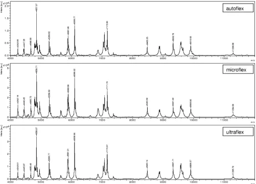
[image:4.585.42.543.68.429.2]To investigate the reproducibility of the spectra generated
with different mass spectrometers, 10 culture collection strains
were randomly chosen to be analyzed with three different mass
spectrometers. For all 10 strains, the log(score) results gave the
same species identification result irrespective of the mass
spec-trometer used, with all log(score) values being above 2.0.
Fig-ure 1 shows representative results for strain Galv12 measFig-ured
with the three different spectrometers. To determine the
in-fluence of different cultivation media on the quality of the
spectra, the 10 cultured strains were also analyzed after
culti-vation on four different media. Furthermore, three strains
were analyzed after up to 7 days storage on Columbia blood
agar at room temperature. In all cases, MS resulted in
identi-cal, correct identification results relative to those in the
refer-ence database.
FIG. 1. Comparison of mass spectra for one exemplary nonfermenting strain (strain Galv12) generated on three different MALDI-TOF MS
instruments (autoflex, microflex, ultraflex). Exemplary masses (in daltons) are depicted. Intens. [a.u.], intensity (in arbitrary units).
on May 16, 2020 by guest
http://jcm.asm.org/
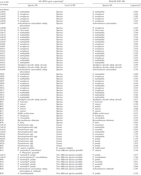
TABLE 2. Identification results for the 80 clinical nonfermenter isolates obtained by MALDI-TOF MS in comparison to those obtained by
partial 16S rRNA gene sequence-based species identification
aLevel of ID and isolate
16S rRNA gene sequencingb
MALDI-TOF MS
Species ID Level of IDc
Species ID Log(score)d
Concordance
Galv01 S. maltophilia Species S. maltophilia 2.481
Galv02 P. aeruginosa Species P. aeruginosa 2.418
Galv03 P. aeruginosa Species P. aeruginosa 2.505
Galv04 P. aeruginosa Species P. aeruginosa 2.477
Galv05 P. aeruginosa Species P. aeruginosa 2.565
Galv07 Achromobacter xylosoxidanssubsp. xylosoxidans
Species Achromobacter xylosoxidans 2.415
Galv08 P. aeruginosa Species P. aeruginosa 2.445
Galv10 P. oleovorans Species P. oleovorans 2.188
Galv12 S. maltophilia Species S. maltophilia 2.364
Galv14 S. maltophilia Species S. maltophilia 2.422
Galv20 S. maltophilia Species S. maltophilia 2.516
Galv24 S. maltophilia Species S. maltophilia 2.438
Galv25 S. maltophilia Species S. maltophilia 2.335
Neb06 P. aeruginosa Species P. aeruginosa 2.502
Neb07 P. aeruginosa Species P. aeruginosa 2.510
Neb15 P. aeruginosa Species P. aeruginosa 2.434
Neb16 P. aeruginosa Species P. aeruginosa 2.516
Neb17 S. maltophilia Species S. maltophilia 2.328
Neb22 S. maltophilia Species S. maltophilia 2.501
Neb23 P. aeruginosa Species P. aeruginosa 2.456
Neb27 S. maltophilia Species S. maltophilia 2.494
Neb32 S. maltophilia Species S. maltophilia 2.418
Neb33 Alcaligenes faecalissubsp.faecalis Species Alcaligenes faecalissubsp.faecalis 2.299 Neb34 Alcaligenes faecalissubsp.faecalis Species Alcaligenes faecalissubsp.faecalis 2.373 Neb40 Achromobacter xylosoxidanssubsp.
xylosoxidans
Species Achromobacter xylosoxidans 2.328
NF02 S. maltophilia Species S. maltophilia 2.489
NF03 P. aeruginosa Species P. aeruginosa 2.587
NF05 S. maltophilia Species S. maltophilia 2.529
NF07 P. aeruginosa Species P. aeruginosa 2.318
NF09 P. aeruginosa Species P. aeruginosa 2.444
NF11 S. maltophilia Species S. maltophilia 2.296
NF16 P. aeruginosa Species P. aeruginosa 2.545
NF17 P. aeruginosa Species P. aeruginosa 2.524
NF19 S. maltophilia Species S. maltophilia 2.519
NF21 S. maltophilia Species S. maltophilia 2.386
NF23 S. maltophilia Species S. maltophilia 2.450
NF24 Alcaligenes faecalissubsp.faecalis Species Alcaligenes faecalissubsp.faecalis 2.417
W01 P. koreensis Species P. koreensis 2.300
W05 P. stutzeri Species P. stutzeri 2.198
W07 P. putida Species P. putida 2.014
W09 P. stutzeri Species P. stutzeri 2.460
W11 P. stutzeri Species P. stutzeri 2.372
W13 Delftia acidovorans Species Delftia acidovorans 2.253
W17 P. aeruginosa Species P. aeruginosa 2.613
W19 P. citronellolis Species P. citronellolis 2.434
W20 Brevundimonas diminuta Species Brevundimonas diminuta 2.292
W30 P. fulva Species P. fulva 2.024
Galv13 Pseudomonasspp. Genus P. savastanoi 1.782
Galv19 Stenotrophomonasspp. Genus S. maltophilia 2.303
Galv21 Pseudomonasspp. Genus P. monteilii 2.034
Galv22 Stenotrophomonasspp. Genus S. maltophilia 2.350
Neb26 Pseudomonasspp. Genus P. savastanoi 1.797
Neb28 Stenotrophomonasspp. Genus S. maltophilia 1.792
Neb36 Pseudomonasspp. Genus P. putida 2.149
NF04 Pseudomonasspp. Genus P. putida 1.832
NF10 Pseudomonasspp. Genus P. putida 1.852
W02 Pseudomonasspp. Genus P. putida 2.446
W35 B. cepaciacomplex B. cepaciacomplex B. multivorans 2.379
W12 P. synxantha/P.mucidolens/
P.libanesis/P.gessardii
Four different species possible P. tolaasii 2.148
Galv17 P. monteilii/P.putida Two different species possible P. putida 2.477
Galv23 P. psychrotolerans/P.oryzohabitans Two different species possible P. oryzihabitans 1.704
W06 P. monteilii/P.putida Two different species possible P. putida 2.127
W10 P. monteilii/P.putida Two different species possible P. monteilii 2.011
W14 P. monteilii/P.putida Two different species possible P. monteilii 1.941
W23 P. monteilii/P.putida Two different species possible P. putida 2.048
W26 Achromobacter xylosoxidanssubsp.
xylosoxidans/A.ruhlandii
Two different species possible Achromobacter ruhlandii 2.395
W29 P. monteilii/putida Two different species possible P. putida 2.124
Continued on facing page
on May 16, 2020 by guest
http://jcm.asm.org/
All 80 nonfermenting strains from the clinical specimens
gave spectra sufficient for species identification. In parallel
with MALDI-TOF MS identification, all strains were analyzed
by partial 16S rRNA gene sequencing, which was used as the
reference method. By sequence comparison analysis, 57
iso-lates were unambiguously identified to the species level (
ⱖ
99%
sequence similarity) and 10 isolates were unambiguously
iden-tified to the genus level (
ⱖ
97% sequence similarity) by partial
16S rRNA gene sequencing and a search for sequence
simi-larity against the sequences in the RIDOM database. Eleven
isolates (isolates W35, W12, NF18, W29, W23, W26, W14,
W10, W06, Galv17, and Galv23) gave multiple species as
re-sults, and these were therefore treated as identified to the
genus level only. The remaining two isolates (isolates NF08
and Neb38) had results below the sequence threshold of 97%
similarity and were therefore excluded from further analysis.
The identification results obtained by MALDI-TOF MS and
16S rRNA gene sequencing, used as the reference method, are
shown in Table 2, together with the achievable levels of
iden-tification. In total, MALDI-TOF MS identified 67 of the 78
isolates (85.9%), concordant with the sequencing results; of
these, 47 of 57 (82.5%) were identified to the species level
[log(score),
ⱖ
2.0] and 20 of 21 (95.2%) were identified to the
genus level [log(score), between
⬍
2.0 and
ⱖ
1.7] (Table 3). Of
the remaining 11 isolates, 4 isolates (isolates Neb14, Neb20,
W15, and W18) had log(score) values of
ⱖ
2.0 and were
cor-rectly identified to the genus level but had discordant species
designations in comparison to the results of the reference
method. For four isolates (isolates W03, W08, W22, and W28),
the correct genus was determined by the MALDI BioTyper
software, with log(score) values of between 1.7 and 2.0,
whereas 16S rRNA gene sequencing gave a species
identifica-tion. For isolate NF18, the genus determination result was
discordant with the 16S rRNA gene sequencing result. Finally,
the two isolates with log(score) values of
⬍
1.7 (isolates Neb37
and W16) were rated as nonidentifiable by MALDI-TOF MS
(Table 3).
To investigate the discriminatory ability of closely related
species by MALDI-TOF MS in comparison to that by 16S
rRNA gene sequencing, nine species of the former
Burkhold-eria cepacia
complex were investigated in detail. By partial 16S
rRNA gene sequencing, three strains,
B. cepacia
DSM 7288 (
B.
cepacia
type strain),
B. cepacia
LMG 2161 (the former
refer-ence strain for
B. cepacia
complex genomovar I), and
Burk-holderia vietnamiensis
LMG 10929 (formerly genomovar V),
were indistinguishable (Fig. 2A). Likewise, three other strains,
[image:6.585.44.541.79.275.2]Burkholderia stabilis
LMG 14294 (formerly genomovar IV),
TABLE 2—
Continued
Level of ID and isolate
16S rRNA gene sequencingb MALDI-TOF MS
Species ID Level of IDc Species ID Log(score)d
Discrepant
Neb14 Elizabethkingia meningoseptica Species Elizabethkingia miricola 2.159
Neb20 Sphingomonas sanguinis Species Sphingomonas paucimobilis 2.182
W03 P. plecoglossicida Species P. putida 1.873
W08 Achromobacter spanius Species Achromobacter denitrificans 1.845
W15 P. putida Species P. fluorescens 2.142
W18 Achromobacter xylosoxidanssubsp.
xylosoxidans
Species Achromobacter ruhlandii 2.333
W22 P. plecoglossicida Species P. putida 1.770
W28 P. plecoglossicida Species P. putida 1.854
Neb37 Chryseobacterium indologenes Species No ID 1.547
W16 Ralstonia insidiosa Species No ID 1.678
NF18 Brevundimonas nasdae/B.
intermedia/B.vesicularis
Three different species possible Arthrobacter castelli 1.800
None
Neb38 No ID No ID possible Acinetobactersp. strain DSM
30009
2.283
NF08 No ID No ID possible Acinetobactersp. strain DSM
30009
1.922
a
Abbreviations:S.,Stenotrophomonas;P.,Pseudomonas;B.,Burkholderia; subsp., subspecies; spp., species; DSM, Deutsche Sammlung von Mikroorganismen und Zellkulturen, Braunschweig, Germany; ID, identification.
b
Sequences were compared by the RIDOM procedure (16).
c
The achievable level of identification derived from 16S rRNA gene sequencing as the reference method is given. Sequence similarities ofⱖ99% were used for identification to the species level, and sequence similarities ofⱖ97% were used for identification to the genus level. A further differentiation to the species level was made between sequences with unique and ambiguous similarity search results (multiple top-scoring results); the latter were rated as identification to the genus level only. Sequence similarities below 97% were rated as not identifiable and the numbers of different species possible are given.
d
Log(score) values ofⱖ2.0 were required for the identification to the species level, and values ofⱖ1.7 were required for identification to the genus level. Log(score) values below 1.7 were rated as not identifiable.
TABLE 3. Aggregated identification results for 78 clinical
nonfermenter isolates obtained by MALDI-TOF MS in
comparison to those obtained by partial 16S rRNA
gene sequencing as the reference method
Level of identification (no. of isolates)
by 16S rRNA gene
No. (%) of isolates with the following MALDI-TOF MS IDa
:
Concordance Discrepant None
Species (57)
47 (82.5)
8 (14.0)
2 (3.5)
Genus (21)
20 (95.2)
1 (4.8)
Total (78)
67 (85.9)
9 (11.5)
2 (2.6)
a
The MALDI-TOF MS identification (ID) was based on the resulting log(score) values after a similarity search against the MALDI-TOF MS reference database. Log(score) values of ⱖ2.0 were required for identification to the species level, and values ofⱖ1.7 were required for identification to the genus level. Log(score) values of⬍1.7 were rated as not identifiable.
on May 16, 2020 by guest
http://jcm.asm.org/
[image:6.585.301.541.604.683.2]Burkholderia pyrrocinia
LMG 14191 (formerly genomovar IX),
and
Burkholderia ambifaria
LMG 11351 (formerly genomovar
VII), were also indistinguishable. In contrast, MALDI-TOF
MS was able to differentiate among all species of the former
B.
cepacia
complex (Fig. 2B).
DISCUSSION
The correct species identification of nonfermenting bacteria
from clinical specimens and from samples from the patient’s
environment is of major importance for optimal patient
man-agement and the establishment of effective infection control
measures. To overcome the problems related to classical
phe-notypic species identification methods, this study evaluated the
capability of MALDI-TOF MS to identify these species. As a
reference method for comparison, partial 16S rRNA gene
se-quencing was chosen. This method has been shown to be a
reliable and universal technique for species identification in
clinical microbiology (6, 35). A reference database of
MALDI-TOF MS spectra comprising well-characterized culture
collec-tion strains only was established and evaluated by using
blind-coded nonfermenter isolates from clinical specimens (Table 2).
With 82.5% isolates correctly identified to the species level and
95.2% isolates correctly identified to the genus level (Table 3),
the results of MALDI-TOF MS-based identification showed a
high concordance to those of the reference method. In
con-trast, the use of two current phenotypic identification methods
(the API 20NE and Vitek 2 systems) resulted in correct
iden-tification rates of only 61% and 54%, respectively, in a different
study with nonfermenters (3).
The identification of species belonging to the former
B.
cepacia
complex, which was reclassified on the basis of
recA
polymorphisms due to variations within the rRNA operon that
are too small (10, 23), is especially problematic and has often
led to misidentifications in the past (21, 26). In this study, one
clinical strain (strain W35) was identified as a member of the
former
B. cepacia
complex. Since the reference method (16S
rRNA gene sequencing) was shown to be unable to
differen-tiate among strains of the former
B. cepacia
complex (Fig. 2A),
identification was achieved only to the level of the
B. cepacia
complex. However, MALDI-TOF MS identified this strain as
Burkholderia multivorans
with a high log(score) of 2.379,
show-ing the possible ability of this method to differentiate the
spe-cies within this complex. Due to the clinical relevance of these
species, a detailed MALDI-TOF MS analysis of the former
B.
cepacia
complex reference strains was performed (Fig. 2B).
FIG. 2. Comparison of discriminatory abilities of partial 16S rRNA gene sequencing and MALDI-TOF MS for strains within the former
Burkholderia cepacia
complex. Nine reference strains of the former
B. cepacia
complex, a
B. cepacia
type strain (DSM 7288), and
Burkholderia
gladioli
DSM 4285 (which was used as the outgroup) are shown on a rooted tree created by the unweighted pair group method with arithmetic
averaging. MEGA software (version 4.0) was used for tree construction (34). (A) Tree based on partial 16S rRNA gene sequences (453 bp,
E. coli
gene positions 54 to 510). The sequence distances are given as the percent difference. (B) Tree based on the MALDI-TOF MS results. The
log(score)-derived distances are given in percent. Abbreviations: LMG, culture collection of the Laboratorium voor Microbiologie, Universiteit
Ghent, Ghent, Belgium; DSM, Deutsche Sammlung von Mikroorganismen und Zellkulturen, Braunschweig, Germany.
on May 16, 2020 by guest
http://jcm.asm.org/
This analysis corroborated the higher discriminatory ability of
MALDI-TOF MS in comparison to that of 16S rRNA gene
sequencing (36) and enabled the valid identification of all
members of the former
B. cepacia
complex.
The proof of principle of the utilization of MALDI-TOF MS
for bacterial species determination was already shown a
de-cade ago (7, 17, 22). However, MALDI-TOF MS has not been
widely used in clinical microbiology due to difficulties with the
reproducibility of the results with different MALDI-TOF mass
spectrometer instruments and with variations in cultivation
conditions and the limited availability of reference data sets.
To address these challenges, we analyzed the spectra
gener-ated by our method from 10 randomly chosen strains with
three different mass spectrometers and four different
cultiva-tion media. Furthermore, the influence of the storage of
cul-tures at room temperature before processing for MALDI-TOF
MS was also investigated. Under all conditions with a defined
culture set, the spectra showed high degrees of uniformity (Fig.
1), and a search of a reference database gave the correct
identification, with log(score) values of
ⱖ
2.0 for all samples
tested. We conclude that by using the established MALDI
BioTyper database, the reproducibility is high and
indepen-dent of the mass spectrometer instrument used and the
con-ditions tested.
In contrast to earlier work (7), which applied a mass range of
550 to 2,200 Da, a range of 2,000 to 20,000 Da was used in this
study. This mass range represents predominantly ribosomal
proteins obtained from whole bacteria or crude bacterial
ex-tracts (24, 32). These proteins are abundant in the cell and are
positively charged, which favors their measurement by
MALDI-TOF MS and which results in a relative robustness
under different culture conditions. Another reason for the
im-proved reproducibility is the use of a dedicated algorithm of
MALDI BioTyper software based on pattern comparisons.
Different approaches for bacterial identification based on
MADLI-TOF MS fingerprint spectra have been described (1,
19, 20, 29). The MALDI BioTyper software applies pattern
matching to compare unknown mass spectra with reference
data stored in a database. In the first step, the software extracts
a list of the mass spectrum peaks after smoothing and baseline
subtraction. This list of peaks is compared with each entry in
the reference database, and thereby, the unknown peak in the
list is aligned with each main spectrum by a dedicated
recali-bration algorithm. Therefore, even suboptimal mass accuracies
of measurements are sufficient for a successful analysis. In
addition, the correlation of intensities of matching peaks is
determined. On the basis of the peak matches and intensity
correlations, a score and the logarithm of this score are
calcu-lated, with values from 0 to 3 indicating 0 to 100% pattern
matches, respectively. After comparison of an unknown
spec-trum with all main spectra in the database, all log(score) values
are ranked. Finally, MALDI-TOF MS-based species
identifi-cation was hampered in the past due to the lack of a
compre-hensive reference database built with data for
well-character-ized strains, e.g., culture collection strains, which is a
prerequisite to reflect at least partially the natural diversity of
bacteria. In this study, a database of spectra for 248 reference
nonfermenting bacteria was initially established and
subse-quently evaluated by using blind-coded clinical isolates.
One advantage to the current study is that the same
refer-ence strains were used both in the 16S rRNA gene database
and in the MALDI-TOF MS database. Comparison of
data-bases with different strains would have prevented an accurate
comparison between the methods. One limitation of the
present study is the exclusive use of 16S rRNA gene
sequenc-ing as the reference method of identification. This method was
not able to identify all clinical isolates to the species level
(
ⱖ
99% sequence similarity). Ideally, a polyphasic approach to
identification, with the combined use of phenotypic and
geno-typic characteristics, should be considered. However, due to
the diversity among the species of nonfermenting bacteria and
the current changes in bacterial nomenclature within this
group, there is no clearly defined strategy for the identification
of nonfermenting bacteria. Moreover, 16S rRNA gene
se-quencing has already been found to be superior to phenotypic
identification techniques for nonfermenting bacteria (3, 14). It
is noteworthy that for 12 of the 21 isolates identified to the
genus level only by 16S rRNA gene sequencing in this study,
MALDI-TOF MS gave species results [with log(score) values
of
ⱖ
2.0]. Although it is difficult to prove, MALDI-TOF MS
most likely outperformed the reference method in these cases.
For nonfermenting bacteria, the MALDI-TOF MS method
achieved a high degree of reproducibility of measurements in
a mass range that included characteristic signals, which are
little influenced by culture conditions. In addition, a new
algo-rithm for pattern matching was developed, and a high-quality
reference database was created. The more stringent control of
cultivation conditions might, however, be necessary for other
bacterial strains that accumulate storage products (36),
sporu-late (e.g., for
Bacillus
species), or show autolysis during
long-time storage (e.g.,
Streptococcus
species).
In summary, a MALDI-TOF MS method that provided
ac-curate and fast species identification of nonfermenting bacteria
and that can be used for routine detection was described.
These results also showed that MALDI-TOF MS is more
ac-curate than partial 16S rRNA gene sequencing for species
identification of members of the
B. cepacia
complex. Further
expansion of the MALDI-TOF MS database with other
bac-terial groups of clinical importance will help enhance the utility
of this methodology for the identification of unknown bacterial
pathogens. In the future, a polyphasic approach with the
com-bined use of 16S rRNA gene sequencing and MALDI-TOF
MS (with its higher discriminatory ability) might be an
attrac-tive alternaattrac-tive for the identification of those bacterial species
that are hard to identify.
ACKNOWLEDGMENTS
The work was partially funded by a grant from the Sa
¨chsische
Auf-baubank (SAB10634) and by the ARUP Institute for Clinical and
Experimental Pathology.
M. Kostrzewa and T. Maier have declared potential conflicts of
interest. They are both employees of Bruker Daltonik GmhH, the
company that produces the MALDI-TOF MS instruments and the
software mentioned in the report. All other authors have declared that
no competing interests exist.
REFERENCES
1.Arnold, R. J., and J. P. Reilly.1998. Fingerprint matching ofE. colistrains with matrix-assisted laser desorption/ionization time-of-flight mass spec-trometry of whole cells using a modified correlation approach. Rapid Com-mun. Mass Spectrom.12:630–636.
2.Becker, K., D. Harmsen, A. Mellmann, C. Meier, P. Schumann, G. Peters,
on May 16, 2020 by guest
http://jcm.asm.org/
and C. von Eiff.2004. Development and evaluation of a quality-controlled ribosomal sequence database for 16S ribosomal DNA-based identification of
Staphylococcusspecies. J. Clin. Microbiol.42:4988–4995.
3.Bosshard, P. P., R. Zbinden, S. Abels, B. Bo¨ddinghaus, M. Altwegg, and E. C. Bo¨ttger.2006. 16S rRNA gene sequencing versus the API 20 NE system and the VITEK 2 ID-GNB card for identification of nonfermenting gram-negative bacteria in the clinical laboratory. J. Clin. Microbiol.44:1359–1366. 4.Bright, J. J., M. A. Claydon, M. Soufian, and D. B. Gordon.2002. Rapid typing of bacteria using matrix-assisted laser desorption ionisation time-of-flight mass spectrometry and pattern recognition software. J. Microbiol. Methods48:127–138.
5.Chen, J. S., K. A. Witzmann, T. Spilker, R. J. Fink, and J. J. LiPuma.2001. Endemicity and inter-city spread ofBurkholderia cepaciagenomovar III in cystic fibrosis. J. Pediatr.139:643–649.
6.Clarridge, J. E. 2004. Impact of 16S rRNA gene sequence analysis for identification of bacteria on clinical microbiology and infectious diseases. Clin. Microbiol. Rev.17:840–862.
7.Claydon, M. A., S. N. Davey, V. Edwards-Jones, and D. B. Gordon.1996. The rapid identification of intact microorganisms using mass spectrometry. Nat. Biotechnol.14:1584–1586.
8.Cloud, J. L., P. S. Conville, A. Croft, D. Harmsen, F. G. Witebsky, and K. C. Carroll.2004. Evaluation of partial 16S ribosomal DNA sequencing for identification ofNocardiaspecies by using the MicroSeq 500 system with an expanded database. J. Clin. Microbiol.42:578–584.
9.Cloud, J. L., H. Neal, R. Rosenberry, C. Y. Turenne, M. Jama, D. R. Hillyard, and K. C. Carroll.2002. Identification ofMycobacteriumspp. by using a commercial 16S ribosomal DNA sequencing kit and additional sequencing libraries. J. Clin. Microbiol.40:400–406.
10.Coenye, T., P. Vandamme, J. R. Govan, and J. J. LiPuma.2001. Taxonomy and identification of theBurkholderia cepaciacomplex. J. Clin. Microbiol.
39:3427–3436.
11.Dugan, K. A., H. S. Lawrence, D. R. Hares, C. L. Fisher, and B. Budowle.
2002. An improved method for post-PCR purification for mtDNA sequence analysis. J. Forensic Sci.47:811–818.
12.Fegan, M., P. Francis, A. C. Hayward, G. H. Davis, and J. A. Fuerst.1990. Phenotypic conversion ofPseudomonas aeruginosain cystic fibrosis. J. Clin. Microbiol.28:1143–1146.
13.Fenselau, C., and P. A. Demirev.2001. Characterization of intact microor-ganisms by MALDI mass spectrometry. Mass Spectrom. Rev.20:157–171. 14.Ferroni, A., I. Sermet-Gaudelus, E. Abachin, G. Quesne, G. Lenoir, P.
Berche, and J. Gaillard.2002. Use of 16S rRNA gene sequencing for iden-tification of nonfermenting gram-negative bacilli recovered from patients attending a single cystic fibrosis center. J. Clin. Microbiol.40:3793–3797. 15.Harmsen, D., S. Dostal, A. Roth, S. Niemann, J. Rothga¨nger, M. Sammeth,
J. Albert, M. Frosch, and E. Richter.2003. RIDOM: comprehensive and public sequence database for identification ofMycobacteriumspecies. BMC Infect. Dis.3:26.
16.Harmsen, D., J. Rothga¨nger, M. Frosch, and J. Albert.2002. RIDOM: ribosomal differentiation of medical micro-organisms database. Nucleic Acids Res.30:416–417.
17.Holland, R. D., J. G. Wilkes, F. Rafii, J. B. Sutherland, C. C. Persons, K. J. Voorhees, and J. O. J. Lay.1996. Rapid identification of intact whole bac-teria based on spectral patterns using matrix-assisted laser desorption/ion-ization with time-of-flight mass spectrometry. Rapid Commun. Mass Spec-trom.10:1227–1232.
18.Hudson, V. L., C. L. Wielinski, and W. E. Regelmann.1993. Prognostic implications of initial oropharyngeal bacterial flora in patients with cystic fibrosis diagnosed before the age of two years. J. Pediatr.122:854–860. 19.Jarman, K. H., S. T. Cebula, A. J. Saenz, C. E. Petersen, N. B. Valentine,
M. T. Kingsley, and K. L. Wahl.2000. An algorithm for automated bacterial identification using matrix-assisted laser desorption/ionization mass spec-trometry. Anal. Chem.72:1217–1223.
20.Jarman, K. H., D. S. Daly, C. E. Petersen, A. J. Saenz, N. B. Valentine, and
K. L. Wahl.1999. Extracting and visualizing matrix-assisted laser desorption/ ionization time-of-flight mass spectral fingerprints. Rapid Commun. Mass Spectrom.13:1586–1594.
21.Kiska, D. L., A. Kerr, M. C. Jones, J. A. Caracciolo, B. Eskridge, M. Jordan, S. Miller, D. Hughes, N. King, and P. H. Gilligan.1996. Accuracy of four commercial systems for identification ofBurkholderia cepacia and other gram-negative nonfermenting bacilli recovered from patients with cystic fibrosis. J. Clin. Microbiol.34:886–891.
22.Krishnamurthy, T., P. L. Ross, and U. Rajamani.1996. Detection of patho-genic and non-pathopatho-genic bacteria by matrix-assisted laser desorption/ion-ization time-of-flight mass spectrometry. Rapid Commun. Mass Spectrom.
10:883–888.
23.Mahenthiralingam, E., T. Coenye, J. W. Chung, D. P. Speert, J. R. Govan, P. Taylor, and P. Vandamme.2000. Diagnostically and experimentally useful panel of strains from theBurkholderia cepaciacomplex. J. Clin. Microbiol.
38:910–913.
24.Maier, T., and M. Kostrzewa.2007. Fast and reliable MALDI-TOF MS-based microorganism identification. Chem. Today25:68–71.
25.McGowan, J. E.2006. Resistance in nonfermenting gram-negative bacteria: multidrug resistance to the maximum. Am. J. Infect. Control34:S29–S37. 26.McMenamin, J. D., T. M. Zaccone, T. Coenye, P. Vandamme, and J. J.
LiPuma.2000. Misidentification ofBurkholderia cepaciain US cystic fibrosis treatment centers: an analysis of 1,051 recent sputum isolates. Chest117:
1661–1665.
27.Mellmann, A., J. L. Cloud, S. Andrees, K. Blackwood, K. C. Carroll, A. Kabani, A. Roth, and D. Harmsen.2003. Evaluation of RIDOM, MicroSeq, and Genbank services in the molecular identification ofNocardiaspecies. Int. J. Med. Microbiol.293:359–370.
28.O’Hara, C. M.2005. Manual and automated instrumentation for identifica-tion of Enterobacteriaceaeand other aerobic gram-negative bacilli. Clin. Microbiol. Rev.18:147–162.
29.Pineda, F. J., J. S. Lin, C. Fenselau, and P. A. Demirev.2000. Testing the significance of microorganism identification by mass spectrometry and pro-teome database search. Anal. Chem.72:3739–3744.
30.Quinn, J. P.1998. Clinical problems posed by multiresistant nonfermenting gram-negative pathogens. Clin. Infect. Dis.27(Suppl. 1):S117–S124. 31.Smith, D. L., L. B. Gumery, E. G. Smith, D. E. Stableforth, M. E. Kaufmann,
and T. L. Pitt.1993. Epidemic ofPseudomonas cepaciain an adult cystic fibrosis unit: evidence of person-to-person transmission. J. Clin. Microbiol.
31:3017–3022.
32.Suh, M., D. Hamburg, S. T. Gregory, A. E. Dahlberg, and P. A. Limbach.
2005. Extending ribosomal protein identifications to unsequenced bacterial strains using matrix-assisted laser desorption/ionization mass spectrometry. Proteomics5:4818–4831.
33.Tablan, O. C., T. L. Chorba, D. V. Schidlow, J. W. White, K. A. Hardy, P. H. Gilligan, W. M. Morgan, L. A. Carson, W. J. Martone, and J. M. Jason.1985.
Pseudomonas cepaciacolonization in patients with cystic fibrosis: risk factors
and clinical outcome. J. Pediatr.107:382–387.
34.Tamura, K., J. Dudley, M. Nei, and S. Kumar.2007. MEGA4: Molecular Evolutionary Genetics Analysis (MEGA) software version 4.0. Mol. Biol. Evol.24:1596–1599.
35.Tang, Y. W., N. M. Ellis, M. K. Hopkins, D. H. Smith, D. E. Dodge, and D. H. Persing.1998. Comparison of phenotypic and genotypic techniques for iden-tification of unusual aerobic pathogenic gram-negative bacilli. J. Clin. Mi-crobiol.36:3674–3679.
36.Vargha, M., Z. Takats, A. Konopka, and C. H. Nakatsu.2006. Optimization of MALDI-TOF MS for strain level differentiation ofArthrobacterisolates. J. Microbiol. Methods66:399–409.
37.Wunschel, S. C., K. H. Jarman, C. E. Petersen, N. B. Valentine, K. L. Wahl, D. Schauki, J. Jackman, C. P. Nelson, and E. V. White.2005. Bacterial analysis by MALDI-TOF mass spectrometry: an inter-laboratory compari-son. J. Am. Soc. Mass Spectrom.16:456–462.