and Reveals Several Putative Novel
Achromobacter
Species
Theodore Spilker,aPeter Vandamme,band John J. LiPumaa
Department of Pediatrics and Communicable Diseases, University of Michigan Medical School, Ann Arbor, Michigan, USA,aand Laboratory of Microbiology, Ghent University, Ghent, Belgiumb
The genusAchromobactercurrently is comprised of seven species, includingAchromobacter xylosoxidans, an opportunistic and nosocomial pathogen that displays broad-spectrum antimicrobial resistance and is recognized as causing chronic respiratory tract infection in persons with cystic fibrosis (CF). To enable strain typing for global epidemiologic investigations, to clarify the taxonomy of “Achromobacter-like” strains, and to elucidate the population structure of this genus, we developed a genus-level multilocus sequence typing (MLST) scheme. We employedin silicoanalyses of whole-genome sequences of several phylogeneti-cally related genera, includingBordetella,Burkholderia,Cupriavidus,Herminiimonas,Janthinobacterium,Methylibium, and Ralstonia, for selecting loci and designing PCR primers. Using this MLST scheme, we analyzed 107 genetically diverse Achromo-bacterisolates cultured from biologic specimens from CF and non-CF patients, 1 isolate recovered from sludge, and an addi-tional 39 strains obtained from culture collections. Sequence data from these 147 strains, plus three recently genome-sequenced Achromobacterstrains, were assigned to 129 sequence types based on seven loci. Calculation of the nucleotide divergence of con-catenated locus sequences within and between MLST clusters confirmed the seven previously namedAchromobacterspecies and revealed 14 additional genogroups. Indices of association showed significant linkage disequilibrium in all of the groups able to be tested, indicating that each group has a clonal population structure. No clear segregation of species/geno-groups between CF and non-CF sources was found.
W
hen the genusAchromobacterwas first proposed, it includedAchromobacter xylosoxidansas its solitary species (17). Ach-romobacter ruhlandiiandAchromobacter piechaudiisubsequently were reassigned to this genus from the genusAlcaligenes(16), and thereafter,Achromobacter xylosoxidanssubsp.denitrificanswas re-classified asAchromobacter denitrificans(4). More recently, Ach-romobacter spanius,Achromobacter insolitus, andAchromobacter marplatensiswere described as novel members of the genus (5,7). TheseAchromobacterspecies vary with respect to their preferred environmental niches and impact on human health.A. xylosoxi-danscan be isolated from various water sources and is well recog-nized as an opportunistic human pathogen capable of causing a variety of infections, including endophthalmitis, keratoconjunc-tivitis, catheter-associated bloodstream infection, endocarditis, pneumonia, meningitis, and peritonitis (1,12,14). The remaining
Achromobacterspecies are considered inhabitants of soil and have been much less frequently associated with human infection (4,5, 7,9).
Achromobacterinfections are particularly noteworthy in per-sons with cystic fibrosis (CF). Although potential virulence factors remain to be elucidated and the role that respiratory tract infec-tion withAchromobacterplays in contributing to lung disease in CF remains unclear, existing data indicate that airway infection in CF patients can be transient or chronic (10,15). The incidence of
Achromobacterinfection in this patient population may be in-creasing; approximately 6.0% of CF patients were reported to have hadAchromobacterinfection in 2005, with the highest rate (8.1%) being seen among persons between the ages of 18 and 25 years (10).
It is a challenge to better understand the epidemiology, ecol-ogy, and clinical impact of human infection withAchromobacter
because the taxonomy and population structure of these species are poorly defined, and consequently, accurate species
identifica-tion is problematic.Achromobacter species are frequently mis-identified by commercial identification systems, being most commonly mistaken for Alcaligenes, Bordetella, Burkholderia,
Cupriavidus,Pandoraea,RalstoniaandStenotrophomonasspecies (2,11). By using repetitive-element PCR (rep-PCR)-based geno-typing employing the BOX A1R primer (BOX-PCR), we have noted remarkable diversity among strains we previously identified asA. xylosoxidansusing a species-specific PCR assay designed to target signature 16S rRNA sequences in this species (11).
The objective of this study was to develop a multilocus se-quence typing (MLST) scheme forAchromobacterto elucidate the taxonomy and population structure of this genus. We took advan-tage of an extensive collection of diverse “Achromobacter-like” strains, as well as several recently available whole-genome se-quences of strains belonging to related species.
MATERIALS AND METHODS
Bacterial strains.The 150 strains included in this study are listed in the supplemental material (see Table S1). A total of 115 isolates recovered from culture of biologic specimens from patients with and without CF
were obtained from the strain collection of theBurkholderia cepacia
Re-search Laboratory and Repository (University of Michigan). All of these
strains tested positive in a PCR assay previously designed to target
Achro-mobacter16S rRNA (11) and were selected, based on repetitive-element
Received27 March 2012Returned for modification4 May 2012
Accepted30 June 2012
Published ahead of print11 July 2012
Address correspondence to John J. LiPuma, [email protected]. Supplemental material for this article may be found athttp://jcm.asm.org/. Copyright © 2012, American Society for Microbiology. All Rights Reserved. doi:10.1128/JCM.00814-12
on May 16, 2020 by guest
http://jcm.asm.org/
PCR genotyping (3), to represent distinct strains (i.e., to avoid including duplicate strains in the sample set). An additional strain was recovered from sludge. Thirty-one reference strains, which included representatives
of the seven named Achromobacter species, were obtained from the
BCCM/LMG (Ghent, Belgium) and ATCC (Manassas, VA) culture
col-lections. DNA sequences from the three remaining Achromobacter
strains were obtained from NCBI (http://www.ncbi.nlm.nih.gov/genomes
/lproks.cgi).
Culture of bacteria and preparation of DNA.All bacterial cultures were incubated aerobically at 32°C for 24 h on Mueller-Hinton agar. For
DNA preparation, a single colony was suspended in 20l of lysis buffer
containing 0.25% (vol/vol) sodium dodecyl sulfate and 0.05 N NaOH.
After heating for 15 min at 95°C, 180l of high-pressure liquid
chroma-tography (HPLC)-grade H2O was added. The suspension was centrifuged
at 13,300 rpm for 5 min, and the supernatant was stored at 4°C.
Selection of MLST loci.At the outset of this project, noAchromobacter
whole-genome sequences were available. Thus, we initially analyzed an-notated DNA sequences of phylogenetically related (based on 16S rRNA sequence analysis) genera available at NCBI, including the following 19
strains, representing seven genera: Bordetella avium 197N, Bordetella
bronchisepticaRB50,Bordetella pertussisTohama I,Bordetella
parapertus-sis12822,Bordetella petriiDSM 12804,Burkholderia ambifariaMC40-6,
Burkholderia cenocepaciaJ2315,Burkholderia malleiATCC 23344, Burk-holderia phytofirmansPsJN,Cupriavidus metalliduransCH34, Cupriavi-dus necator H16, Cupriavidus pinotubonensis JMP134, Cupriavidus taiwanensisLMG 19424,Herminiimonas arsenicoxydansULPAs1, Janthi-nobacteriumsp. Marseille, Methylibium petroleiphilumPM1,Ralstonia pickettii 12D, Ralstonia pickettii 12J, and Ralstonia solanacearum
GMI1000.
DNA sequences were aligned with MAUVE 2.3.0 build 98(c) using default parameters with MUSCLE 3.6 to identify large contiguous blocks (LCBs) that were present in all genomes and that had approximately 70%
homology. LCBs were analyzed, using the annotated genome ofB.
bron-chisepticaRB50 as a reference, to identify candidate MLST genes. Candi-date genes from all 19 sequenced strains were aligned using Clustal W, and the resulting dendrograms were each compared to a dendrogram based on the respective 16S rRNA genes from these strains. If the two dendrograms indicated different phylogenies, then the gene was eliminated as a possible MLST candidate. Candidate genes were limited to those with only a single copy in the genome, and in the case of species with multiple chromosomes
(e.g.,Burkholderiaspp.), candidate genes were also limited to those
lo-cated on the primary chromosome. Genes were removed as candidates if
a paralog or homolog with a nucleotide identity ofⱖ80% was identified in
any one of the 19 sequenced strains. Ultimately, this process identified
seven genes for inclusion in the MLST scheme:eno,gltB,lepA,nrdA,nuoL,
nusA, andrpoB.
Amplification and DNA sequencing.The MLST locus within each of
the selected genes ranged in length from 214 bp (eno) to 449 bp (nrdA)
(Table 1). PCR primers for each locus were manually designed to target
sequences⬃50 to 200 bases upstream or downstream from the sequence
of interest and then checked for hairpin turns and primer dimers using Primer Select (DNAStar, Madison, WI). Amplification of targeted DNA
was carried out in 25-l reaction volumes, each containing final
concen-trations of the following reagents: 2 mM MgCl2, 50 mM KCl, 20 mM
Tris-Cl (pH 8.4), 250M (each) deoxynucleoside triphosphates (ISC
Bio-Express, Kaysville, UT), 0.4M (each) primer (IDT Technology,
Coralville, IA), 5% (vol/vol) dimethyl sulfoxide (DMSO) (Sigma, St.
Louis, MO), 1 M betaine monohydrate (Sigma), 2 U ofTaqpolymerase
(Invitrogen, Carlsbad, CA), and 2l of whole-cell bacterial lysate,
ad-justed to 25l by the addition of HPLC-grade H2O. Amplification was
carried out in a PTC-100 (Bio-Rad, Hercules, CA) thermal controller. After an initial denaturation for 2 min at 95°C, 30 cycles were completed, with each consisting of 30 s at 94°C, 30 s at the appropriate annealing
temperature (Table 1), and 60 s at 72°C. A final extension of 5 min at 72°C
was applied with an infinite hold at 8°C. Amplified PCR products were purified using the Qiagen QIAquick PCR purification kit (Qiagen Inc., Valencia, CA) by following the manufacturer’s instructions. DNA se-quencing was carried out with an Applied Biosystems ABI model 3730 sequencer using the protocols provided by the manufacturer (PE Applied Biosystems, Foster City, CA) by using the BigDye Terminator cycle se-quencing ready reaction kit.
Sequence chromatograms were visualized and edited with Chromas version 2.31 (Technelysium Pty. Ltd.). All sequences were aligned and appropriate cutoffs were applied using MegAlign (DNAStar); trimmed sequences were saved individually. Concatenation of sequences was per-formed with SeqEdit (DNAStar).
Allele and ST assignment.Alignments of sequences from each gene were performed using Clustal V. A different number was assigned to each unique allele, and a sequence type (ST) was assigned to each unique allelic
profile. These are available in theAchromobacterMLST database (http:
//pubmlst.org/achromobacter/).
Phylogenetic analyses.Alignment of concatenated MLST sequences (2,249 bp) for each isolate was performed using MegAlign with Clustal W to generate a phylogenetic tree, with 1,000 bootstrap replications using the default parameters in MegAlign. Statistical analyses of allelic profiles, number of polymorphic sites, frequencies of alleles, GC content, and the
ratio of nonsynonymous to synonymous substitutions (dN/dS) were
[image:2.585.39.550.79.245.2]cal-culated using START (http://pubmlst.org/software/analysis/start/). The
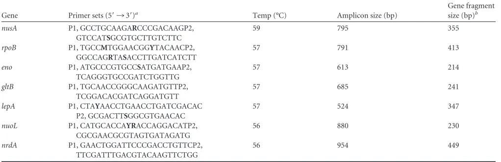
TABLE 1Genes used for the MLST scheme with primers used for amplification
Gene Primer sets (5=¡3=)a Temp (°C) Amplicon size (bp)
Gene fragment
size (bp)b
nusA P1, GCCTGCAAGARCCCGACAAGP2,
GTCCATSGCGTGCTTGTCTTC
59 795 355
rpoB P1, TGCCMTGGAACGGYTACAACP2,
GGCCAGRTASACCTTGATCATCTT
57 791 413
eno P1, ATGCCCGTGCCSATGATGAAP2,
TCAGGGTGCCGATCTGGTTG
57 613 214
gltB P1, TGCAACCGGGCAAGATGTTP2,
TCGGACACGATCAGGATGTT
57 685 241
lepA P1, CTAYAACCTGAACCTGATCGACAC
P2, GCGACTTSGGCGTGAACAC
57 524 347
nuoL P1, CATGCACCAYRACCAGGACATP2,
CGCGAACGCGTAGTGATAGATG
56 880 230
nrdA P1, GAACTGGATTCCCGACCTGTTCP2,
TTCGATTTGACGTACAAGTTCTGG
56 954 449
aStandard mixed oligonucleotide bases for primer sequences are listed in bold. b
Sequence length used in MLST analysis for each locus.
on May 16, 2020 by guest
http://jcm.asm.org/
standardized index of association (IA) was calculated using LIAN 3.5 (http://pubmlst.org), and significance (P⫽0.05) was established under
the null hypothesis of linkage equilibrium (H0:VD⫽Ve) by performing
1,000 Monte Carlo resamplings. Simpson’s index of diversity was
calcu-lated using the comparing partitions program (available athttp://darwin
.phyloviz.net/ComparingPartitions/index.php?link⫽Tool).
For validation of distinctness of sequence similarity clusters in the
population, thekparameter, which is the ratio of the intergroup
diver-gence to the mean of the intragroup diverdiver-gence, was calculated as
previ-ously described (13). Ratios greater than 2 were considered distinct
se-quence similarity clusters. Subgroups within clusters were analyzed separately and designated “a” or “b.”
All sequence data and related information were deposited in the
Ach-romobacterMLST database (http://pubmlst.org/achromobacter/).
RESULTS
Allelic variation inAchromobacter.Analysis of all 150 strains re-vealed 129 STs (see Table S1 in the supplemental material). The number of alleles at each locus ranged from 53 (gltB) to 70 (rpoB), while the number of polymorphic sites ranged from 45 (gltB) to 93 (lepA) (Table 2). Simpson’s index of diversity ranged from 0.959 (nuoL) to 0.980 (nusA), indicating a high level of discrimination. LowdN/dSvalues indicated the absence of strong positive selec-tion pressure at these loci and supported their suitability for use in population genetic analyses.
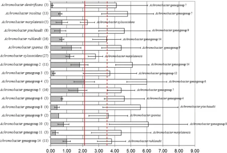
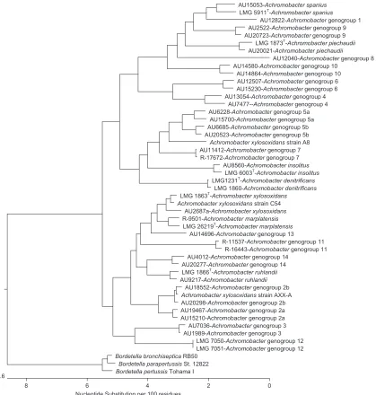
Achromobacterspecies and genogroup delineations. Align-ment of concatenated MLST sequences from all 150 isolates re-vealed interspecies/genogroup divergence rates ranging from 1.6% to 5.3%. Therefore, a rate of 1.6% was used as the cutoff for defining the lower limit of any single species or genogroup (Fig. 1). Maximum intraspecies divergence was 2.1%. Applying this as the cutoff in the concatenated MLST dendrogram (rooted with Bor-detellareference strains) identified at least 14 additional geno-groups, most of which had distinctness of population ratio (k) parameters ofⱖ2 (Fig. 2).A. xylosoxidansandA. marplatensis
could not be considered separate sequence similarity clusters, with
[image:3.585.39.286.79.184.2]kvalues of 1.68, despite both being taxonomically validly named species. The only other genogroup failing to meet the distinctness of population ratio wasAchromobactergenogroup 5 (k⫽1.58); however, the closest genogroup, Achromobacter genogroup 7, contained only 2 strains. The five remaining previously named TABLE 2Analysis of the seven MLST loci in the 150 strains studied
Gene
No. of alleles
No. of polymorphic sites
% GC content
Simpson’s
index (D) dN/dS
nusA 62 87 64.6 0.973 0.0136
rpoB 70 52 62.8 0.959 0.0448
eno 64 47 60.6 0.973 0.0469
gltB 53 45 62.9 0.963 0.0350
lepA 69 93 64.9 0.978 0.0946
nuoL 58 54 62.2 0.976 0.1042
nrdA 65 77 58.2 0.980 0.0473
FIG 1Nucleotide divergence of concatenated MLST loci. Shaded bars represent divergence within a species/genogroup; open bars represent the divergence between species/genogroup and its closest neighbor (listed to the right), as defined by percentage similarity. Error bars represent the standard errors of the shaded and open bars. The solid line represents maximum intraspecies divergence; the dashed line represents the average interspecies divergence.
on May 16, 2020 by guest
http://jcm.asm.org/
[image:3.585.68.520.388.693.2]Achromobacterspecies and new genogroups hadkparameters in-dicating distinctness (range, 2.36 to 40.00).
Presence of distinct subgroups.The concatenated MLST tree revealed thatAchromobactergenogroups 2 and 5 contained dis-tinct subgroups, each labeled a and b. Using bootstrapping with 1,000 replicates, Achromobactergenogroups 2a and 2b showed 100% support that distinct nodes would be maintained. Similarly,
Achromobactergenogroups 5a and 5b showed 93.8% support that distinct nodes would be maintained. Intradivergence and interdi-vergence levels, as well askparameters, indicated that genogroups 2a and 2b and genogroups 5a and 5b could be considered separate clusters, withkparameters of 3.27 and 2.62, respectively. These data provide evidence that these two genogroups contain distinct diverging subgroups.
Linkage disequilibrium.Assessment of the population
struc-ture ofAchromobacterwas investigated by calculating theIAfor each species or genogroup. All species/genogroups showedIA
val-ues differing from zero, indicating that all were in linkage disequi-librium. However, five groups (genogroups 1, 7, 9, 12, and 13) did not include enough strains for reliable testing of significance be-tween the observed variance and maximum trial variance based on the criteria proposed by Kaiser et al. (8). Among the remaining 16 species/genogroups, all showed significant evidence of linkage disequilibrium based on the number of strains analyzed, while 14 maintained significance when analyzed for number of STs only (see Table S2 in the supplemental material).
DISCUSSION
Achromobacterspecies are increasingly recognized as nosocomial and opportunistic human pathogens, particularly in persons with FIG 2Dendrogram of concatenated MLST loci sequences containing two strains of eachAchromobacterspecies or genogroup (unless only a single strain was
available) and rooted withBordetellaspecies.
on May 16, 2020 by guest
http://jcm.asm.org/
[image:4.585.86.505.65.505.2]CF, in whom respiratory tract infection likely contributes to pro-gressive lung disease (10). The broad-spectrum antibiotic resis-tance of most clinical isolates limits effective therapy of infection, and prevention of infection and identification of virulence factors are challenged by an incomplete understanding of the ecology, epidemiology, and taxonomy ofAchromobacterspecies. Unfortu-nately, little progress has been made in these areas during the past decade. The novel speciesA. insolitusandA. spaniuswere added to the genus in 2003, andA. marplatensiswas proposed as yet another novel species in 2010, bringing the total number of validly named species in this genus to seven (7). However, our ongoing analyses of bacterial isolates recovered primarily from human specimens have identified a variety of “Achromobacter-like” strains that can-not be placed into one of these seven species with confidence, suggesting the existence of several additional distinct taxa. These strains, along with strains from each of the seven named species, tested positive in a previously described PCR assay targeting 16S rRNA gene sequences intended to be specific forA. xylosoxidans
(11). Genotyping analyses of such strains using a rep-PCR typing method (3) indicated a high level of genetic diversity and identi-fied several clusters, again suggestive of distinct species.
In order to delineate the taxonomy and epidemiology of Ach-romobacterspecies, we sought to devise an MLST scheme that would not only provide strain genotyping but also enable reliable genus-level phylogenetic analyses. We took advantage of the whole-genome sequences available for 19 strains from several re-lated genera and employed a strategy to select candidate MLST genes that would predict phylogenetic relationships among these genera concordant with those predicted by analysis of their com-plete 16S rRNA gene sequences. Limiting MLST genes to those located on the primary chromosome in species with multiple chromosomes (e.g.,Burkholderiaspp.) avoided genes with poten-tially greater rates of evolutionary change that may be found on secondary chromosomes (6).
We applied this MLST scheme to examine a large collection of putativeAchromobacterstrains, primarily recovered from human sources and selected based on previous genotyping analysis to represent a diverse strain set. The analysis reiterated the high level of diversity in this set, reflected by a large number of alleles and polymorphic sites, as well as a Simpson index of diversity of at least 0.959 in all seven loci. Our results revealed the existence of 14 distinct genogroups in addition to the seven currently named spe-cies. Analysis of the concatenated MLST sequences from the 150 strains included in the study demonstrated that these 21 species/ genogroups had low (maximum, 2.1%) intragroup and high (av-erage, 3.54%) intergroup sequence divergence levels. Thek pa-rameter, a measure of distinctness between two groups, was high (⬎2) for most pairwise sets; the value between genogroup 5 and genogroup 7 was limited by the inclusion of only two strains in genogroup 7. Interpretation of the lowkvalue (1.68%) betweenA. xylosoxidansandA. marplatensiswas similarly limited by the pres-ence of only two distinct strains in the latter species. This species has been shown, nevertheless, to be a distinctAchromobacter spe-cies on the basis of polyphasic analyses, including DNA-DNA hy-bridization values and phenotypic characteristics (7). The concat-enated MLST analysis also identified two distinct subgroups in both genogroup 2 and genogroup 5. Sequence divergence levels, as well askvalues, indicated that these four subgroups most likely represent distinct taxa. Confirmation of this will require further
taxonomic analyses and would corroborate that the design of our MLST scheme provides phylogenetically sound data.
TheIA, a summary statistic based on the variance of pairwise
distances between strains in a set, revealed values different from zero for all 21 species/genogroups. However, because the expected value ofIAdepends on sample size, testing of the significance of theIAwas not reliable in the five genogroups that each contained
fewer than three strains. We found significant evidence for linkage disequilibrium in all remaining groups, including the subgroups in genogroups 2 and 5, consistent with a clonal epidemic popula-tion structure.
Our results have important implications in efforts to better understand the epidemiology, ecology, clinical microbiology, and virulence ofAchromobacterspecies. Advances in each of these ar-eas are predicated on a clearer appreciation of the taxonomy and population structure of this genus. For example, we have found that, based on a preliminary analyses of a large set of Achromobac-terisolates recovered from respiratory cultures from CF patients in the United States, approximately 1/3 are notA. xylosoxidans, although essentially all were initially identified as this species by commercial identification systems. It appearsA. ruhlandii will comprise a considerable minority ofAchromobacterspecies recov-ered from CF patients. Our analysis also shows that, among theA. xylosoxidansstrains for which genome sequencing has been per-formed, only strain C54 is actually a member of this species; strain AXX-A is placed in genogroup 2b, while strain A8 cannot be con-fidently placed in any of the 21 species/genogroups identified in our study.
Our MLST scheme provides a sequence database and method for genotypingAchromobacter strains for global epidemiologic studies and for assigning strains to defined or new genogroups. Ongoing work is aimed at developing less labor-intensive meth-ods to delineate each of the species/genogroups in this genus.
ACKNOWLEDGMENT
This work was supported by the Cystic Fibrosis Foundation.
REFERENCES
1.Aisenberg G, Rolston KV, Safdar A.2004. Bacteremia caused by Achro-mobacterandAlcaligenesspecies in 46 patients with cancer (1989 –2003).
Cancer101:2134 –2140.
2.Brisse S, et al.2002. Comparative evaluation of the BD Phoenix and
VITEK 2 automated instruments for identification of isolates of the
Burk-holderia cepaciacomplex. J. Clin. Microbiol.40:1743–1748.
3.Coenye T, Spilker T, Martin A, LiPuma JJ.2002. Comparative
assess-ment of genotyping methods for epidemiologic study ofBurkholderia
ce-paciagenomovar III. J. Clin. Microbiol.40:3300 –3307.
4.Coenye T, et al.2003.Kerstersia gyiorumgen. nov., sp. nov., a novel
Alcaligenes faecalis-like organism isolated from human clinical samples,
and reclassification ofAlcaligenes denitrificansRuger and Tan 1983 as
Ach-romobacter denitrificanscomb. nov. Int. J. Syst. Evol. Microbiol.53:1825– 1831.
5.Coenye T, Vancanneyt M, Falsen E, Swings J, Vandamme P. 2003.
Achromobacter insolitussp. nov. andAchromobacter spaniussp. nov., from
human clinical samples. Int. J. Syst. Evol. Microbiol.53:1819 –1824.
6.Cooper VS, Vohr SH, Wrocklage SC, Hatcher PJ. 2010. Why genes evolve faster on secondary chromosomes in bacteria. PLoS Comput. Biol.
6:e1000732.
7.Gomila M, et al.2011.Achromobacter marplatensissp. nov., isolated from
a pentachlorophenol-contaminated soil. Int. J. Syst. Evol. Microbiol.61:
2231–2237.
8.Kaiser S, Biehler K, Jonas D. 2009. A Stenotrophomonas maltophilia
multilocus sequence typing scheme for inferring population structure. J.
Bacteriol.191:2934 –2943.
9.Kiredjian M, Holmes B, Kersters K, Guilvout I, De Ley J.1986.
on May 16, 2020 by guest
http://jcm.asm.org/
igenes piechaudii, a new species from human clinical specimens and the
environment. Int. J. Syst. Evol. Microbiol.36:282–287.
10. LiPuma JJ.2010. The changing microbial epidemiology in cystic fibrosis.
Clin. Microbiol. Rev.23:299 –323.
11. Liu L, et al.2002. Ribosomal DNA-directed PCR for identification of
Achromobacter(Alcaligenes)xylosoxidansrecovered from sputum samples
from cystic fibrosis patients. J. Clin. Microbiol.40:1210 –1213.
12. Oh JY, Shin YJ, Wee WR.2005. A case of epidemic keratoconjunctivitis
complicated byAlcaligenes xylosoxidansinfection. Korean J. Ophthalmol.
19:233–234.
13. Palys T, Nakamura LK, Cohan FM.1997. Discovery and classification of ecological diversity in the bacterial world: the role of DNA sequence data.
Int. J. Syst. Bacteriol.47:1145–1156.
14. Robert PY, et al.2008.Alcaligenes xylosoxidansendophthalmitis
follow-ing phacoemulsification and intraocular lens implantation. Ophthalmic
Surg. Lasers Imaging39:500 –504.
15. Rønne Hansen C, Pressler T, Høiby N, Gormsen M.2006. Chronic
infection withAchromobacter xylosoxidansin cystic fibrosis patients; a
ret-rospective case control study. J. Cyst. Fibros.5:245–251.
16. Yabuuchi E, Kawamura Y, Kosako Y, Ezaki T.1998. Emendation of
genusAchromobacterandAchromobacter xylosoxidans(Yabuuchi and
Yano) and proposal ofAchromobacter ruhlandii(Packer and Vishniac)
comb. nov.,Achromobacter piechaudii(Kiredjian et al.) comb. nov., and
Achromobacter xylosoxidanssubsp.denitrificans(Ruger and Tan) comb.
nov. Microbiol. Immunol.42:429 – 438.
17. Yabuuchi E, Yano I.1981.Achromobactergen. nov. andAchromobacter xylosoxidans(ex Yabuuchi and Ohyama 1971) nom. rev. Int. J. Syst. Evol.
Microbiol.31:477– 478.