Internal-Process-Controlled Real-Time PCR Assay for Sensitive and
Specific Detection of
Blastocystis
spp.
Christen Rune Stensvold, Umran Nisar Ahmed, Lee O’Brien Andersen, and Henrik Vedel Nielsen
Laboratory of Parasitology, Department of Microbiological Diagnostics, Statens Serum Institut, Copenhagen, Denmark
Blastocystis
is a common intestinal parasite of unsettled clinical significance, which is not easily detected by standard
parasito-logical methods. The genus comprises at least 13 subtypes (STs) (which likely represent separate species), 9 of which have been
found in humans. Recent data indicate that at least one of the subtypes is associated with intestinal disease. A quantitative
Taq-Man 5
=
nuclease real-time PCR (TaqMan PCR) including an internal process control (IPC) was developed for the detection of
Blastocystis
and shown to be applicable to genomic DNAs extracted directly from feces. The assay enabled successful
amplifica-tion of DNAs from all relevant subtypes within the genus (ST1 to ST9). For assay evaluaamplifica-tion, 153 samples previously tested by
xenic
in vitro
culture (XIVC) were screened by the TaqMan assay. A total of 49/51 samples positive by XIVC and 13/102 samples
negative by XIVC were positive by the TaqMan assay; samples positive by the TaqMan assay and negative by XIVC were
subse-quently tested by conventional PCR, and amplicons could be identified to the subtype level by sequencing in 69% of the cases.
Compared to the TaqMan assay, XIVC had a sensitivity of 79%. This is the first time that a genus-specific, probe-based,
internal-process-controlled real-time PCR assay for the detection
Blastocystis
has been introduced.
B
lastocystis
is a single-celled intestinal parasite of humans and a
vast array of animals. Based on small-subunit (SSU)
ribo-somal DNA (rDNA) analysis, the genus comprises at least 13
sub-types (STs), 9 of which have been found in humans (
29
,
32
,
34
,
36
); it is very likely that each subtype represents a separate species
(
36
). In humans, ST3 appears to be the most common subtype,
followed in prevalence by ST1, ST2, and ST4 (
19
,
23
,
30
–
32
,
35
–
38
). There apparently is a geographical component to variation in
global subtype distribution; for instance, ST4 is rarely reported
outside Europe.
Besides
Blastocystis
being associated with irritable bowel
syn-drome (IBS) (
32
), recent data indicate that at least one subtype
may be associated with gastrointestinal illness (
6
,
31
). In studies
aiming to further explore and clarify the epidemiology and
patho-genicity of
Blastocystis
, accurate identification of carriers and
non-carriers in screening situations is essential (
34
). Recently, various
diagnostic methods, including conventional PCR, xenic
in vitro
culture (XIVC), permanently stained preparations of fixed feces,
and microscopy of fecal concentrates, were compared, and PCR
and culture were found to be the most sensitive methods and to be
almost equally sensitive (
30
). However, culture results are
avail-able only 48 to 72 h after sample submission. Although it involves
DNA extraction, molecular detection is faster and enables
subse-quent subtyping by analysis of sequences obtained from specific
PCR products.
The incentive for the application of real-time PCR-based
screening platforms in diagnostic parasitology is strong (
33
). Such
assays are advantageous in many ways, primarily due to high
spec-ificity and sensitivity and the facts that real-time PCRs are
oper-ated in a closed-tube system with minimal risk of contamination
and that a cutoff can be set to automatically distinguish positive
from negative samples, thus eliminating subjective bias. Only two
real-time PCR assays for
Blastocystis
have been published so far.
One targeted an unknown gene and was shown to enable
ampli-fication of DNAs from ST1, ST3, and ST4 (
11
); it is unknown
whether the assay enables the detection of
Blastocystis
strains
be-longing to other subtypes, and since the gene target is unknown, it
is impossible theoretically to determine specificity and sensitivity
based on gene copy numbers. Another assay was reported by
Poirier et al. (
22
) and was designed as a genus-specific PCR
tar-geting the SSU rRNA gene, enabling amplification of DNAs from
Blastocystis
strains belonging to all subtypes so far identified in
humans. However, the amplicon was 339 bp long, and generally,
significantly shorter amplicons are wanted in diagnostic PCRs to
increase sensitivity. Moreover, the assay was based on SYBR green
detection of double-stranded DNA and had only 95% specificity.
Neither of these two assays included an internal amplification
control; for diagnostic PCR assays, testing for potential PCR
inhi-bition in fecal DNA samples that are PCR negative is essential.
The aim of the present study was to design and evaluate a
genus-specific TaqMan assay for
Blastocystis
with an internal
am-plification control.
(This work was carried out as part of a B.Sc. project performed
by Umran Nisar Ahmed [Technical University of Denmark]).
MATERIALS AND METHODS
Primer Design.Complete SSU rDNA sequences ofBlastocystissp. ST1 to ST10, otherBlastocystisspecies, and species of taxonomic and differential diagnostic relevance, namely,Proteromonas lacertae,Candida albicans, andSaccharomyces cerevisiae, were aligned (Fig. 1) using MegAlign in DNASTAR (DNASTAR, Madison, WI) and MultAlin (2), and target se-quences for genus-specific primer and probes were identified and
de-Received2 January 2012 Returned for modification31 January 2012 Accepted2 March 2012
Published ahead of print14 March 2012
Address correspondence to Christen Rune Stensvold, [email protected].
Copyright © 2012, American Society for Microbiology. All Rights Reserved.
doi:10.1128/JCM.00007-12
on May 16, 2020 by guest
http://jcm.asm.org/
signed by eye and using Primer Express 2.0 (Applied Biosystems) and Generunner 3.01 (http://www.generunner.net/). All sequences were downloaded from GenBank (http://www.ncbi.nlm.nih.gov/GenBank/), except for the sequence ofBlastocystissp. ST10, which was kindly provided by Graham Clark. Oligonucleotides are shown inTable 1. The forward primer had a mismatch range of 0 to 1 bp inBlastocystissubtypes, 1 to 2 bp in otherBlastocystisspecies, and 10 to 11 bp in non-Blastocystisspecies. The probe exhibited a mismatch range of 1 to 2 bp inBlastocystissubtypes, 0 to 2 bp in otherBlastocystisspecies, and 4 to 8 bp in non-Blastocystis
species. Finally, the reverse primer showed a mismatch range of 0 to 1 bp inBlastocystissubtypes, 0 bp in otherBlastocystisspecies, and 0 to 2 bp in non-Blastocystisspecies (Fig. 1).
Real-time PCR assay, standard curves, and controls.To enable the detection ofTaqDNA polymerase inhibitors or suboptimal reaction con-ditions, an internal process control (IPC) was constructed as described previously (7,8). Briefly, primers for amplification of parts of the phage lambda genome were synthesized with a tail that included the sequence of each of theBlastocystisprimers added to the 5=end of the corresponding phage lambda primer (Table 1). PCR products of 190 bp thus containing the binding sites of theBlastocystisprimers were obtained by amplification of 1 ng of purified phage lambda DNA. The amplicons were gel purified, and a 10-fold titration of the IPC was added to separate master mixtures. A dilution of the IPC that had no influence on the cycle threshold (CT) number for purifiedBlastocystisDNA was used in the assay. The optimum dilution of the IPC was found to be 1⫻10⫺8. The IPC probe (TAG
Copenhagen, Copenhagen, Denmark) was 5=labeled with 6-carboxyte-tramethylrhodamine (TAMRA) and quenched with Black Hole Quencher
2 (Table 1).BlastocystisDNA from a strain available in culture was used to
generate standard curves.
Real-time PCRs were performed in 50-l volumes, and the following reagents were used: 1M each of the primers (Table 1), 300 nM each of the two probes (Blastocystisand IPC), 5 U/l PlatinumTaqpolymerase (Invitrogen, Taastrup, Denmark) in 20 mM Tris-HCl (pH 8.0), 40 mM sodium chloride, 2 mM sodium phosphate, 0.1 mM dithiothreitol (DTT), stabilizers, 50% (vol/vol) glycerol, 10⫻PCR buffer minus MgCl2(200
mM Tris [pH 8.4], 500 mM KCl) (Invitrogen, Taastrup, Denmark), 5 mM MgCl2, dUTP mix (12.5 mM dUTP, 50 mM dGTP, 50 mM dATP, 50 mM
dCTP), 5l of the appropriate dilution of IPC, 50% glycerol, and water. Samples were processed on an ABI 7500 real-time PCR system instrument with a 96-well block (Applied Biosystems, Nærum, Denmark). The PCR profile consisted of 50°C for 1 s, 95°C for 2 min, and 50 cycles of denatur-ation at 95°C for 15 s followed by annealing and extension at 60°C for 1 min.
Evaluation and validation of real-time PCR.DNAs from various sub-types (ST1 to ST9) were tested in the assay to enable confirmation of genus sensitivity. Assay sensitivity was determined by testing a 10-fold dilution series of DNA extracted from 1 millionBlastocystisorganisms isolated from xenicin vitroculture by gradient centrifugation (38) and eluted in
200l (Table 2). Hence, 5l of DNA from the undiluted sample was
equivalent to 25,000 organisms.
FIG 1Alignment ofBlastocystis-specific oligonucleotides (forward, probe, and reverse) and SSU rDNAs fromBlastocystissp. ST1 to ST10, otherBlastocystisspp.,
[image:2.585.62.525.66.236.2]Proteromonas lacertae,Candida albicans, andSaccharomyces cerevisiae. Polymorphic bases are highlighted in gray. Dashes indicate missing or nonexisting bases.
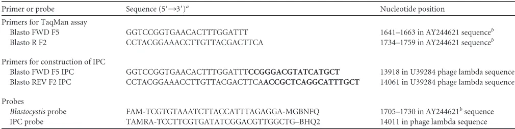
TABLE 1Primers and probes used in the TaqMan assay for amplification ofBlastocystisand the internal process control
Primer or probe Sequence (5=¡3=)a Nucleotide position
Primers for TaqMan assay
Blasto FWD F5 GGTCCGGTGAACACTTTGGATTT 1641–1663 in AY244621 sequenceb Blasto R F2 CCTACGGAAACCTTGTTACGACTTCA 1734–1759 in AY244621 sequenceb
Primers for construction of IPC
Blasto FWD F5 IPC GGTCCGGTGAACACTTTGGATTTCCGGGACGTATCATGCT 13918 in U39284 phage lambda sequence Blasto REV F2 IPC CCTACGGAAACCTTGTTACGACTTCAACCGCTCAGGCATTTGCT 14061 in U39284 phage lambda sequence Probes
Blastocystisprobe FAM-TCGTGTAAATCTTACCATTTAGAGGA-MGBNFQ 1705–1730 in AY244621bsequence
IPC probe TAMRA-TCCTTCGTGATATCGGACGTTGGCTG–BHQ2 14011 in phage lambda sequence
aBoldface corresponds to the phage lambda sequence (GenBank accession number J02459). MGBFQ, minor groove binder and nonfluorescent quencher; and BHQ2, Black Hole
Quencher 2 (nonfluorescent quencher).
bSequence may exhibit polymorphism compared to oligonucleotide.
on May 16, 2020 by guest
http://jcm.asm.org/
[image:2.585.39.544.570.697.2]The assay was specificity tested against panel dilutions of fungal DNAs fromCandida albicans(ATCC 64548),Candida glabrata(ATCC 90030),
Candida parapsilosis (ATCC 22019), Candida tropicalis (UKNEQAS 0527),Candida krusei(ATCC 6258),Geotrichum candidum(UKNEQAS 1911), andSaccharomyces cerevisiae(ATCC 8258). DNAs from the follow-ing bacterial ATCC strains were also used for specificity testfollow-ing:Bacillus cereus(ATCC 14579),Bacillus subtilis(ATCC 6633),Campylobacter coli
(ATCC 33559), Enterobacter cloacae (ATCC 13047), Escherichia coli
(ATCC 25922), andProteus mirabilis(ATCC 12453). Tested DNAs from non-ATCC bacterial strains representedAeromonas caviae,Bacteroides fragilis, Campylobacter jejuni, Campylobacter upsaliensis, Citrobacter freundii,Clostridium difficile,Clostridium perfringens,Clostridium sordelli,
Hafnia alvei,Klebsiella pneumoniae,Listeria monocytogenes,Plesiomonas shigelloides,Pseudomonas aeruginosa, Salmonella enteritidis,Salmonella paratyphi, Serratia marcescens, Shigella dysenteriae, Shigella flexneri,
Staphylococcus aureus,Staphylococcus pyogenes,Vibrio choleraeserotype Ogawa,Vibrio parahaemolyticus, andYersinia enterocolitica.
DNAs used for diagnostic validation of the real-time PCR assay rep-resented 51 samples positive and 102 samples negative forBlastocystisby XIVC from Danish patients submitting stools for parasitological analysis. Culture analyses had been carried out as previously described using Jones’ medium supplemented with 10% horse serum (30,38). Of the 102 XIVC-negative samples, 42 were positive forDientamoeba fragilis, 1 for Crypto-sporidium, and 1 forEntamoeba disparby in-house real-time PCR assays used for detection ofGiardia,Cryptosporidium,Entamoeba histolytica,E. dispar, and D. fragilis(33), and the prevalence of intestinal parasites among the XIVC-positive samples was comparable.
Genomic DNAs from aliquots of fecal samples tested by XIVC were extracted from fresh fecal samples using the NucliSENS easyMAG proto-col (bioMérieux, Herlev, Denmark) according to the recommendation of the manufacturer.
In order to validate results obtained by real-time PCR, samples posi-tive by the TaqMan assay and negaposi-tive by XIVC were subjected to con-ventional PCR using primers described by Scicluna et al. (25) and Stensvold et al. (38). All products were sequenced unidirectionally. The obtained nucleotide sequences were assigned to subtypes by using BLAST against theBlastocystisdatabase available atwww.pubmlst.org
/blastocystis(10,28).
Cohen’s kappa index and comparison of means.Means and medians of cycle threshold (CT) values were calculated and a two-tailed Studentttest for comparison of means carried out using software available athttp://qudata
.com/online/statcalc/andhttp://studentsttest.com/. Cohen’s kappa index for
intertest agreement was calculated (http://olmosantonio.com/diagnostics
/kappa/online/calculator.html).
RESULTS
The TaqMan assay allowed amplification of all subtypes included
in the study, and no amplification of fungal or bacterial DNA was
detected. DNA from 25,000 parasites per reaction was detected at
a
C
Tvalue of 21.89/21.90 in duplicate determinations, and
repro-ducible
C
Tvalues were obtained down to a 10
⫺4dilution, which is
equivalent to template DNA from 2.5 parasites per reaction (
Table
2
). DNA from a lower number of parasites was detectable;
how-ever, since the number of SSU rRNA gene copies per cell is not
known, the absolute detection level of the PCR cannot currently
be ascertained.
Forty-nine samples were positive by both XIVC and the
Taq-Man assay (
Table 3
), with
C
Tvalues ranging from 14.03 to 39.52,
a mean
C
Tvalue of 20.48 (standard deviation [SD], 5.85) and a
median
C
Tvalue of 18.76 (interquartile range [IQR], 17.23 to
21.46). Thirteen samples negative by XIVC were positive by the
TaqMan assay (
Table 3
), with
C
Tvalues ranging from 16.25 to
40.26, a mean
C
Tvalue of 28.93 (SD, 4.99) and a median
C
Tvalue
of 29.33 (IQR, 26.02 to 31.89). A comparison of the two means
gave a
P
value of 0.00067, which means that samples negative by
XIVC and positive by the TaqMan assay were generally
character-ized by having a smaller amount of
Blastocystis
-specific DNA than
that present in samples positive by both methods.
The sensitivity and specificity of XIVC compared to the
Taq-Man assay were 79% and 98%, respectively. Cohen’s kappa index
was 0.79, indicating substantial intertest agreement.
The 13 samples positive by real-time PCR and negative by
XIVC were tested by conventional PCR and sequencing. Using
primers amplifying a product of
⬃
600 bp (
25
) or
⬃
300 bp (
38
), it
was possible to amplify 9/13 samples by conventional PCR (ST1, 3
samples; ST2, 1 sample; and ST3, 5 samples); the mean
C
Tvalue
for the 4 samples not amplifiable by conventional PCR was 33.48.
In total, unambiguous sequences were obtained in 56/62
real-time PCR-positive cases. ST1 was seen in 21 cases, ST2 in 14, ST3
in 16, and ST4 in 5. The mean
C
Tvalues (SDs) for individual
subtypes were 19.33 (3.64) for ST1, 19.76 (6.80) for ST2, 23.52
(6.03) for ST3, and 24.36 (9.06) for ST4. Samples positive for ST1
had lower
C
Tvalues than samples positive for ST3 (
P
⫽
0.022).
The two samples positive by XIVC and negative by real-time
PCR tested negative by the conventional PCR after repeated
ef-TABLE 2Cycle threshold values for the 10-fold dilution row of DNA from 1 millionBlastocystisorganisms/200l elution buffer
Probe
CTvalues for duplicates at dilution ofa:
1⫻100 1⫻10⫺1 1⫻10⫺2 1⫻10⫺3 1⫻10⫺4 1⫻10⫺5 1⫻10⫺6 1⫻10⫺7 1⫻10⫺8
Blasto 21.90/21.89 25.15/25.22 28.67/28.67 31.04/30.97 34.84/35.11 36.82/40.91 39.21/38.12 40.93/UD UD/UD IPC UD/UD UD/UD UD/47.26 36.56/38.64 35.17/35.11 35.18/35.70 36.76/36.03 36.70/34.95 35.98/37.15
[image:3.585.40.544.78.132.2]aDNAs were tested in duplicates to test for reproducibility. UD, undetermined (i.e., signal absent).
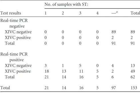
TABLE 3Comparison of test results for real-time PCR and XIVC and distribution ofBlastocystissubtypes
Test results
No. of samples with ST:
1 2 3 4 —a Total
Real-time PCR negative
XIVC negative 0 0 0 0 89 89
XIVC positive 0 0 0 0 2 2
Total 0 0 0 0 91 91
Real-time PCR positive
XIVC negative 3 1 5 0 4 13
XIVC positive 18 13 11 5 2 49
Total 21 14 16 5 6 62
Total 21 14 16 5 97 153
a—, not applicable.
on May 16, 2020 by guest
http://jcm.asm.org/
[image:3.585.298.543.554.717.2]forts with multiple DNA dilutions. Results from IPC analysis
showed that inhibition in these two samples was not an issue.
DISCUSSION
Accurate diagnostic tools are of vital significance in clinical and
epidemiological studies of
Blastocystis
. So far, PCR has been used
mostly for characterization purposes (
1
,
3
–
6
,
12
–
14
,
16
–
21
,
23
–
27
,
29
,
31
,
32
,
35
,
37
,
40
–
42
), although a few diagnostic PCRs have
been published, two of which are based on real-time PCR
technol-ogy (
11
,
22
).
A major challenge in the development of genus-specific
Blas-tocystis
PCRs is the genetic diversity seen within the genus, which
limits the number of potential targets in the SSU rRNA gene. The
pairwise genetic distance of
Blastocystis
subtypes amounts to at
least 14.8% across the SSU rRNA gene (
29
), and some conserved
regions are likely to be conserved in other genera as well, which
hampers identification of oligonucleotide target regions.
Compared to previously published real-time PCR assays (
11
,
22
), the present one has the advantage of probe-based detection,
which increases assay specificity. Based on confirmatory
sequenc-ing, the TaqMan assay did not produce any false positives, and this
is probably due to the fact that primers and probe sequences were
highly specific. The real-time PCR assay developed by Poirier et al.
(
22
) generated 8/186 false positives and had a specificity of 95%.
Specificity testing of previously published diagnostic PCRs has
included testing against other intestinal parasites, such as
Entam-oeba
,
Dientamoeba
,
Giardia
, and
Cryptosporidium
(
22
), or even
bacteria (
11
), but it is also highly relevant to evaluate the assay
against a panel of fungi such as
Candida
,
Geotrichum
, and
Saccha-romyces
, which are common components of the fecal flora (
9
,
15
)
and which differ from
Blastocystis
by only about 20% at the SSU
rDNA level.
A previous comparison between XIVC and conventional PCR
(amplifying 550 bp) revealed a nonsignificant difference in
sensi-tivity in favor of PCR (
30
). Although the sensitivity of the TaqMan
assay is higher than that of the XIVC, it is not immediately
com-parable to the data presented by Poirier et al. (
22
), who found that
the sensitivity of XVIC was only 53% compared to their SYBR
green assay. Importantly, Poirier et al. (
22
) used Jones’ medium
supplemented with antibiotics (100 IU/ml penicillin and 100
g/ml streptomycin), while we used Jones’ medium without
add-ing antibiotics. It is not unlikely that these antibiotics will
indi-rectly suppress the growth of
Blastocystis
by a reduction of
bacte-ria, despite the fact that anaerobic chambers were used.
Samples with
C
Tvalues of
ⱕ
35 are most likely indicative of
active, ongoing infestation.
C
Tvalues of
⬎
35 possibly represent
samples with relatively few
Blastocystis
organisms, and it could be
speculated that there is no active
Blastocystis
infection going on in
the patients from whom those samples came. It is possible that
these patients had been exposed to nonviable
Blastocystis
(detect-able by real-time PCR but not by XIVC) or that they were clearing
an infection.
C
Tvalues of
⬎
40 may primarily reflect unspecific
amplification of a target present in the DNA samples that is of
non-
Blastocystis
origin.
Whether parasite intensity is linked to clinical outcome of
Blas-tocystis
infections remains unclear. It is known that
Blastocystis
shedding exhibits day-to-day variation (
39
). The present data
ob-tained by real-time PCR analysis confirmed that
Blastocystis
-pos-itive fecal samples exhibit a range in
C
Tvalues from 12 to 40. Such
a span of
C
Tvalues likely reflects vast differences in relative
para-site load. Using real-time PCR, Poirier et al. (
22
) did not find any
correlation between high intensity and symptoms, but the study
was limited with regard to sample size. In the present study, all
samples were from patients submitting stools for parasitological
analysis due to travel-associated or persistent diarrhea. Future
studies should aim to investigate whether differences in symptoms
and the severity of these are associated with differences in
C
Tval-ues. If low
C
Tvalues are associated with diarrhea and/or other
symptoms, epidemiological cutoff values could be determined
and used in the clinical management of
Blastocystis
-positive
pa-tients.
The overall subtype distribution reflected the usual subtype
distribution seen in Danish cohorts (
23
,
30
,
32
,
35
,
37
,
38
) and
indicates that assay detection is independent of subtype. ST1
sam-ples had lower
C
Tvalues than ST3 samples, indicating that ST3
infections might be lighter in parasite load. However, larger data
sets are needed to confirm this hypothesis and allow speculation
on its clinical implications.
The present assay has a built-in IPC, which distinguishes it
from previously published PCR assays. In the current evaluation,
inhibition or suboptimal conditions appeared not to be a
prob-lem. The assay does not enable accurate subtyping by sequencing
of PCR products; although the amplicon spans a hypervariable
region, it is relatively small compared to the amplicon size usually
recommended for subtyping (
25
,
30
).
In conclusion, we have developed a highly applicable TaqMan
assay for sensitive and specific screening for
Blastocystis
ST1 to ST9
of large numbers of DNAs extracted directly from human fecal
samples. We believe that this method will prove to be an
invalu-able tool in all studies aiming at accurately identifying carriers and
noncarriers of
Blastocystis
. Once DNAs have been found to be
positive, these can be subjected to the genus-specific PCR
pub-lished by Scicluna et al. (
25
) for subtyping.
ACKNOWLEDGMENTS
We thank Lis Lykke Wassmann, Gitte Jensen, and Birthe Dohn (Statens Serum Institut) for excellent technical assistance, Graham Clark (London School of Hygiene and Tropical Medicine) for providing the ST10 se-quence, Maiken Cavling Arendrup, Rasmus Hare Jensen, and Søren Persson (Statens Serum Institut) for providing fungal and bacterial DNAs for specificity testing, and Jørgen Skov Jensen (Statens Serum Institut) for professional advice.
REFERENCES
1.Abd-Alla MD, Wahib AA, Ravdin JI. 2000. Comparison of antigen-capture ELISA to stool-culture methods for the detection of asymptom-atic Entamoeba species infection in Kafer Daoud, Egypt. Am. J. Trop. Med. Hyg.62:579 –582.
2.Corpet F.1988. Multiple sequence alignment with hierarchical clustering. Nucleic Acids Res.16:10881–10890.
3.Dogruman-Al F, Dagci H, Yoshikawa H, Kurt O, Demirel M.2008. A possible link between subtype 2 and asymptomatic infections of Blasto-cystis hominis. Parasitol. Res.103:685– 689.
4.Dogruman-Al F, et al.2009. Blastocystis subtypes in irritable bowel syn-drome and inflammatory bowel disease in Ankara, Turkey. Mem. Inst. Oswaldo Cruz104:724 –727.
5.Dogruman-Al F, Yoshikawa H, Kustimur S, Balaban N.2009. PCR-based subtyping of Blastocystis isolates from symptomatic and asymp-tomatic individuals in a major hospital in Ankara, Turkey. Parasitol. Res.
106:263–268.
6.Domínguez-Márquez MV, Guna R, Muñoz C, Gómez-Muñoz MT, Borrás R.2009. High prevalence of subtype 4 among isolates of Blasto-cystis hominis from symptomatic patients of a health district of Valencia (Spain). Parasitol. Res.105:949 –955.
on May 16, 2020 by guest
http://jcm.asm.org/
7.Jensen JS, Björnelius E, Dohn B, Lidbrink P.2004. Use of TaqMan 5=
nuclease real-time PCR for quantitative detection of Mycoplasma genita-lium DNA in males with and without urethritis who were attendees at a sexually transmitted disease clinic. J. Clin. Microbiol.42:683– 692. 8.Jensen JS, Borre MB, Dohn B.2003. Detection of Mycoplasma
genita-lium by PCR amplification of the 16S rRNA gene. J. Clin. Microbiol.
41:261–266.
9.Jobst D, Kraft K.2006. Candida species in stool, symptoms and com-plaints in general practice—a cross-sectional study of 308 outpatients. Mycoses49:415– 420.
10. Jolley KA, Maiden MC.2010. BIGSdb: scalable analysis of bacterial ge-nome variation at the population level. BMC Bioinformatics11:595. 11. Jones MS, et al.2008. Detection of Blastocystis from stool samples using
real-time PCR. Parasitol. Res.103:551–557.
12. Jones MS, et al.2009. Association of Blastocystis subtype 3 and 1 with patients from an Oregon community presenting with chronic gastrointes-tinal illness. Parasitol. Res.104:341–345.
13. Li LH, et al.2007. Cross-sectional surveys and subtype classification of human Blastocystis isolates from four epidemiological settings in China. Parasitol. Res.102:83–90.
14. Li LH, et al.2007. Molecular epidemiology of human Blastocystis in a village in Yunnan province, China. Parasitol. Int.56:281–286.
15. Macura AB, Witalis J.2010. Fungi isolated from the stool in patients with gastrointestinal disorders in 2005-2009. Przegl. Epidemiol.64:313–317. 16. Meloni D, et al.2011. Molecular subtyping of Blastocystis sp. isolates
from symptomatic patients in Italy. Parasitol. Res.109:613– 619. 17. Navarro C, et al.2008. High prevalence of Blastocystis sp. in pigs reared
under intensive growing systems: frequency of ribotypes and associated risk factors. Vet. Parasitol.153:347–358.
18. Noël C, et al.2005. Molecular phylogenies of Blastocystis isolates from different hosts: implications for genetic diversity, identification of species, and zoonosis. J. Clin. Microbiol.43:348 –355.
19. Ozyurt M, et al.2008. Molecular epidemiology of Blastocystis infections in Turkey. Parasitol. Int.57:300 –306.
20. Parkar U, et al.2007. Direct characterization of Blastocystis from faeces by PCR and evidence of zoonotic potential. Parasitology134:359 –367. 21. Parkar U, et al.2010. Molecular characterization of Blastocystis isolates
from zoo animals and their animal-keepers. Vet. Parasitol.169:8 –17. 22. Poirier P, et al.2011. Development and evaluation of a real-time PCR
assay for detection and quantification of blastocystis parasites in human stool samples: prospective study of patients with hematological malignan-cies. J. Clin. Microbiol.49:975–983.
23. Rene BA, Stensvold CR, Badsberg JH, Nielsen HV. 2009. Subtype analysis of Blastocystis isolates from Blastocystis cyst excreting patients. Am. J. Trop. Med. Hyg.80:588 –592.
24. Santín M, Gómez-Muñoz MT, Solano-Aguilar G, Fayer R.2011. Devel-opment of a new PCR protocol to detect and subtype Blastocystis spp. from humans and animals. Parasitol. Res.109:205–212.
25. Scicluna SM, Tawari B, Clark CG.2006. DNA barcoding of blastocystis. Protist157:77– 85.
26. Souppart L, et al.2010. Subtype analysis of Blastocystis isolates from symptomatic patients in Egypt. Parasitol. Res.106:505–511.
27. Souppart L, et al.2009. Molecular epidemiology of human Blastocystis isolates in France. Parasitol. Res.105:413– 421.
28. Stensvold CR, Alfellani M, Clark CG.2012. Levels of genetic diversity vary dramatically between Blastocystis subtypes. Infect. Genet. Evol.12: 263–273.
29. Stensvold CR, et al.2009. Subtype distribution of Blastocystis isolates from synanthropic and zoo animals and identification of a new subtype. Int. J. Parasitol.39:473– 479.
30. Stensvold CR, Arendrup MC, Jespersgaard C, Mølbak K, Nielsen HV.
2007. Detecting Blastocystis using parasitologic and DNA-based methods: a comparative study. Diagn. Microbiol. Infect. Dis.59:303–307. 31. Stensvold CR, Christiansen DB, Olsen KE, Nielsen HV.2011.
Blasto-cystis sp. subtype 4 is common in Danish BlastoBlasto-cystis-positive patients presenting with acute diarrhea. Am. J. Trop. Med. Hyg.84:883– 885. 32. Stensvold CR, et al.2009. Blastocystis: unravelling potential risk factors
and clinical significance of a common but neglected parasite. Epidemiol. Infect.137:1655–1663.
33. Stensvold CR, Nielsen HV.2012. Comparison of microscopy and PCR for the detection of intestinal parasites in Danish patients supports incen-tive for molecular screening platforms. J. Clin. Microbiol.50:540 –541. 34. Stensvold CR, Nielsen HV, Mølbak K, Smith HV.2009. Pursuing the
clinical significance of Blastocystis— diagnostic limitations. Trends Para-sitol.25:23–29.
35. Stensvold CR, et al.2011. The prevalence and clinical significance of intestinal parasites in HIV-infected patients in Denmark. Scand. J. Infect. Dis.43:129 –135.
36. Stensvold CR, et al. 2007. Terminology for Blastocystis subtypes—a consensus. Trends Parasitol.23:93–96.
37. Stensvold CR, et al.2007. Blastocystis: subtyping isolates using pyrose-quencing technology. Exp. Parasitol.116:111–119.
38. Stensvold R, Brillowska-Dabrowska A, Nielsen HV, Arendrup MC.
2006. Detection of Blastocystis hominis in unpreserved stool specimens by using polymerase chain reaction. J. Parasitol.92:1081–1087.
39. Vennila GD, et al.1999. Irregular shedding of Blastocystis hominis. Parasitol. Res.85:162–164.
40. Yoshikawa H, et al.2004. Polymerase chain reaction-based genotype classification among human Blastocystis hominis populations isolated from different countries. Parasitol. Res.92:22–29.
41. Yoshikawa H, Wu Z, Nagano I, Takahashi Y.2003. Molecular compar-ative studies among Blastocystis isolates obtained from humans and ani-mals. J. Parasitol.89:585–594.
42. Yoshikawa H, et al.2009. Molecular characterization of Blastocystis iso-lates from children and rhesus monkeys in Kathmandu, Nepal. Vet. Para-sitol.160:295–300.