S197 A glossary of specialist terms used in this chapter appears at the end of the text section.
Introduction and historical background
Evolving concepts of disease mechanisms for rheumatoid arthritis (RA) have provided a paradigm for understanding the pathogenesis of autoimmune disease. This paradigm proposes that genetic and environmental factors shape a complex series of molecular and cellular interactions leading to a chronic inflammatory response. CD4+T
lym-phocytes have featured prominently because the genetic elements most strongly associated with RA susceptibility or severity are encoded within the MHC class II region (discussed in this issue in the chapters by H McDevitt, and G Sønderstrup). Precisely how effector T cells initiate
and promote the inflammatory process in RA, however, remains far from clear. Much effort has focussed on estab-lishing the molecular nature of antigenic reactivity, in the belief that the established chronic phase of the disease is antigen-driven. Animal models of inflammatory arthritis would certainly lend support to this view. However, the results of detailed phenotypic and functional analyses of chronically activated T cells derived from inflamed joints are difficult to reconcile with traditional models of carti-lage-antigen-driven inflammatory disease in patients with RA. This chapter aims to explore this theme in more depth, beginning with an outline of the molecular events that
Supplement Review
Studies of T-cell activation in chronic inflammation
Andrew P Cope
The Kennedy Institute of Rheumatology Division, Faculty of Medicine, Imperial College, London, UK
Correspondence:Andrew P Cope, The Kennedy Institute of Rheumatology Division, Faculty of Medicine, Imperial College, Arthritis Research Campaign Building, 1 Aspenlea Road, Hammersmith, London W6 8LH, UK. Tel: +44 (0)20 8383 4444; fax: +44 (0)20 8383 4499; e-mail: andrew.cope@ic.ac.uk
Chapter summary
The strong association between specific alleles encoded within the MHC class II region and the development of rheumatoid arthritis (RA) has provided the best evidence to date that CD4+T cells play
a role in the pathogenesis of this chronic inflammatory disease. However, the unusual phenotype of synovial T cells, including their profound proliferative hyporesponsiveness to TCR ligation, has challenged the notion that T-cell effector responses are driven by cognate cartilage antigens in inflamed synovial joints. The hierarchy of T-cell dysfunction from peripheral blood to inflamed joint suggests that these defects are acquired through prolonged exposure to proinflammatory cytokines such as tumour necrosis factor (TNF)-α. Indeed, there are now compelling data to suggest that chronic cytokine activation may contribute substantially to the phenotype and effector function of synovial T cells. Studies reveal that chronic exposure of T cells to TNF uncouples TCR signal transduction pathways by impairing the assembly and stability of the TCR/CD3 complex at the cell surface. Despite this membrane-proximal effect, TNF selectively uncouples downstream signalling pathways, as is shown by the dramatic suppression of calcium signalling responses, while Ras/ERK activation is spared. On the basis of these data, it is proposed that T-cell survival and effector responses are driven by antigen-independent, cytokine-dependent mechanisms, and that therapeutic strategies that seek to restore T-cell homeostasis rather than further depress T-cell function should be explored in the future.
Keywords:inflammation, rheumatoid arthritis, signal transduction, T cells, TNF Received: 3 January 2002
Accepted: 21 January 2002 Published: 9 May 2002
Arthritis Res2002, 4 (suppl 3):S197-S211 © 2002 BioMed Central Ltd
S198
dictate the differentiation of T helper (Th) cells at the outset of adaptive immune responses in regional lymph nodes. Much of the remainder of the discussion focuses on the different ways in which, in the longer term, the chronic inflammatory process influences maturation, differ-entiation, and function of effector T cells at sites of inflam-mation. I conclude by speculating about how our understanding of T-cell activation in chronic inflammation may influence future therapy, and discuss this in the context of the prevailing view that in a susceptible host, chronic inflammatory disease occurs through a failure of regulatory T cells to downregulate the inflammatory process.
Acquisition of transcriptional competence
during differentiation of T helper cells
There is now good evidence that there exists a coordi-nated programme of molecular events initiated at the outset of T-cell differentiation that leads to the generation of CD4+Th effector cells [1]. This process of
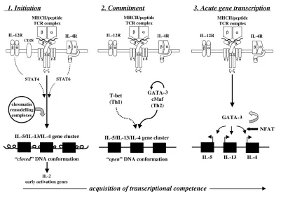
differentia-tion is characterised by a distinctive pattern of cytokine production and is important because its outcome dictates the host response to foreign pathogens such as Listeria monocytogenesinfection or to parasitic infestation [2]. For cytokine genes, at least three stages are thought to be required for the acquisition of transcriptional competence in T cells: an initiation phase, a commitment phase, and a phase of acute gene transcription (Fig. 1). The existence of these stages has been deduced largely from experi-ments in which monospecific T cells are stimulated in vitro
in bulk cultures from naïve precursors [3,4]. During the ini-tiation phase, naïve T cells are engaged through their T-cell receptors (TCRs) by MHC/peptide complexes expressed on the surface of dendritic cells. Only those T cells that form a functional immunological synapse are likely to differentiate [5]. At this point, intracellular sig-nalling pathways emanating from stable clusters of TCR/CD3 complexes are integrated with those from cytokine receptors following engagement by cytokines. For example, IL-12 and IL-18 are important for differentia-tion of Th1 cells but also play a key role in innate immunity. In polarised Th cells, some cytokines activate families of transcription factors called ‘signal transducers and activa-tors of transcription’, or STATs, such as STAT4 for IL-12 (Th1), or STAT6 for IL-4 (Th2) [6]. The initial engagement phase may last from hours to days, and in general will result in the production of IL-2 and entry of the cell into the cell cycle. At this stage, cells are incapable of producing Th1 (IFN-γ) or Th2 (IL-4) cytokines, despite optimal stimu-lation by antigenic peptide and cytokines. Moreover, sur-prisingly few T cells progress beyond this early stage of maturation.
The commitment phase is characterised at the molecular level by the induction and recruitment of Th-subset-spe-cific transcription factors. Those considered to be lineage-specific include GATA-3 and c-Maf for Th2 cells [7,8], and
T-bet and ERM for Th1 cells [9,10]. Once these factors are expressed, differentiation is stabilised and maintained even in the absence of further TCR stimulation (see Fig. 1, middle panel). The third phase, that of acute gene tran-scription, is determined by secondary contact with antigen and necessitates the recruitment of nuclear factor of acti-vated T cells (NFAT) together with subset-specific tran-scription factors to the transcriptosome complex (see Fig. 1, right panel). This process is thought to be monoal-lelic and stochastic, probably because it depends upon chromatin accessibility [1,6]. Thus, specific loci become transcriptionally active through a series of changes to chromatin structure, including chromatin decondensation and remodelling, and recruitment of complexes to the nuclear matrix [11]. Within 48 hours of stimulation, new clusters of DNase-hypersensitive sites can be detected, as demonstrated for the IL-4 gene [12], possibly through the coordinated action of STATS and other transcription factors such as the binding of p300 and of calcium-binding proteinC/EBP (CCAAT/enhancer calcium-binding protein) to DNA elements [1,6]. These sites are markers of stable, differentiated T cells. Coincident with these changes in the nucleus are overall increases in histone acetylation, histone phosphorylation, and DNA demethylation [13,14], which occur during the S phase of the cell cycle [15].
Full commitment to a specific lineage is established gradu-ally and in most cases takes place in regional lymph nodes. According to this model, Th cells can be thought of as being in ‘antigen mode’, since the transcriptional pro-gramme required for effector function is absolutely deter-mined by antigen and TCR signalling pathways. It follows from this that the expression and stability of the TCR on the T-cell surface, its avidity for MHC/peptide complexes, the signal strength, and the integrity of protein tyrosine kinases and signalling adaptor molecules would be essen-tial in determining both qualitative and quantitative charac-teristics of the immune response [16].
T-cell differentiation in chronic inflammation –
clues from the rheumatoid joint
While these molecular events go some way towards explaining why some CD4+ T cells differentiate into Th1
S199 What do the available data tell us about T-cell activation
and differentiation in established chronic inflammation? The evidence for chronic immune activation is unambigu-ous (Table 1). This is best illustrated by histological analy-ses of sections of synovial tissue in which, in subsets of patients with more severe RA, there exist perivascular fol-licular lymphoid-like structures resembling germinal centres of lymphoid organs [17,18]. Lymphoid aggregates in synovial tissue are rich in T cells, B cells expressing MHC class II, and dendritic cells, and their precise cellular organisation is thought to depend on the local expression of cytokines and chemokines [19]. Ex vivo,flow cytometric analysis reveals a memory phenotype for synovial tissue and fluid T cells expressing CD45RO but low levels of CD45RB [20,21], suggestive of past or persistent anti-genic stimulation. Their cell surface carries other markers of activation, such as CD69, CD44, and HLA-DR, as well as the chemokine receptors CCR4, CCR5, CXCR3, and CX3CR1, whose selective expression may facilitate homing to synovial joints [20,22]. Synovial T cells persist in the joint, possibly through an environment that favours cell survival. The expression of stromal cell derived survival
factors such as IFN-α may contribute [23]. The demon-stration of both significant and premature telomere short-ening would also suggest that these cells undergo progressive self-replication in situ[24], and so by the time the inflammatory process is established, subsets of syn-ovial T cells may already be approaching the stage of ter-minal differentiation or senescence.
[image:3.612.108.509.104.385.2]While there is a general consensus that in inflamed syn-ovial joints there is enrichment of Th1 T cells [25], the demonstration of significant populations of polarised Th1 subsets has been difficult, even with the advent of intracel-lular staining techniques [26,27]. Indeed, expression of cytokines cannot be detected easily without stimulation, and even after stimulation with anti-CD3 and anti-CD28, the frequency of cytokine-expressing cells may be very low, necessitating pharmacological stimulants (e.g. phorbol ester and ionophore) to demonstrate the pres-ence of cytokine-producing T cells. Nonetheless, the finding of a paucity of Th2-cytokine-expressing T cells by many laboratories has been more consistent [25,28], and recently there are data to suggest that naïve RA peripheral
Figure 1
Acquisition of transcriptional competence during differentiation of T helper cells. Th cells become productive effectors of immunoinflammatory responses following a complex series of molecular events dependent upon membrane-proximal TCRs and cytokine receptor signals. Chromatin remodelling is an essential step in the process leading to a switch from the ‘closed’ to ‘open’ DNA conformation. This in turn permits accessibility of Th-subset-specific transcription factors and accessory factors to the promoter elements of the Th2 gene cluster, as illustrated here. Ultimately, NFAT is recruited to the transcriptosome, after which cytokine gene transcription proceeds. c-Maf, transcription factor specific for Th2 cells;
S200
blood T cells may be refractory to Th2 differentiation [29]. According to these data, there exists in vivoan imbalance between proinflammatory cytokines and anti-inflammatory cytokines whereupon a deficiency of anti-inflammatory cytokines might be expected to favour the failure of immunoregulatory mechanisms [30]. Taken together, these experimental observations go some way towards supporting the idea that in inflamed synovial joints, the T cell is in a state of chronic immune activation, if not to say a state of chronic stress or exhaustion, and that the default regulatory mechanisms operating in the nonsusceptible host are absent or insufficient.
Synovial T cells are hyporesponsive to TCR
engagement
The low levels of constitutive cytokine expression in syn-ovial T cells is puzzling, given their activated phenotype. More puzzling is the finding that they proliferate very poorly
in vitro in response to either mitogen, recall antigens, or CD3 ligation with specific agonistic monoclonal antibodies (Table 1, and [31–33]). Indeed, suppression of proliferative and cytokine responses has led some workers in the field to conclude that terminally differentiated T cells may not contribute to the established inflammatory response [34]. However, this does not seem compatible with the histopathological features of inflamed joints outlined above.
There are several possible reasons for the unusual pheno-type of synovial T cells, which may have to do with both spatial and temporal parameters. Firstly, the anatomy and cellular environment of inflamed synovial tissue are differ-ent from the architecture and cellular constitudiffer-ents of lymph nodes, with hypoxia and extremes of extracellular pH imposing significant pathophysiological effects (dis-cussed in this issue in chapters by P Taylor and E Pale-olog). Secondly, extensive analysis of the inflammatory environment demonstrates that the cytokine milieu in RA synovial tissue is different from that expressed in a lymph node during primary interactions between dendritic cells and precursors of Th cells, with a strong predominance of macrophage products [30]. Accordingly, the expression of
cytokine receptors and the acquisition of chronic cytokine responsiveness is likely to be a distinguishing feature of chronically activated T cells in inflamed joints. Historically, it has been assumed that T-cell activation in the rheuma-toid joint would be driven by peptide fragments of carti-lage antigens presented by disease-associated MHC-class-II molecules [35,36]. However, this model now has to take into account the fact that synovial T cells are hyporesponsive to TCR engagement. These findings raise the possibility that during the evolution of immune and inflammatory responses, the balance of stimulation shifts from ‘antigen mode’, in which T cells are engaged through the TCR/CD3 complex during the early phases of Th dif-ferentiation, to ‘inflammation mode’, in which T-cell activa-tion and effector responses are driven by proinflammatory cytokines (Fig. 2).
A model of T-cell activation in chronic
inflammation
Almost a decade ago, we began to think about ways to explore how the chronic inflammatory process might influ-ence T-cell autoreactivity and effector responses, in the belief that this might contribute to an understanding of the immunopathogenic processes involved in the chronic phase of RA. Our approach was influenced to a great extent by a series of observations arising from a larger pro-gramme of work in the laboratory and by others, which sought to document in depth the broad range of cytokines expressed in rheumatoid joints. The observations of partic-ular importance included the findings that tumour necrosis factor (TNF)-αbioactivity persists in synovial joint cell cul-tures and in vivo[37–40]; that both high-affinity p55 and p75 tumour-necrosis-factor receptors (TNFRs) are upreg-ulated on synovial joint T cells [41]; that TNFRs are expressed in lymphoid aggregates and colocalise with ligand [42]; and that expression of the naturally occurring TNF inhibitors, the soluble TNFRs, is also increased in synovial fluid but is insufficient to completely neutralise bioactive TNF in vivo [43]. The implication of these find-ings was that synovial mononuclear-cell infiltrates, includ-ing T cells, are chronically exposed to TNF in vivo. We
Table 1
Characteristics of chronically activated T lymphocytes in the synovium of patients with rheumatoid arthritis
1 T lymphocytes are found in follicular lymphoid aggregates
2 The cell-surface phenotype is suggestive of chronic immune activation, e.g. expression of CD45RO, CD69, and subsets of chemokine receptors 3 T cells are terminally differentiated, with significant telomere loss
4 Synovial T cells are hyporesponsive to TCR ligation 5 Synovial T cells exist in an environment favouring cell survival
S201 therefore predicted that the environment generated in
chronically inflamed joints must be very different from that provided by an acute inflammatory or infectious episode (Table 1), and that chronic exposure to inflammatory cytokines might have effects distinct from those induced after short-term exposure.
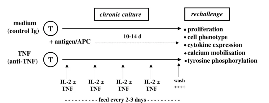
We set out to mimic chronic exposure to cytokines by cul-turing antigen-activated T cells in the presence of TNF. This in vitromodel was very similar to that used in many laboratories to explore the effects of cytokines such as IFN-γ, IL-4, and IL-12 on T-cell differentiation [3,4], with the exception that recombinant TNF was added repeat-edly to T-cell cultures to mimic better the sustained TNF signalling we believed existed in inflamed joints (Fig. 3) [44]. The principal finding that chronic, as opposed to acute, exposure to TNF suppressed T-cell activation was unambiguous, and could not have been predicted from published data at that time, which suggested that TNF was costimulatory and a growth factor for T cells [45]. These results have been confirmed in other laboratories [46–48] and have been supported further through exten-sive analyses of both human and murine T-cell lines and clones in vitro,as well as from experiments in vivo under-taken in T-cell receptor transgenic mice treated with recombinant TNF, anti-TNF, or after intercrossing to
human-TNF-globin transgenic mice [49,50]. The findings are summarised in Table 2. Two lines of evidence con-vinced us that these unexpected findings were potentially important and worthy of further investigation. The first was the observation that peripheral blood T-cell responses from patients with RA were dramatically and rapidly restored after treatment with anti-TNF (infliximab, Remi-cade™) [44] and that these immunological parameters closely followed clinical improvement [51]. The second line of evidence came from a series of studies undertaken in TCR transgenic mice. These experiments revealed that repeated injections of otherwise healthy transgenic mice with anti-TNF enhanced T-cell responses to cognate peptide antigen, and implied that physiological concentra-tions of TNF had immunomodulatory properties in vivo
[49].
[image:5.612.101.523.100.324.2]The immunomodulatory effects of TNF in the
initiation and resolution of autoimmunity
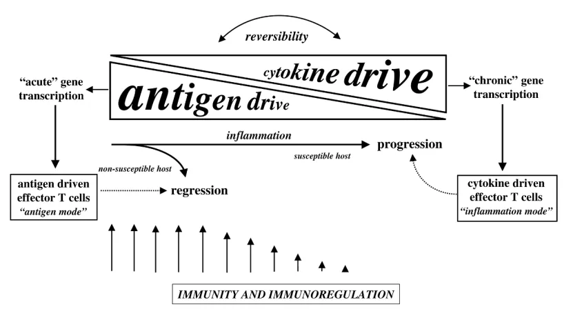
Until the late 1980s, it was assumed that TNF was both proinflammatory and co-stimulatory [30]. How could these initial observations be reconciled with the immunosuppres-sive effects outlined above? An extenimmunosuppres-sive series of studies in mice deficient for TNF or TNFR have confirmed that TNF has potent immunomodulatory effects in vivo, capable of regulating T-cell autoreactivity and autoimmunity whenFigure 2
A model for the role of CD4+T cells in the pathogenesis of chronic inflammation. Antigen drive predominates during the early phase of
S202
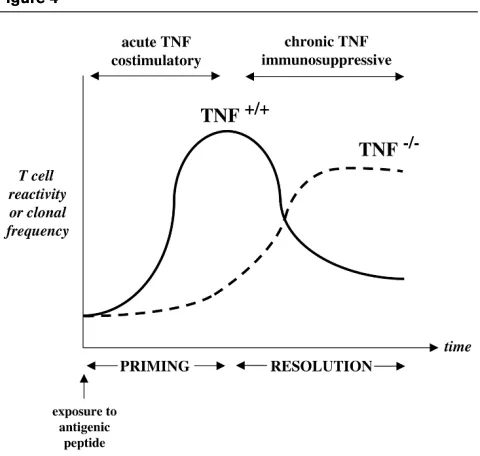
studied in autoimmune-susceptible strains of mice [50]. Indeed, the suppressive effects of chronic TNF were entirely consistent with the studies of Jacob and McDevitt [52], as well as Gordon and colleagues [53], who first demonstrated the disease-protecting effects of chronic TNF therapy in the NZB/W F1lupus-prone mouse. Similar effects were reported subsequently in murine models of type I diabetes [54,55]. Using a mouse model of multiple sclerosis, Kollias and colleagues demonstrated that while acute TNF exposure is important for T-cell priming to cognate antigen, chronic TNF is required for resolution of T-cell reactivity to myelin antigens [56]. They found that the initiation of T-cell reactivity to myelin basic protein or myelin oligodendrocyte glycoprotein in TNF-deficient mice of the H-2b strain, which is normally resistant to
experi-mental autoimmune encephalomyelitis, was dramatically impaired, consistent with the idea that TNF was an absolute pre-requisite for T-cell priming by antigen. However, by following the T-cell responses to self-anti-gens over time, it became apparent that while antigen reactivity peaked and then declined to concentrations no
longer detectable in wild-type mice, responses gradually increased and became sustained in TNF-deficient litter-mates many weeks after immunisation. This sustained and uncontrolled autoreactivity to myelin oligodendrocyte gly-coprotein correlated closely with the development of a chronic demyelinating disease in an otherwise disease-resistant strain [56]. These data provide compelling evi-dence to suggest that while short-term TNF is important for antigen priming, sustained TNF expression is neces-sary for resolution of T-cell responses (Fig. 4).
How does chronic TNF attenuate T-cell
activation?
[image:6.612.82.538.100.275.2]The potent immunodulatory effects of prolonged TNF exposure in vitro and in vivo in both mouse and man has prompted us to explore in more depth the molecular and biochemical basis for these findings, in the belief that an understanding of the processes involved might unravel one of nature’s immunosuppressive mechanisms. There-fore, we began to study TNF effects on T-cell hybridomas, since these cells could be propagated in the absence of accessory cells. Using this model, we found that the sup-pression of IL-2 production (to 10% of that in control T cells) was the most profound that we had observed to date. Our first series of experiments revealed that chronic TNF stimulation increased the threshold for T-cell activa-tion through the TCR, such that more peptide/MHC com-plexes were required for longer periods of time for TNF-treated T cells to commit to IL-2 production [57]. Closer scrutiny of TNF-treated T cells revealed both dose-and time-dependent reductions in expression of the TCR/CD3 complex at the cell surface, as determined by flow cytometry or by cell surface immunoprecipitation experiments. In contrast, levels of expression of CD3ε in
Figure 3
Model for studying the effects of TNF on T-cell differentiation and maturation. T cells are stimulated with cognate antigen in the presence of irradiated antigen-presenting cells for periods of up to 14 days in the presence or absence of recombinant TNF. Cytokines are added to cultures every 2 or 3 days. At the end of the culture period, T cells are washed extensively and then rechallenged with specific antigen or anti-CD3 mAb in the absence of TNF. APC, antigen-presenting cell; T, T cell; TNF, tumour necrosis factor-α.
Table 2
Characteristics of CD4+T cells chronically exposed to tumour
necrosis factor
1 Upregulation of activation antigens such as CD69 2 Induction of nondeletional, proliferative hyporesponsiveness 3 Suppression of cytokine production
S203 whole-cell lysates from the same cells were unimpaired
[57]. An understanding of the process of TCR/CD3 complex assembly provided the first clues as to the most likely mechanism for this unexpected observation.
TNF impairs assembly and stability of the
TCR/CD3 complex at the cell surface
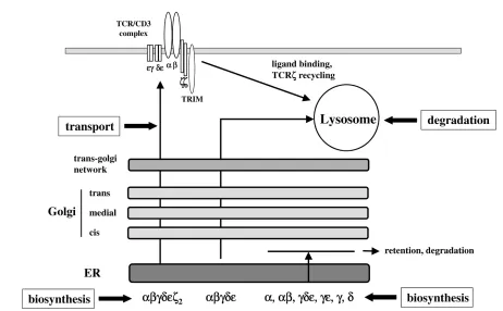
Current concepts of TCR/CD3 complex assembly have been based largely on detailed molecular analyses in T-cell hybridomas using metabolic labelling and pulse-chase experiments. They describe a process where, for full func-tion, the polymorphic TCRαβ chains associate with the invariant chains (CD3 γ, δ, and εand TCRζ) consisting of noncovalently linked γε and δε heterodimers and disul-phide-linked ζ–ζ homodimers, which transmit signals inside the cell (Fig. 5). Association of TCRζ dimers with newly synthesized hexameric complexes (αβγεδε) results in the transport and subsequent expression of the com-plete TCR/CD3 complex (αβγεδεζ2) at the cell surface. Studies in T-cell hybridomas have revealed that TCRζ is synthesized at ~10% of the rate of other components [58], and therefore the amount of TCRζ available in a given T cell is thought to regulate TCR/CD3 expression at the cell surface.
Our results predicted that expression of TCRζmay be one target of chronic TNF stimulation. Indeed, following closely
the kinetics of IL-2 downregulation, western blotting analy-sis of whole-cell lysates revealed that chronic stimulation with TNF suppressed the expression of TCRζ in a dose-and time-dependent fashion, while concentrations of the protein tyrosine kinases ZAP-70, p56Lck, and p59Fyn were not altered [57]. Furthermore, immunoprecipitation of CD3ε-containing complexes revealed normal concen-trations of CD3ε, γ and δ, indicating that TNF had selec-tive effects on TCRζ expression. Given that TCRζ might be rate limiting for TCR/CD3 assembly, the data were consistent with a model in which TNF appeared to disrupt the assembly of TCR/CD3 complexes through its effects on TCRζ expression. Profound reduction in concentra-tions of cell-surface biotinylated TCRζ in TNF-treated T cells strongly supported this notion [57].
A second unexpected experimental observation provided further evidence that persistent TNF signalling in T cells could perturb TCR/CD3 expression at the cell surface. Immunoblotting analysis of unstimulated and TNF-stimu-lated T cells revealed that the expression of the novel transmembrane adaptor protein TRIM (T-cell-receptor-interacting molecule) was markedly downregulated by TNF treatment [59; Isomäki and Cope, unpublished data]. Closer examination revealed that TRIM expression was reduced by TNF before changes in TCRζ expression could be detected, and that reconstitution of both TRIM and TCRζ expression was required to fully restore TCR responsiveness in TNF-treated cells. The implications of these findings have only recently become apparent, through studies of TRIM expression in human peripheral blood and Jurkat T cells. In collaborative studies with Dr Burkhart Schraven, it was found that the half-life of TCR/CD3 complexes in stable Jurkat clones overexpress-ing TRIM is increased [60]. This in turn leads to increased cell-surface expression of TCR and enhanced signalling responses as determined by intracellular calcium mobilisa-tion. We can conclude from these experiments that sus-tained TNF signals in T cells impair TCR/CD3 assembly not only through its effects on TCRζexpression, but also by reducing the half-life of assembled complexes at the cell surface by downregulating the expression of TRIM (see Fig. 5). The kinetics of these changes, as well as the precise interactions between TCRζ and TRIM, are now being studied. Nevertheless, the findings provided a mole-cular basis for the profound hyporesponsiveness of T cells after TNF stimulation and predicted that downstream TCR signalling pathways might be significantly attenuated as a consequence of these structural constraints.
TNF attenuates membrane-proximal TCR
signalling pathways
[image:7.612.59.299.93.321.2]One of the earliest events detected after TCR ligation is the phosphorylation of tandemly arranged tyrosine residues within immunoreceptor tyrosine-based activation motifs (ITAMs) of TCRζchain and CD3γ, δ, and ε chains
Figure 4
The immunomodulatory effects of TNF during the evolution of the immune response. After TCR ligation, TNF is costimulatory and required for antigen priming. As the immune response proceeds over time, TNF is required to suppress subsequent clonal expansion (TNF+/+; unbroken
line). In TNF-deficient animals (TNF–/–; dotted line), immune responses
S204
by Src family kinases, notably Lck and Fyn [61]. In con-trast to CD3 chains, which contain just one ITAM, TCRζ carries three, providing the TCR/CD3 complex with a signal sensor and amplification module [62,63]. In addi-tion, TCRζ plays a role in proofreading extracellular signals, since differences in the quality, intensity, and dura-tion of the antigenic stimulus are translated into specific patterns of TCRζ phosphorylation [64]. Once phosphory-lated, TCRζ ITAMs function as docking sites for protein tyrosine kinases of the Syk family, such as ZAP-70 [65]. The phosphorylation of several adaptor proteins by ZAP-70 and Src kinases then serves as a link between mem-brane-proximal phosphorylation events and the activation of downstream signalling pathways leading to IL-2 produc-tion, T-cell proliferaproduc-tion, and effector responses [61].
Given that TCRζ functions as a signal-amplification module as well as a key component of TCR/CD3 complex assembly, we reasoned that reduced expression of TCRζ homodimers by TNF might impair membrane-proximal tyro-sine phosphorylation events. A comprehensive analysis of signalling pathways in control and TNF-treated T cells has shown that concentrations of phospho-TCRζare reduced in TNF-treated T cells after TCR ligation [57]. Furthermore, in spite of normal Lck kinase activity, the recruitment of
[image:8.612.69.531.101.387.2]ZAP-70 to phospho-TCRζthrough its SH2 domains and its subsequent phosphorylation were also impaired. The transmembrane adaptor protein linker for activation of T cells (LAT) is an in vivosubstrate for ZAP-70 kinase, and plays a key role in linking membrane-proximal events with both calcium and Ras/MAPK (mitogen-activated protein kinase) pathways [66,67]. LAT phosphorylation was sub-stantially reduced in TNF-treated cells, and, as predicted, intracellular calcium mobilisation was also dramatically attenuated [57]. The precise mechanisms for the down-regulation of TCRζ expression by TNF are not clear. However, TCRζmRNA is reduced in T cells treated with higher concentrations of TNF (2.5 ng/ml). Furthermore, TNF may also reduce TCRζ concentrations indirectly through the generation of reactive oxygen species, since culture of TNF-treated T cells with the glutathione precur-sor, N-acetylcysteine, reverses some but not all of the sig-nalling defects we had documented in TNF-treated T cells, possibly by restoring TCRζexpression [57]. Regardless of the mechanisms, the data were consistent with a model in which proximal signalling was impaired as a direct result of the effects of TNF on TCRζ expression and phosphoryla-tion and suggested a novel mechanism whereby the inflammatory process might suppress T-cell reactivity in RA synovial joints.
Figure 5
S205
Selective uncoupling of downstream
signalling by TNF
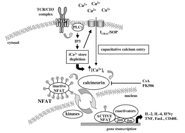
Since the transmembrane adaptor protein LAT functions as a pivotal bifurcation point for downstream Ras/ERK (extracellular signal-regulated kinase) and calcium sig-nalling pathways [67], the reductions in concentrations of phosphorylated LAT would predict that these downstream pathways should be attenuated in TNF-treated T cells. Several lines of preliminary evidence suggest that this pre-diction may be too simplistic. We have been struck by the extent to which TCR-induced calcium responses are attenuated in TNF-treated T cells [49,57]. However, very recent experiments have documented additional defects in calcium signalling that arise through mechanisms indepen-dent of the effects of TNF on membrane-proximal phos-phorylation events and TCR/CD3 expression. For example, while TNF depletes to only a modest extent the thapsigargin-depletable intracellular calcium pool, TNF attenuates to a much greater extent the influx of calcium through store-operated ICRAC (calcium-release-activated calcium current) channels ([68], and Fig. 6). The activation of ICRAC calcium channels and influx of extracellular calcium contributes significantly to the amplitude, dura-tion, and kinetics of the total calcium signal, which in turn is known to profoundly influence gene expression in T cells [69]. We believe that this may provide an additional mechanism through which TNF attenuates gene transcrip-tion through calcium/NFAT-dependent pathways, and may go some way to explain the profound proliferative and cytokine hyporesponsiveness following TCR ligation that we have observed after chronic TNF exposure.
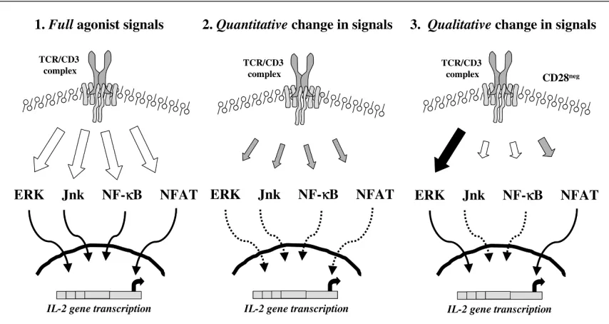
We next undertook a systematic analysis of the Ras/ERK pathway in control and TNF-treated T cells, expecting to document similar degrees of attenuation. The results were unexpected. While concentrations of TCR-induced GTP-Ras are only modestly reduced in TNF-treated T cells, phosphorylation of Raf-1, activation of ERK1/2, induction of c-Fos, and TCR-induced expression of CD69 are unam-biguously preserved. These results would predict that AP (activator protein)-1 transactivation is likely to be spared in TNF-treated T cells. If this is found to be the case, the data define a novel biochemical basis for acquired suppression of T-cell activation based upon selective attenuation of the calcium but not of the Ras/ERK pathways (Fig. 7). At the biochemical level, this result is of particular interest given the observations that anergic T cells have reciprocal defects, namely, reductions in TCR-induced Ras/ERK acti-vation, while calcium responses are spared [70,71].
Aberrant signal transduction pathways in
synovial T cells in rheumatoid arthritis
The effects of chronic TNF described above have pro-vided an experimental framework for exploring the role of inflammatory cytokines in regulating the phenotype of syn-ovial T cells from inflamed joints of patients with RA. Many
of the features of T cells chronically exposed to TNF resemble RA synovial T cells (see Tables 1 and 2). Most notable among these are the nondeletional, reversible T-cell hyporesponsiveness [31–33], the upregulation of T- cell-surface antigens [20], and the repression of CD28 gene expression [72]. These similarities suggest that TNF may play a role in driving this phenotype, and raise the intrigu-ing possibility that there may also be similarities in terms of the aberrations of intracellular signalling pathways that might account for this phenotype.
The pioneering studies of Verweij and colleagues in this field have perhaps most comprehensively and systemati-cally documented proximal TCR signalling in synovial-fluid T cells [73]. The most relevant to this discussion is the downregulation of TCRζchain expression in synovial-fluid T cells in comparison with peripheral blood, as determined by flow cytometry, immunoblotting, or immunohistochem-istry [73,74]. Expression and phosphorylation of p36LAT and its recruitment to the plasma membrane is also reduced in synovial-fluid T cells [75], and both this phe-nomenon and the loss of TCRζ expression can be reversed at least partially by restoring concentrations of glutathione by culturing T cells ex vivo with N -acetylcys-teine [73,75]. This is an important finding, since it indi-cates that TCR signalling pathways are sensitive to reactive oxygen species and redox potential. More recent data indicate that the generation of such oxygen species is regulated by Ras, which is itself expressed in a constitu-tively active GTP bound form in synovial T cells, and that increased concentrations of these oxygen species may influence the tertiary structure and conformation of LAT at the plasma membrane [76].
The similarities between synovial T cells and T cells gener-ated in vitroafter repeated TNF stimulation extend further to calcium responses, including influx through ICRAC chan-nels, which are also attenuated in peripheral blood and joint T cells from patients [77,78], as well as in Jurkat T cells treated with TNF [79]. Using a completely different approach, Isaacs and colleagues have compared the char-acteristics of anergic CD4+T cells and RA synovial T cells
at the mRNA level by differential display RT-PCR [80]. One striking transcriptional event common to both sets of T cells was the downregulation of calmodulin, a gene whose product plays an important role in coupling calcium responses to downstream pathways. Indeed, transcription of calmodulin in RA synovial T cells was less than 1% that in synovial samples from patients with reactive arthritis, who served as the controls in these studies. Expression in synovial T cells was lower than that observed in paired peripheral blood T cells. Interestingly, calmodulin tran-scripts increased 5- to 10-fold after TNF blockade in vivo
S206
selective uncoupling of TCR signalling in chronically active T cells in vivo, similar to that observed in TNF-treated T cells (see Fig. 7). Moreover, direct comparisons of periph-eral blood and joint T cells have established hierarchical attenuation of signalling pathways from peripheral blood to the joint, with the most profound defects being observed in the joint [73–75], suggesting that these signalling anomalies are not inherited but are likely to be acquired through chronic exposure to the environment in inflamed joints over prolonged periods of time.
Are there any clues suggesting selective activation of downstream signalling pathways leading to specific tran-scriptional events at the nuclear level? Data up to now are scarce, but there are isolated reports of constitutive NF-κB activation in RA synovial T cells [81]. The finding of constitutive activation of NF-κB in synovial T cells is of particular interest given the recent studies demonstrating the beneficial therapeutic effects of a T-cell selective NF-κB inhibitory compound (SP100030) in collagen-induced arthritis [82]. The same group have documented attenua-tion of adjuvant arthritis in rats using dominant negative
IKKβ(inhibitor of NF-κB kinase β) delivered by adenoviral vector [83], although when this approach is used, inhibi-tion of NF-κB would not be confined to T cells. In addiinhibi-tion, a recent analysis of constitutive MAPK activation in syn-ovial tissue suggests that there may be preferential activa-tion of ERK in lymphoid aggregates in perivascular tissue [84]. It is perhaps premature to draw any firm conclusions about which transcription factors are active in synovial T cells, other than to state that there is evidence for consti-tutive activation of pathways in vivothat exert a potential for promoting the inflammatory process, and perhaps cell survival.
Implications for the pathogenesis of chronic
inflammatory disease
[image:10.612.120.498.108.381.2]The effects of TNF on downstream TCR signalling path-ways outlined above, together with the studies of calcium signalling in synovial T cells from patients with RA, predict that transactivation of NFAT should be dramatically reduced in the synovial joint. If this indeed is the case, then it provides a molecular framework for exploring further how the inflammatory process might influence
Figure 6
The calcium/calcineurin/NFAT signalling pathway in T cells. After TCR ligation and PLCγ1 activation, newly synthesized IP3 binds to tetrameric IP3 receptor complexes inducing the release of intracellular calcium stores from the sarco-endoplasmic reticulum. Store depletion leads directly to the opening of ICRACor store-operated channels (SOC) in the plasma membrane through mechanisms that are unclear. This leads ultimately to activation of the serine phosphatase calcineurin, dephosphorylation of NFAT, and translocation of this transcription factor to the nucleus. For many genes, NFAT binds cooperatively to AP-1 complexes for optimal gene transcription. AP, activator protein; CsA, cyclosporin A; iCa2+, intracellular
calcium; ICRAC, calcium-release-activated calcium current; IP, inositol phosphate; P, phosphate group; PLCγ, phospholipase Cγ; TCR, T-cell
S207 immunity and inflammation in vivo. For example, NFAT is
required for the transcription of many genes involved in the initiation of the immune response, cell growth and differen-tiation, the induction of immunoregulatory cytokines, host defence, and resolution of the immune response through activation-induced cell death [85]. Accordingly, defects in this pathway would lead not only to depressed immunity and the failure to generate productive Th effector responses, but also to the failure of tolerance by impaired TCR-induced expression of FasL (Fas ligand), attenuation of activation-induced cell death, and the failure to mount significant immunoregulatory responses.
Our own studies of the effects of chronic TNF signalling emphasise the potential for cytokine-dependent, antigen-independent effector mechanisms, driven perhaps through chronic stimulation of the κB pathway. Sustained NF-κB activation would promote trafficking to sites of inflam-mation, as well as enhance the survival of cells in the inflamed joint, thereby promoting effector responses dependent on cell-to-cell interactions [86,87]. Preliminary phenotyping and genotyping analyses in our laboratory suggest that a number of potential candidates may be upregulated on the cell surface of TNF-treated T cells as a direct consequence of chronic NF-κB activation. These include RANK (receptor activator of nuclear factor κB)
ligand, whose upregulation enhances osteoclastogenesis and bone resorption; β integrins, which promote T-cell trafficking to inflamed joints; and CD69, a surface antigen that has been shown to promote signalling between macrophages and T cells and inflammatory cytokine pro-duction [87]. This switch from ‘antigen mode’ to ‘inflam-mation mode’ and the generation of antigen-independent effector responses in T cells (see Fig. 2) suggests that conventional therapeutic approaches for modulating T-cell reactivity may need to be revised.
Future prospects for therapy
[image:11.612.91.518.98.325.2]It has long been recognised that cellular immunity and, in particular, T-cell activation are restored after treatment with remission-inducing therapy, regardless of the disease-modifying agent. We now know that anti-TNF treatment is no exception [44]. The question of whether inflammatory disease remits as a consequence of the recovery of immune competence or in spite of it has never been addressed in depth. On the basis of the available data, we can only conclude that recovery of T-cell reactiv-ity is compatible with attenuation of the disease process and does not seem to exacerbate the inflammatory process. The possibility that normalisation of function of a subset of specific T cells with anti-inflammatory activity occurs is consistent with this conclusion.
Figure 7
S208
Results from several laboratories, including our own, suggest that therapeutic strategies aimed at restoring T-cell homeostasis should be given serious consideration, and should in addition take into account the effects of the inflammatory process on thymic function [50,80,88,89]. This therapeutic approach is in line with the thesis propos-ing that susceptibility to autoimmunity arises not through clonal expansion of autoaggressive effector T cells as a primary event, but more through the failure of the adaptive immune system to regulate an inflammatory response [90].
How could recovery of T-cell regulatory activity be achieved in man? A major challenge in the short term will be to define more precisely a phenotype for regulatory T-cell subsets so that their frequency can be studied in peripheral blood and at sites of inflammation. It would be of particular interest to establish whether their TCR sig-nalling responses and regulatory function correlate inversely with disease activity. Such studies might include analyses of the new generation of ‘suppressor’ T cells such as IL-10-producing, Tr1-like CD4+ cells,
CD4+CD25+regulatory T cells, or IL-16-producing CD8+
T cells [91–94]. If, in the longer term, antigenic specificity can be established, T-cell responsiveness could be restored towards ‘normal’ (but notbeyond) by combining peptide therapy with anti-TNF. Precise knowledge of the specific TCR signalling defects could facilitate the moni-toring of such therapy, so that any potential for rebound hyper-reactivity of bystander T cells and its deleterious consequences, including systemic autoimmune disease, could be substantially reduced [95,96]. Anti-TNF in com-bination with nondepleting anti-CD4 or anti-CD3 mAb might have similar beneficial therapeutic effects.
There exists an alternative to the hypothesis that the acquisition of T-cell hyporesponsiveness promotes the inflammatory process. For example, defective T-cell reac-tivity at sites of inflammation may turn out to be an essen-tial adaptive response for suppressing autoreactivity, as suggested by studies in mice (50,88). In this event, it would be important to understand how the inflammatory process uncouples signalling pathways, since this might facilitate the development of novel immunosuppressive agents. Rather than target pathways that are already sup-pressed, such as the calcium/calcineurin pathway, these strategies might attenuate those that are dominant and that drive the inflammatory process. According to this model, the Ras/ERK and NF-κB signalling pathways would be good candidates.
Concluding remarks
The molecular events that shape the early phase of Th cell differentiation in regional lymph nodes are quite likely to be distinct from those imposed by the environment of an inflamed joint. It follows from this that the design of strate-gies for manipulating T-cell effector responses should be
governed by the chronicity of the immune response. Until now, therapies that target T cells have been based largely upon the potency of agents in acute immunoinflammatory responses in the laboratory. With a growing knowledge base of the characteristics and phenotype of chronically activated T cells, we can look forward to a new generation of therapeutics targeting selective intracellular pathways involved directly in promoting the chronic inflammatory process. Whether such strategies will necessitate targeted immunoablation or restoration of T-cell homeostasis remains to be seen. However, when considering the available options, it should be borne in mind that protection against foreign pathogens is the primary function of the immune system and that this immunity is provided to the host at the expense of a huge propensity for cross-reactivity to self tissue antigens. My own bias, which is derived in part from the working model illustrated in Fig. 2, would be to focus on methods of reconstituting the immune system of patients with the regulatory networks that keep this cross-reactivity in check and the vast majority of individuals in good health.
Glossary of terms
c-Maf = a transcription factor specific for Th2 cells; ERM = a transcription factor specific for Th1 cells; ICRAC = calcium-release-activated calcium current; ITAM = immunoreceptor tyrosine-based activation motifs; GATA-3 = a transcription factor specific for Th2 cells; T-bet = a transcription factor specific for Th1 cells.
Acknowledgements
I gratefully acknowledge Professors Marc Feldmann, David Wallach, and Hugh McDevitt, in whose laboratories much of this work was undertaken, my mentor Professor Tiny Maini for guidance and support throughout my career, and members of the ‘Cope lab’, who are contin-uing with this work. These studies have been funded by The Wellcome Trust and the Arthritis Research Campaign.
References
1. Avni O, Rao A: T cell differentiation: a mechanistic view.Curr Opin Immunol2000, 12:654-659. [key review]
2. Mossman TR, Coffman RL: TH1 and TH2 cells: different pat-terns of lymphokine secretion lead to different functional properties.Annu Rev Immunol1989, 7:145-173. [key review] 3. Seder RA, Paul WE, Davis MM, Fazekas de St Groth B: The
presence of interleukin 4 during in vitro priming determines the lymphokine-producing potential of CD4+ T cells from T cell receptor transgenic mice. J Exp Med 1992, 176:1091-1098. [general reference]
4. Hosken NA, Shibuya K, Heath AW, Murphy KM, O’Garra A: The effect of antigen dose on CD4+ T helper cell phenotype development in a T cell receptor-alpha beta-transgenic model.J Exp Med1995, 182:1579-1584. [general reference] 5. Dustin ML, Cooper JA: The immunological synapse and the
actin cytoskeleton: molecular hardware for T cell signaling. Nat Immunol2000, 1:23-29. [key review]
6. Murphy KM, Ouyang W, Farrar JD, Yang J, Ranganath S, Asnagli H, Afkarian M, Murphy TL: Signaling and transcription in T helper development.Annu Rev Immunol2000, 18:451-494. [key review] 7. Ouyang W, Ranganath SH, Weindel K, Bhattacharya D, Murphy
TL, Sha WC, Murphy KM: Inhibition of Th1 development medi-ated by GATA-3 through an IL-4-independent mechanism. Immunity1998, 9:745-755. [archival research]
S209 9. Szabo SJ, Kim ST, Costa GL, Zhang X, Fathman CG, Glimcher
LH: A novel transcription factor, T-bet, directs Th1 lineage commitment. Cell2000, 100:655-669. [archival research] 10. Ouyang W, Jacobson NG, Bhattacharya D, Gorham JD, Fenoglio
D, Sha WC, Murphy TL, Murphy KM: The Ets transcription factor ERM is Th1-specific and induced by IL-12 through a Stat4-dependent pathway.Proc Natl Acad Sci U S A1999, 96:3888-3893. [general reference]
11. Vignali M, Hassan AH, Neely KE, Workman JL: ATP-dependent chromatin-remodeling complexes. Mol Cell Biol 2000, 20: 1899-1910. [key review]
12. Ouyang W, Lohning M, Gao Z, Assenmacher M, Ranganath S, Radbruch A, Murphy KM: Stat6-independent GATA-3 autoacti-vation directs IL-4-independent Th2 development and com-mitment.Immunity2000, 12:27-37. [general reference] 13. Blackwood EM, Kadonaga JT: Going the distance: a current
view of enhancer action.Science1998, 281:61-63. [general ref-erence]
14. Fitzpatrick DR, Shirley KM, McDonald LE, Bielefeldt-Ohmann H, Kay GF, Kelso A: Distinct methylation of the interferon gamma (IFN-gamma) and interleukin 3 (IL-3) genes in newly activated primary CD8+ T lymphocytes: regional IFN-gamma promoter demethylation and mRNA expression are heritable in CD44(high)CD8+ T cells. J Exp Med 1998, 188:103-117. [general reference]
15. Bird JJ, Brown DR, Mullen AC, Moskowitz NH, Mahowald MA, Sider JR, Gajewski TF, Wang CR, Reiner SL: Helper T cell differ-entiation is controlled by the cell cycle.Immunity1998, 9:229-237. [general reference]
16. Germain RN, Stefanova I: The dynamics of T cell receptor sig-naling: complex orchestration and the key roles of tempo and cooperation.Annu Rev Immunol1999, 17:467-522. [key review] 17. Young CL, Adamson TC, Vaughan JH, Fox RI: Immunohistologic characterization of synovial membrane lymphocytes in rheumatoid arthritis.Arthritis Rheum1984, 27:32-39. [general reference]
18. Wagner UG, Kurtin PJ, Wahner A, Brackertz M, Berry DJ, Goronzy JJ, Weyand CM: The role of CD8+ CD40L+ T cells in the for-mation of germinal centers in rheumatoid synovitis.J Immunol 1998, 161:6390-6397. [general reference]
19. Takemura S, Braun A, Crowson C, Kurtin PJ, Cofield RH, O’Fallon WM, Goronzy JJ, Weyand CM: Lymphoid neogenesis in rheumatoid synovitis. J Immunol 2001, 167:1072-1080. [general reference]
20. Cush JJ, Lipsky PE: Phenotypic analysis of synovial tissue and peripheral blood lymphocytes isolated from patients with rheumatoid arthritis. Arthritis Rheum 1988, 31:1230-1238. [general reference]
21. Thomas R, McIlraith M, Davis LS, Lipsky PE: Rheumatoid syn-ovium is enriched in CD45RBdim mature memory T cells that are potent helpers for B cell differentiation.Arthritis Rheum 1992, 35:1455-1465. [general reference]
22. Katschke KJ Jr, Rottman JB, Ruth JH, Qin S, Wu L, LaRosa G, Ponath P, Park CC, Pope RM, Koch AE: Differential expression of chemokine receptors on peripheral blood, synovial fluid, and synovial tissue monocytes/macrophages in rheumatoid arthritis. Arthritis Rheum 2001, 44:1022-1032. [general refer-ence]
23. Salmon M, Scheel-Toellner D, Huissoon AP, Pilling D, Sham-sadeen N, Hyde H, D’Angeac AD, Bacon PA, Emery P, Akbar AN: Inhibition of T cell apoptosis in the rheumatoid synovium. J Clin Invest1997, 99:439-446. [general reference]
24. Koetz K, Bryl E, Spickschen K, O’Fallon WM, Goronzy JJ, Weyand CM: T cell homeostasis in patients with rheumatoid arthritis. Proc Natl Acad Sci U S A2000, 97:9203-9208. [general refer-ence]
25. Simon AK, Seipelt E, Sieper J: Divergent T-cell cytokine pat-terns in inflammatory arthritis.Proc Natl Acad Sci U S A1994, 91:8562-8566. [general reference]
26. Morita Y, Yamamura M, Kawashima M, Harada S, Tsuji K, Shibuya K, Maruyama K, Makino H: Flow cytometric single-cell analysis of cytokine production by CD4+ T cells in synovial tissue and peripheral blood from patients with rheumatoid arthritis. Arthritis Rheum1998, 41:1669-1676. [general reference] 27. Ronnelid J, Berg L, Rogberg S, Nilsson A, Albertsson K,
Klareskog L: Production of T-cell cytokines at the single-cell level in patients with inflammatory arthritides: enhanced
activ-ity in synovial fluid compared to blood. Br J Rheumatol1998, 37:7-14. [general reference]
28. Dolhain RJ, van der Heiden AN, ter Haar NT, Breedveld FC, Mil-tenburg AM: Shift toward T lymphocytes with a T helper 1 cytokine-secretion profile in the joints of patients with rheumatoid arthritis. Arthritis Rheum 1996, 39:1961-1969. [general reference]
29. Davis LS, Cush JJ, Schulze-Koops H, Lipsky PE: Rheumatoid synovial CD4+ T cells exhibit a reduced capacity to differenti-ate into IL-4-producing T-helper-2 effector cells.Arthritis Res 2001, 3:54-64. [general reference]
30. Feldmann M, Brennan FM, Maini RN: Role of cytokines in rheumatoid arthritis. Annu Rev Immunol 1996, 14:397-440. [key review]
31. Malone DG, Wahl SM, Tsokos M, Cattell H, Decker JL, Wilder RL: Immune function in severe, active rheumatoid arthritis. A rela-tionship between peripheral blood mononuclear cell prolifera-tion to soluble antigens and synovial tissue immunohistologic characteristics.J Clin Invest 1984, 74:1173-85. [general refer-ence]
32. Emery P, Panayi GS, Nouri AM: Interleukin-2 reverses deficient cell-mediated immune responses in rheumatoid arthritis. Clin Exp Immunol1984, 57:123-129. [general reference]
33. Emery P, Panayi GS, Welsh KI, Cole BC: Relationship of HLA-DR4 to defective cellular immunity in rheumatoid arthritis using PPD, and mycoplasma and lectin mitogens.J Rheumatol 1985, 12:859-864. [general reference]
34. Firestein GS, Xu WD, Townsend K, Broide D, Alvaro-Gracia J, Glasebrook A, Zvaifler NJ: Cytokines in chronic inflammatory arthritis. I. Failure to detect T cell lymphokines (interleukin 2 and interleukin 3) and presence of macrophage colony-stimu-lating factor (CSF-1) and a novel mast cell growth factor in rheumatoid synovitis. J Exp Med 1988, 168:1573-1586. [general reference]
35. Stastny P. Association of the B-cell alloantigen DRw4 with rheumatoid arthritis. N Engl J Med 1978, 298: 869-871. [archival research]
36. Panayi GS, Wooley PH, Batchelor JR: HLA-DRw4 and rheuma-toid arthritis.Lancet1979, 1:730. [key review]
37. Buchan G, Barrett K, Turner M, Chantry D, Maini RN, Feldmann M: Interleukin-1 and tumour necrosis factor mRNA expression in rheumatoid arthritis: prolonged production of IL-1αα. Clin Exp Immunol1988, 73:449-455. [general reference]
38. Saxne T, Palladino MA Jr, Heinegard D, Talal N, Wollheim FA: Detection of tumor necrosis factor alpha but not tumor necro-sis factor beta in rheumatoid arthritis synovial fluid and serum. Arthritis Rheum1988, 31:1041-1045. [archival research] 39. Brennan FM, Chantry D, Jackson A, Maini R, Feldmann M: Inhibitory effect of TNF alpha antibodies on synovial cell inter-leukin-1 production in rheumatoid arthritis. Lancet1989, 2: 244-247. [archival research]
40. Chu CQ, Field M, Feldmann M, Maini RN: Localization of tumor necrosis factor alpha in synovial tissues and at the cartilage-pannus junction in patients with rheumatoid arthritis.Arthritis Rheum1991, 34:1125-1132. [archival research]
41. Brennan FM, Gibbons DL, Mitchell T, Cope AP, Maini RN, Feld-mann M: Enhanced expression of tumor necrosis factor recep-tor mRNA and protein in mononuclear cells isolated from rheumatoid arthritis synovial joints.Eur J Immunol1992, 22: 1907-1912. [archival research]
42. Deleuran BW, Chu CQ, Field M, Brennan FM, Mitchell T, Feld-mann M, Maini RN: Localization of tumor necrosis factor recep-tors in the synovial tissue and cartilage-pannus junction in patients with rheumatoid arthritis. Implications for local actions of tumor necrosis factor alpha.Arthritis Rheum1992, 35:1170-1178. [archival research]
43. Cope AP, Aderka D, Doherty M, Engelmann H, Gibbons D, Jones AC, Brennan FM, Maini RN, Wallach D, Feldmann M: Increased levels of soluble tumor necrosis factor receptors in the sera and synovial fluid of patients with rheumatic diseases.Arthritis Rheum1992, 35:1160-1169. [archival research]
S210
45. Yokota S, Geppert TD, Lipsky PE: Enhancement of antigen- and mitogen-induced human T lymphocyte proliferation by tumor necrosis factor-alpha.J Immunol1988, 140:531-536. [general reference]
46. Lorenz HM, Antoni C, Valerius T, Repp R, Grunke M, Schwerdtner N, Nusslein H, Woody J, Kalden JR, Manger B: In vivo blockade of TNF-alpha by intravenous infusion of a chimeric mono-clonal TNF-alpha antibody in patients with rheumatoid arthri-tis. Short term cellular and molecular effects.J Immunol1996, 156:1646-1653. [general reference]
47. Maurice MM, van der Graaff WL, Leow A, Breedveld FC, van Lier RA, Verweij CL: Treatment with monoclonal anti-tumor necro-sis factor alpha antibody results in an accumulation of Th1 CD4+ T cells in the peripheral blood of patients with rheuma-toid arthritis.Arthritis Rheum1999, 42:2166-2173. [general ref-erence]
48. Berg L, Lampa J, Rogberg S, van Vollenhoven R, Klareskog L: Increased peripheral T cell reactivity to microbial antigens and collagen type II in rheumatoid arthritis after treatment with soluble TNFalpha receptors. Ann Rheum Dis 2001, 60:133-139. [general reference]
49. Cope AP, Liblau RS, Yang XD, Congia M, Laudanna C, Schreiber RD, Probert L, Kollias G, McDevitt HO: Chronic tumor necrosis factor alters T cell responses by attenuating T cell receptor signaling.J Exp Med1997, 185:1573-1584. [general reference] 50. Cope AP. Regulation of autoimmunity by proinflammatory
cytokines.Curr Opin Immunol1998, 10:669-676. [key review] 51. Elliott MJ, Maini RN, Feldmann M, Kalden JR, Antoni C, Smolen
JS, Leeb B, Breedveld FC, Macfarlane JD, Bijl H, Woody J: Ran-domised double-blind comparison of chimeric monoclonal antibody to tumour necrosis factor alpha (cA2) versus placebo in rheumatoid arthritis.Lancet1994, 344:1105-1110. [archival research]
52. Jacob CO, McDevitt HO: Tumour necrosis factor-ααin murine autoimmune ‘lupus’ nephritis. Nature 1988, 331:356-358. [archival research]
53. Gordon C, Ranges GE, Greenspan JS, Wofsy D: Chronic therapy with recombinant tumor necrosis factor-alpha in autoimmune NZB/NZW F1 mice.Clin Immunol Immunopathol 1989, 52:421-434. [general reference]
54. Satoh J, Seino H, Abo T, Tanaka SI, Shintani S, Ohta S, Tamura K, Sawai T, Nobunaga T, Ohteki T, Kumagai K, Toyota T: Recombi-nant human tumor necrosis factor ααsuppresses autoimmune diabetes in non-obese diabetic mice.J Clin Invest1989, 84: 1345-1348. [general reference]
55. Jacob CO, Aiso S, Michie SA, McDevitt HO, Acha-Orbea H: Pre-vention of diabetes in nonobese diabetic mice by tumor necrosis factor (TNF): similarities between TNF-alpha and interleukin 1. Proc Natl Acad Sci U S A 1990, 87:968-972. [general reference]
56. Kassiotis G, Kollias G: Uncoupling the proinflammatory from the immunosuppressive properties of tumor necrosis factor (TNF) at the p55 TNF receptor level.Implications for pathogene-sis and therapy of autoimmune demyelination. J Exp Med2001, 193:427-434. [general reference]
57. Isomäki P, Panesar M, Annenkov A, Clark JM, Foxwell BM, Cher-najovsky Y, Cope AP: Prolonged exposure of T cells to TNF down-regulates TCRzeta and expression of the TCR/CD3 complex at the cell surface.J Immunol2001, 166:5495-5507. [general reference]
58. Minami Y, Weissman AM, Samelson LE, Klausner RD: Building a multichain receptor: synthesis, degradation, and assembly of the T-cell antigen receptor.Proc Natl Acad Sci U S A1987, 84: 2688-2692. [general reference]
59. Bruyns E, Marie-Cardine A, Kirchgessner H, Sagolla K, Shevchenko A, Mann M, Autschbach F, Bensussan A, Meuer S, Schraven B: T cell receptor (TCR) interacting molecule (TRIM), a novel disulfide-linked dimer associated with the TCR-CD3-zeta complex, recruits intracellular signaling proteins to the plasma membrane.J Exp Med1998, 188:561-575. [general ref-erence]
60. Kirchgessner H, Dietrich J, Scherer J, Isomäki P, Korinek V, Hilgert I, Bruyns E, Leo A, Cope AP, Schraven B: The transmembrane adaptor protein TRIM regulates T cell receptor (TCR) expres-sion and TCR-mediated signaling via an association with the TCR zeta chain.J Exp Med2001, 193:1269-1284. [general ref-erence]
61. Weiss A, Littman DR: Signal transduction by lymphocyte antigen receptors. Cell1994, 76:263-274. [key review] 62. Irving BA, Weiss A: The cytoplasmic domain of the T cell
receptor zeta chain is sufficient to couple to receptor-associ-ated signal transduction pathways. Cell 1991, 64:891-901. [archival research]
63. Letourneur F, Klausner RD: Activation of T cells by a tyrosine kinase activation domain in the cytoplasmic tail of CD3 epsilon.Science1992, 255:79-82. [archival research]
64. Sloan-Lancaster J, Shaw AS, Rothbard JB, Allen PM: Partial T cell signaling: altered phospho-zeta and lack of zap70 recruit-ment in APL-induced T cell anergy. Cell 1994, 79:913-922. [general reference]
65. Chan AC, Iwashima M, Turck CW, Weiss A: ZAP-70: a 70 kd protein-tyrosine kinase that associates with the TCR zeta chain. Cell1992, 71:649-662. [archival research]
66. Zhang W, Sloan-Lancaster J, Kitchen J, Trible RP, Samelson LE: LAT: the ZAP-70 tyrosine kinase substrate that links T cell receptor to cellular activation. Cell 1998, 92:83-92. [archival research]
67. Finco TS, Kadlecek T, Zhang W, Samelson LE, Weiss A: LAT is required for TCR-mediated activation of PLCgamma1 and the Ras pathways. Immunity1998, 9:617-626. [general reference] 68. Clark JM, Isomäki P, Panesar M, Anenkov A, Chernajowsky Y,
Cope AP: Prolonged stimulation by TNF uncouples T-cell receptor (TCR) signalling pathways at multiple levels [abstract].Immunol2001, 104:OP269. [general reference] 69. Feske S, Giltnane J, Dolmetsch R, Staudt LM, Rao A: Gene
regu-lation mediated by calcium signals in T lymphocytes. Nat Immunol2001, 2:316-324. [general reference]
70. Kang SM, Beverly B, Tran AC, Brorson K, Schwartz RH, Lenardo MJ: Transactivation by AP-1 is a molecular target of T cell clonal anergy. Science 1992, 257:1134-1138. [general refer-ence]
71. Schwartz RH. T cell clonal anergy.Curr Opinion Immunol1997, 9:351-357. [key review]
72. Bryl E, Vallejo AN, Weyand CM, Goronzy JJ: Down-regulation of cd28 expression by TNF-alpha. J Immunol 2001, 167:3231-3238. [general reference]
73. Maurice MM, Lankester AC, Bezemer AC, Geertsma MF, Tak PP, Breedveld FC, van Lier RA, Verweij CL: Defective TCR-mediated signaling in synovial T cells in rheumatoid arthritis.J Immunol 1997, 159:2973-2978. [general reference]
74. Berg L, Ronnelid J, Klareskog L, Bucht A: Down-regulation of the T cell receptor CD3zeta chain in rheumatoid arthritis (RA) and its influence on T cell responsiveness.Clin Exp Immunol 2000, 120:174-182. [general reference]
75. Gringhuis SI, Leow A, Papendrecht-Van Der Voort EA, Remans PH, Breedveld FC, Verweij CL: Displacement of linker for acti-vation of T cells from the plasma membrane due to redox balance alterations results in hyporesponsiveness of synovial fluid T lymphocytes in rheumatoid arthritis. J Immunol2000, 164:2170-2179. [general reference]
76. Remans PHJ, van Laar JM, Reedquist KA, Papendrecht EAM, Levarht NEW, Bos JI, Breedveld FC, Verweij CL: Deregulated Ras and Rap1 signalling in rheumatoid arthritis synovial fluid T lymphocytes leads to persistent reactive oxygen species production and chronic oxidative stress [abstract]. Arthritis Rheum2001, 44:S2034. [general reference]
77. Allen ME, Young SP, Michell RH, Bacon PA: Altered T lympho-cyte signaling in rheumatoid arthritis.Eur J Immunol1995, 25: 1547-1554. [general reference]
78. Carruthers DM, Arrol HP, Bacon PA, Young SP: Dysregulated intracellular Ca2+ stores and Ca2+ signaling in synovial fluid T lymphocytes from patients with chronic inflammatory arthri-tis. Arthritis Rheum2000, 43:1257-1265. [general reference] 79. Church LD, Goodall JE, Rider DA, Bacon PA, Young SP: TNF-αα
modulates TCR-induced intracellular calcium signalling in T cells: implications for sphingomyelinase activation [abstract]. Immunology2001, 104:OP165. [general reference]
80. Ali M, Ponchel F, Wilson KE, Francis MJ, Wu X, Verhoef A, Boyl-ston AW, Veale DJ, Emery P, Markham AF, Lamb JR, Isaacs JD: Rheumatoid arthritis synovial T cells regulate transcription of several genes associated with antigen-induced anergy.J Clin Invest2001, 107:519-528. [general reference]
S211 patients with rheumatic diseases: a preliminary report. Ann
Rheum Dis1998, 57:738-741. [general reference]
82. Gerlag DM, Ransone L, Tak PP, Han Z, Palanki M, Barbosa MS, Boyle D, Manning AM, Firestein GS: The effect of a T cell-spe-cific NF-kappa B inhibitor on in vitro cytokine production and collagen-induced arthritis. J Immunol 2000, 165:1652-1658. [general reference]
83. Tak PP, Gerlag DM, Aupperle KR, van de Geest DA, Overbeek M, Bennett BL, Boyle DL, Manning AM, Firestein GS: Inhibitor of nuclear factor kappaB kinase beta is a key regulator of syn-ovial inflammation. Arthritis Rheum 2001, 44:1897-1907. [general reference]
84. Schett G, Tohidast-Akrad M, Smolen JS, Schmid BJ, Steiner CW, Bitzan P, Zenz P, Redlich K, Xu Q, Steiner G: Activation, differ-ential localization, and regulation of the stress-activated protein kinases, extracellular signal-regulated kinase, c-JUN N-terminal kinase, and p38 mitogen-activated protein kinase, in synovial tissue and cells in rheumatoid arthritis. Arthritis Rheum2000, 43:2501-2512. [general reference]
85. Rao A, Luo C, Hogan PG: Transcription factors of the NFAT family: regulation and function. Annu Rev Immunol1997, 15: 707-747. [key review]
86. Lacraz S, Isler P, Vey E, Welgus HG, Dayer JM: Direct contact between T lymphocytes and monocytes is a major pathway for induction of metalloproteinase expression. J Biol Chem 1994, 269:22027-22033. [general reference]
87. McInnes IB, Leung BP, Sturrock RD, Field M, Liew FY: Inter-leukin-15 mediates T cell-dependent regulation of tumor necrosis factor-alpha production in rheumatoid arthritis. Nature Med1997, 3:189-195. [general reference]
88. Cope A, Ettinger R, McDevitt H: The role of TNF alpha and related cytokines in the development and function of the autoreactive T-cell repertoire. Res Immunol 1997, 148:307-312. [key review]
89. Goronzy JJ, Weyand CM: Thymic function and peripheral T-cell homeostasis in rheumatoid arthritis. Trends Immunol 2001, 22:251-255. [key review]
90. Mason D, Powrie F: Control of immune pathology by regulatory T cells.Curr Opin Immunol 1998, 10:649-655. [key review] 91. Groux H, O’Garra A, Bigler M, Rouleau M, Antonenko S, de Vries
JE, Roncarolo MG: A CD4+ T-cell subset inhibits antigen-spe-cific T-cell responses and prevents colitis.Nature1997, 389: 737-742. [general reference]
92. Stephens LA, Mottet C, Mason D, Powrie F: Human CD4(+)CD25(+) thymocytes and peripheral T cells have immune suppressive activity in vitro.Eur J Immunol2001, 31: 1247-1254. [general reference]
93. Jonuleit H, Schmitt E, Stassen M, Tuettenberg A, Knop J, Enk AH: Identification and functional characterization of human CD4(+)CD25(+) T cells with regulatory properties isolated from peripheral blood. J Exp Med 2001, 193:1285-1294. [general reference]