Telomerase mutations in smokers with severe
emphysema
Susan E. Stanley, … , Kathleen C. Barnes, Mary Armanios
J Clin Invest.
2015;125(2):563-570. https://doi.org/10.1172/JCI78554.
Mutations in the essential telomerase genes
TERT
and
TR
cause familial pulmonary
fibrosis; however, in telomerase-null mice, short telomeres predispose to emphysema after
chronic cigarette smoke exposure. Here, we tested whether telomerase mutations are a risk
factor for human emphysema by examining their frequency in smokers with chronic
obstructive pulmonary disease (COPD). Across two independent cohorts, we found 3 of 292
severe COPD cases carried deleterious mutations in
TERT
(1%). This prevalence is
comparable to the frequency of alpha-1 antitrypsin deficiency documented in this
population. The
TERT
mutations compromised telomerase catalytic activity, and mutation
carriers had short telomeres. Telomerase mutation carriers with emphysema were
predominantly female and had an increased incidence of pneumothorax. In families,
emphysema showed an autosomal dominant inheritance pattern, along with pulmonary
fibrosis and other telomere syndrome features, but manifested only in smokers. Our findings
identify germline mutations in telomerase as a Mendelian risk factor for COPD susceptibility
that clusters in autosomal dominant families with telomere-mediated disease including
pulmonary fibrosis.
Research Article
Pulmonology
Find the latest version:
Introduction
Chronic obstructive pulmonary disease (COPD) is the third lead-ing cause of death in the United States (1). Aside from cigarette smoke, age is the major risk factor. However, only a subset of smokers, approximately 10%, develops COPD, and the cluster-ing of emphysema in families has suggested that genetic factors explain a significant portion of this susceptibility (2). Alpha-1 anti-trypsin deficiency is the known Mendelian cause for emphysema (2). It manifests autosomal recessive inheritance because of bial-lelic mutations in the SERPINA1 gene and accounts for young- onset, severe COPD in 0.5%–1% of smokers of European descent (3). The other monogenic factors that underlie COPD susceptibil-ity are not fully known (2, 4).
Telomeres are the DNA-protein structures that protect chro-mosome ends. Telomeres shorten with cell division and advanc-ing age, and short dysfunctional telomeres signal a DNA damage response that provokes cellular senescence and apoptosis (5, 6). Telomerase is the specialized polymerase that synthesizes new
telomere repeats (7–9). It has two core components: TERT, the telomerase reverse transcriptase, and TR, the telomerase RNA that provides the template for telomere repeat addition (9, 10). Mutations in TERT and TR cause telomerase haploinsufficiency, and the consequent short telomere defect is most frequently rec-ognized in clinical settings as autosomal dominant pulmonary fibrosis (6). Even though lung disease is the primary life-threaten-ing presentation in these patients, the telomere defect is systemic and can manifest concurrently in a predictable, syndromic pattern that includes bone marrow failure, liver disease, and osteoporosis (11, 12). Because extrapulmonary telomere-mediated disease can cause significant morbidity, recognizing this subset of pulmonary fibrosis patients has been shown to be relevant for treatment deci-sions in several settings (6, 12, 13).
We recently sought to understand the mechanisms by which telomere dysfunction causes pulmonary fibrosis by studying telomerase-null mice with short telomeres. Although these mice have no obvious de novo lung defects, they develop emphysema after chronic cigarette smoke exposure (14). Coincident with these findings, we identified a family with a deleterious TR muta-tion that included two female siblings who developed premature emphysema, and emphysema combined with fibrosis, after mod-est smoking histories (14). Another family was recently reported that had a similar clustering of emphysema with fibrosis in associ-ation with a TERT mutassoci-ation (15). The observassoci-ations in mice, in light of these anecdotal cases, led us to systematically test the hypoth-esis that mutations in telomerase may be a risk factor for emphy-sema in unselected populations. Using a candidate gene approach, Mutations in the essential telomerase genes TERT and TR cause familial pulmonary fibrosis; however, in telomerase-null
mice, short telomeres predispose to emphysema after chronic cigarette smoke exposure. Here, we tested whether telomerase mutations are a risk factor for human emphysema by examining their frequency in smokers with chronic obstructive pulmonary disease (COPD). Across two independent cohorts, we found 3 of 292 severe COPD cases carried deleterious mutations in TERT (1%). This prevalence is comparable to the frequency of alpha-1 antitrypsin deficiency documented in this population. The TERT mutations compromised telomerase catalytic activity, and mutation carriers had short telomeres. Telomerase mutation carriers with emphysema were predominantly female and had an increased incidence of pneumothorax. In families, emphysema showed an autosomal dominant inheritance pattern, along with pulmonary fibrosis and other telomere syndrome features, but manifested only in smokers. Our findings identify germline mutations in telomerase as a Mendelian risk factor for COPD susceptibility that clusters in autosomal dominant families with telomere-mediated disease including pulmonary fibrosis.
Telomerase mutations in smokers with
severe emphysema
Susan E. Stanley,1,2 Julian J.L. Chen,3 Joshua D. Podlevsky,3 Jonathan K. Alder,1 Nadia N. Hansel,4 Rasika A. Mathias,4
Xiaodong Qi,3 Nicholas M. Rafaels,4 Robert A. Wise,4 Edwin K. Silverman,5 Kathleen C. Barnes,4,6 and Mary Armanios1,6
1Department of Oncology and 2Medical Scientist Training Program, Johns Hopkins University School of Medicine, Baltimore, Maryland, USA. 3Department of Chemistry and Biochemistry, Arizona State
University, Tempe, Arizona, USA. 4Department of Medicine, Johns Hopkins University School of Medicine, Baltimore, Maryland, USA. 5Channing Division of Network Medicine and Department of Medicine,
Brigham and Women’s Hospital and Harvard Medical School, Boston, Massachusetts, USA. 6McKusick-Nathans Institute of Genetic Medicine, Johns Hopkins University School of Medicine,
Baltimore, Maryland, USA.
Conflict of interest: None of the authors have a direct conflict of interest that is relevant
to this study. Outside the content of the study, Robert A. Wise has received consulting fees from the following: AstraZeneca, Boehringer Ingelheim, GlaxoSmithKline, Novartis, Forest Laboratories, Sunovion, Pfizer, Merck, Genentech, MedImmune, Mylan, Janssen, Spiration, and Pulmonx. He has received grant support from GlaxoSmithKline, Boehringer Ingelheim, Forest Laboratories, and Pearl Therapeutics. In the past three years, Edwin K. Silverman has received honoraria and consulting fees from Merck and grant support and consulting fees from GlaxoSmithKline.
Submitted: August 15, 2014; Accepted: November 25, 2014.
DNA from the COPDGene parent study and confirmed the presence of these TERT variants by Sanger sequencing (Figure 1A).
We next tested the frequency of telo-merase mutations in a second cohort of smok-ers with severe COPD. We selected cases from the Lung Health Study (18) that matched the criteria of age and forced expiratory volume in 1 second (FEV1) used in the COPD Gene exome sequencing study and examined the TERT and TR sequences along with the severe alpha-1 antitrypsin deficiency SER-PINA1 alleles (Table 1). Among 83 cases that fulfilled these criteria, we found no homozy-gous SERPINA1 mutations, but we identified an individual with a heterozygous missense TERT mutation: His925Gln (Figure 1A). This mutation was previously reported in several members of a family with pulmonary fibrosis and liver disease (19). In total, across the two COPD cohorts, there were three TERT vari-ants among the severe COPD cases (3 of 292, 1%), and all of them fell in conserved motifs within the telomerase reverse transcriptase domain (Supplemental Figure 3). The TERT gene contains few deviations from reference sequence (20, 21), and none of these variants were found in 2,020 controls, including 1,092 from the 1000 Genomes Project (http://www.1000genomes. org/), 528 published controls (21), and 400 individuals of similar European ancestry whom we additionally sequenced (Supplemen-tal Table 1). These data suggested a clustering of rare telomerase variants in smokers with severe emphysema.
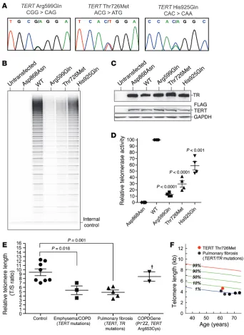
TERT variants compromise telomerase function and telomere length. To test the functional significance of the rare variants, we reconstituted each of the TERTs in cells. We measured telomerase enzyme activity by quantifying the telomere products using the direct primer-extension assay. The three emphysema-associated TERT variants substantially compromised enzyme activity com-pared with wild-type telomerase as evidenced by the decreased intensity of the telomere repeat ladder (P < 0.001, Student’s t test, Figure 1, B–D). The reduction in activity was similar to that resulting from pathogenic TERT and TR mutations documented in pulmonary fibrosis (12, 22). In contrast, the enzyme activity of the TERT variant from the control group was comparable to that of wild-type telomerase (Supplemental Figure 2). These data indi-cated that the clinical phenotype of severe emphysema enriched for individuals with rare, deleterious TERT mutations and that this clustering was statistically significant (3 of 292 COPD subjects [1%] vs. 0 of 2,226 controls [2,020 healthy controls and 206 con-trol smokers], P = 0.002, Fisher’s exact test).
We examined the functional impact of the variants in vivo by measuring telomere length. In two deceased subjects we mea-sured telomere length using archived DNA by quantitative PCR and found it was short relative to that in age-matched healthy controls (P = 0.018, Student’s t test, Figure 1E) and comparable to that in TERT and TR mutation carriers with idiopathic pulmonary in two cohorts of smokers with severe emphysema/COPD, along
with pedigree data from a Johns Hopkins–based study, we show that deleterious telomerase mutations are a risk factor for COPD and may occur at a frequency that is similar to alpha-1 antitrypsin deficiency in individuals with severe, early-onset disease.
Results
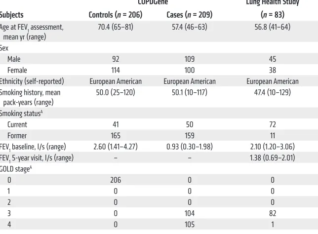
[image:3.585.37.359.88.325.2]Rare TERT variants cluster with the severe COPD phenotype. To test whether telomerase mutations are a risk factor for emphysema, we examined the telomerase gene sequences in exome data from the COPDGene cohort, a study designed to understand the genetic determinants of COPD susceptibility. These data have been recently made accessible through the National Heart Lung and Blood Institute (NHLBI) Exome Sequencing Project (Lung GO) (16). The clinical characteristics of the control group (n = 206) and emphysema subjects (n = 209) are summarized in Table 1. We designed a filtering strategy to identify novel or rare variants that could then be functionally examined (Supplemental Figure 1; sup-plemental material available online with this article; doi:10.1172/ JCI78554DS1). In the control group, one rare TERT variant, Arg-653Cys, was identified; however, this missense substitution did not affect telomerase catalytic activity (97% ± 5% of wild-type telomerase activity ± SEM, P = 0.49, Student’s t test, Supplemental Figure 2). In contrast, among the smokers with emphysema, there were two heterozygous missense variants in TERT: Arg599Gln and Thr726Met (2 of 209, 1%) that we subsequently found to be func-tionally deleterious, as shown below. The Arg599Gln variant was, to our knowledge, novel, and Thr726Met was previously reported in a child with bone marrow failure (17). We obtained archived
Table 1. Clinical characteristics of subjects analyzed for telomerase gene sequences
COPDGene Lung Health Study Subjects Controls (n = 206) Cases (n = 209) (n = 83)
Age at FEV1 assessment,
mean yr (range) 70.4 (65–81) 57.4 (46–63) 56.8 (41–64) Sex
Male 92 109 45
Female 114 100 38 Ethnicity (self-reported) European American European American European American Smoking history, mean
pack-years (range) 50.0 (25–120) 50.1 (10–117) 47.4 (10–129) Smoking statusA
Current 41 50 72
Former 165 159 11 FEV1 baseline, l/s (range) 2.60 (1.41–4.27) 0.93 (0.30–1.98) 2.10 (1.20–3.06) FEV1 5-year visit, l/s (range) – – 1.38 (0.69–2.01) GOLD stageA
0 206 0 0
1 0 0 0
2 0 0 0
3 0 104 82
4 0 105 1
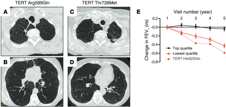
COPDGene subjects showed apical airspace destruction, and one subject had additional interstitial lung abnormalities and bron-chiectasis (Figure 2, A–D). The dyspnea was severe, requiring sup-plemental oxygen support, and one subject died within 5 years of enrollment (Supplemental Table 2). Lung Health Study subjects had spirometry documented over 5 years, and, in the subject with the TERT His925Gln mutation, lung function declined faster than the lowest quartile of this 5,887-person cohort of smokers (18), suggesting an accelerated disease course (Figure 2E). Family his-tory was not detailed in study records, but we learned that the only living subject reported her mother, who was a smoker, was oxygen- dependent for the diagnosis of COPD.
Female gender and telomerase-associated emphysema. Mutations in telomerase and short telomeres are a risk factor for pulmo-nary fibrosis (20), and we sought to understand the predictors of emphysema onset compared with fibrosis. We examined the clini-cal features of 50 telomere syndrome cases with lung disease that fibrosis (P = 0.66). Telomere length in the telomerase-associated
emphysema cases was also short compared with that in the con-trol with the functionally intact TERT variant as well as the SER-PINA1 mutation carrier with alpha-1 antitrypsin deficiency from the COPD Gene Study (Figure 1E). We additionally measured lymphocyte telomere length by flow cytometry and FISH in the one living subject we could contact and found it fell near the 10th age-adjusted percentile, similar to that in mutation carriers with idiopathic pulmonary fibrosis who were also included in the quan-titative PCR telomere length analysis (Figure 1, E and F). These data supported the observation that the emphysema-associated TERT variants we identified were functionally deleterious.
[image:4.585.40.382.53.523.2]Telomerase mutations may be associated with a more severe emphysema phenotype. We examined the clinical data available from COPDGene and the Lung Health Study records and found all three mutation carriers were female with a mean age of 48 years at diagnosis (Supplemental Table 2). Chest CT scans in the
Figure 1. Functional consequences of telomerase variants identified by chronic obstructive pulmonary disease subjects. (A)
Chromatograms of PCR-amplified products of variants identified by next-generation sequencing. (B) Gel image of telomere
repeat ladder generated from wild-type and mutant telomerases reconstituted in vivo and immunopurified. The decreased intensity of the DNA repeat products reflects impaired enzymatic activity of TERT Arg599Gln, Thr726Met, and His925Gln. TERT Asp868Asn is a negative control, catalytically defective in one of the aspartic acid residues essential for reverse transcription. 32P end-labeled 18-mer oligonucleotide was included as an internal control for the recovery of DNA products. (C)
Northern blot for TR levels from immunopuri-fied telomerases (top). Western blot for TERT expression in cells (bottom) was performed with anti-FLAG and anti-GAPDH antibodies for ectopically expressed FLAG-tagged TERT and endogenous GAPDH, respectively. (D)
Mean telomerase activity was derived from 4 activity assays from cell lysates prepared from two separate transfections. (E) Relative
telomere length as measured by quantitative PCR in age-matched controls (ages 37–64,
n = 8), TERT mutation carriers (ages 46–57,
n = 3) from the COPDGene Study and the Lung Health Study (LHS), telomerase mutation carriers with pulmonary fibrosis (ages 45–63,
TERTn = 2, TRn = 3), and COPDGene controls: a homozygous SERPINA1 Glu366Lys mutation carrier (formerly coded Glu342Lys, rs28929474, PI*ZZ genotype, age 46) and the control TERT
Arg653Cys variant (age 68). T/S ratio, ratio of telomere repeat number to single gene copy number. †PI*ZZ mutation carrier. (F) Lymphocyte telomere length by flow cytom-etry and FISH of a TERT mutation carrier and telomerase mutation carriers with pulmonary fibrosis relative to a nomogram of 400 con-trols. Error bars represent SEM, and 2-sided P
one had classic features of a telomere syndrome (11). The emphy-sema phenotype in these pedigrees showed an autosomal domi-nant inheritance pattern with pulmonary fibrosis and other telo-mere phenotypes, including bone marrow failure and liver disease (Figure 3A). The severity of the telomere defect did not predict the lung disease phenotype, as both emphysema and fibrosis patients had equally short telomere length (Figure 3B). Notably, even within a single family that shared a telomerase mutation, emphy-sema appeared in female smokers who had an apical bleb distribu-tion, while never-smokers developed pulmonary fibrosis (Figure 3, A–C). These data indicated that telomere-mediated emphysema were consecutively recruited as part of a Johns Hopkins–based
study (Table 2). Among never-smokers, there were no cases of emphysema (0 of 39, 0%). However, among 11 smokers, we iden-tified 7 emphysema cases (64%). Notably, the emphysema cases were predominantly female, with all the female smokers (6 of 6, 100%) developing either emphysema alone (n = 2) or combined with fibrosis (n = 4). In contrast, only 1 in 5 male smokers had radiographic evidence of emphysema (P = 0.015, Fisher’s exact test for enrichment of the emphysema phenotype in female smok-ers). These data suggested that short telomeres mediate a unique genetic-environmental interaction that predisposes to emphy-sema, but only in smokers; this interaction seems to manifest pre-dominantly in females.
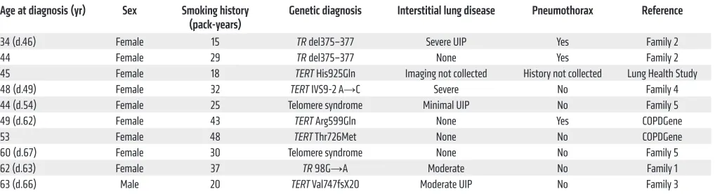
We analyzed the clinical features of telomere-associated emphysema phenotype by combining the Johns Hopkins cases with those we identified in the COPD cohorts (Table 3). In aggre-gate, 9 of the 10 cases were female. The average smoking history was 30 pack-years (range, 15–48), and of those who died, the mean age at the time of death from lung disease was 62 years (range, 46–68, n = 7, Table 3). Notably, 3 of 9 subjects had spontaneous pneumothorax (33%), a rare, life-threatening complication of COPD that normally affects 5% of cases (23). Relative to historic estimates, the likelihood of recurrent spontaneous pneumothorax in this genetically uniform subset occurring by chance alone is low (P = 0.016, Fisher’s exact test). These clinical observations, albeit in a relatively small number, suggested that the telomere-asso-ciated COPD phenotype is assotelomere-asso-ciated with an increased risk for spontaneous air leaks.
Emphysema shows autosomal dominant inheritance with pul-monary fibrosis. We examined the pattern of inheritance in the 5 pedigrees of the Johns Hopkins emphysema cases. Four families carried known deleterious mutations in telomerase (12, 14, 22), including our originally reported family with the mutant TR, and
Figure 2. Radiographic and pulmonary function studies of telomerase mutation carriers with COPD. (A–D) High-resolution inspiratory CT images from
[image:5.585.86.484.57.245.2]COPDGene subjects with telomerase mutations. The panels are labeled with the subject’s mutation. Images from TERT mutation carriers show apical centrilobular emphysema (A–D). In the subject with TERT Arg599Gln, bronchiectasis and a reticular, subpleural interstitial lung abnormality could also be appreciated (B). (E) Rate of change in forced expiratory volume in 1 second (FEV1) from baseline across the 5 years of the Lung Health Study in the subject with a TERT His925Gln mutation. The rate of change is graphed relative to the highest and lowest quartiles of the 5,887 study population and based on longitudinal data available through dbGaP from 163 and 174 participants, respectively. Error bars represent SEM.
Table 2. Characteristics of consecutive telomere syndrome cases with parenchymal lung disease (n = 50)A
Male Female
Number of subjects 25 25 Age at diagnosis, mean yr (range) 53.1 (32–77) 54.2 (34–68) Smoking status
Never-smokers 20 19
Smokers 5 6
Lung disease type to smoking history
Smokers with emphysema 1 6B
Smokers with fibrosis 4 0 Never-smokers with emphysema 0 0 Never-smokers with pulmonary fibrosis 20 19 Smoking history, mean pack-years
(range) 24 (10–41) 30 (25–37)
subset of cases we identified. First, the coverage for TERT in COPDGene included only 75% of the coding sequence, similar to what has been seen previously (25). Moreover, in addition to TERT and TR, a number of other telomerase and telomere genes have been implicated in the monogenic telomere disorders (26, 27). It is therefore possible that although the telomerase genes may account for a small subset, other mutant telomere pathway genes will collectively explain a larger proportion of susceptibility. Even when telomerase is wild-type, telomere length is genetically determined, and short telomeres are sufficient to predispose to degenerative disease in the lung and elsewhere (28, 29). The prev-alence of telomerase and telomere gene mutations in smokers with emphysema will require future confirmation in other cohorts.
Our findings are relevant for patient care because individuals with telomerase mutations are at risk for recurrent syndromic fea-tures including liver disease, osteoporosis, and certain malignan-cies (6). Some of these same telomere syndrome morbidities are known to occur at higher frequency in patients with severe emphy-sema (30). Our data suggest the inherited telomere defect may play a role in simultaneously predisposing to these systemic comorbid-ities along with the lung disease. A telomere-mediated sub-pheno-type of COPD may thus require individualized clinical care algo-rithms. Identifying telomere syndrome patients at the bedside is particularly important in the setting of lung transplant, since some of these patients may be at increased risk for serious toxicities of immunosuppressive medications because of limited reserves in the bone marrow, gastrointestinal tract, and elsewhere (13). Rela-tives of telomerase mutation carriers may also be at risk for telo-mere syndrome complications, which may occur at an earlier age in successive generations because of genetic anticipation (26). Given the public health burden of COPD, our report suggests that emphy-sema may be a recurrent manifestation of telomere syndromes in populations where smoking remains prevalent.
Methods
Subjects
COPDGene study. We accessed the Database of Genotypes and Phe-notypes (dbGaP; http://www.ncbi.nlm.nih.gov/gap) on March 1, 2013, after review and approval from the Johns Hopkins Medicine
Insti-manifests as an autosomal dominant trait, along with pulmonary fibrosis, but only appears in smokers.
Discussion
We report here that germline mutations in telomerase are a risk fac-tor for severe emphysema in smokers. Because telomere dysfunc-tion lowers the threshold to emphysema in animal models, we tested whether telomerase mutations predispose to human emphysema and found, in two independent cohorts, 1% of cases carried deleteri-ous mutations in TERT. This frequency, although it constitutes a rel-atively small subset, is similar to that reported for alpha-1 antitrypsin deficiency in matched COPD populations (3). The emphysema- associated TERT variants compromised telomerase catalytic activ-ity, and mutation carriers had abnormally short telomeres. Although a family history of pulmonary fibrosis was not detailed in the COPD cohorts we studied, in the families we fully characterized, emphy-sema showed autosomal dominant inheritance with pulmonary fibrosis and other telomere phenotypes. The familial clustering of emphysema with fibrosis suggests that these two lung phenotypes, heretofore considered distinct pathologies, may in some cases rep-resent a continuum of degenerative lung disease that shares telo-mere dysfunction as a genetic susceptibility. For emphysema, in con-trast to fibrosis, cigarette smoke exposure is a necessary second hit.
The evidence we document in human emphysema is compel-ling because short telomeres are a determinant of emphysema susceptibility in telomerase-null mice (14). In these animals, short telomeres lower the threshold to damage caused by ciga-rette smoke in epithelial cells (14). The additive damage of these “two hits” provokes a DNA damage response that causes epithelial senescence (6, 14). Senescence and the resultant loss of regener-ative capacity may thus be critical events that drive the airspace destruction in telomere-mediated emphysema (J.K. Alder and M. Armanios, unpublished observations). Telomere length is normal in emphysema patients with alpha-1 antitrypsin deficiency (24). Our data, in light of these observations, indicate that telomere dysfunction may be a second, independent mechanism of emphy-sema susceptibility that is distinct from the protease imbalance characteristic of alpha-1 antitrypsin deficiency.
[image:6.585.42.551.82.220.2]Several pieces of evidence suggest that short telomeres may play a broader role in emphysema susceptibility beyond the small
Table 3. Clinical characteristics of telomerase mutation carriers with emphysema alone or combined with fibrosis (n = 10)
Age at diagnosis (yr) Sex Smoking history
(pack-years) Genetic diagnosis Interstitial lung disease Pneumothorax Reference
34 (d.46) Female 15 TR del375–377 Severe UIP Yes Family 2 44 Female 29 TR del375–377 None Yes Family 2 45 Female 18 TERT His925Gln Imaging not collected History not collected Lung Health Study 48 (d.49) Female 32 TERT IVS9-2 A→C Severe No Family 4 44 (d.54) Female 25 Telomere syndrome Minimal UIP No Family 5 49 (d.62) Female 43 TERT Arg599Gln None Yes COPDGene 53 Female 48 TERT Thr726Met None No COPDGene 60 (d.67) Female 30 Telomere syndrome None No Family 5 62 (d.63) Female 37 TR 98G→A Moderate No Family 1 63 (d.66) Male 20 TERT Val747fsX20 Moderate UIP No Family 3
tutional Review Board and the NHLBI Data Access Committee. We analyzed the dbGaP clinical and exome data for the COPDGene study (32) and examined the TERT, TR, and SERPINA1 gene sequences (phs000179.v3.p2 and pht002239.v2.p2.c1). Supplemental Figure 4 summarizes the depth of coverage for the coding and exon-flanking sequences of the candidate genes.
The COPDGene subjects who underwent exome sequencing (total n = 415) were smokers who were selected for extreme phenotypes (33). Case subjects were selected to enrich for a severe, early-onset pheno-type: younger than 63 years, with severe or very severe obstruction
(FEV1 less than 50% of predicted values), and greater than 15%
emphy-sema on CT scan (n = 209) (32). Controls were older than 65 years, had
FEV1 greater than 80%, and showed less than 5% emphysema on CT
(n = 206). Quantitative chest CT scan assessment was based on the per-centage of the lung with low attenuation areas below –950 Hounsfield units. Although subjects with known severe alpha-1 antitrypsin defi-ciency were excluded from COPDGene based on protein phenotyping, one PI*ZZ subject was inadvertently included. To verify exome
vari-ant calls, we obtained and sequenced archived DNA after review and approval of the COPDGene Ancillary Study Committee.
Lung Health Study. To test the hypothesis in a second cohort, we selected Lung Health Study subjects who fulfilled criteria similar to those in the COPDGene study for candidate gene sequencing.
Smok-ers had FEV1 less than 50% of predicted values and were younger than
65 years (n = 83). Lung Health Study participants had mild or moder-ate obstruction at the time of study entry (33), so we used selection cri-teria to identify subjects who developed severe obstruction at 5 years, the last time point at which pulmonary function was documented. The Lung Health Study longitudinal pulmonary function data were gen-erated from dbGAP files (phs000291.v2.p1 and pht002273.v1.p1.c1) accessed on March 1, 2013.
Johns Hopkins registry. Families were recruited through the Johns Hopkins Telomere Syndrome Registry from July 1, 2005, to June 30, 2014. This study aims at understanding the genetics and natural his-tory of telomere-mediated disease (34). The study was approved by the Johns Hopkins Medicine Institutional Review Board, and all
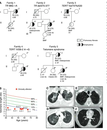
sub-Figure 3. Pedigrees of telomere syndrome cases with emphysema. (A)
Pedigrees of emphysema cases with telomere defects and their relatives’ clinical history. The asterisk denotes individuals with DNA sequence data available and/or telomere length measurement performed. Boldface indicates individuals who carried the mutant gene and/or had very short telomeres (shown in B). DC,
dyskerato-sis congenita, a telomere syndrome defined by mucocutaneous features; CS, positive smoking history; NS, never-smoker; BMF, bone marrow failure; d., age at death from lung disease; ds, disease. (B) Measurement
of lymphocyte telomere length by flow cytometry and FISH shows the short telomere defect in affected members relative to age-matched controls. The nomogram was based on data from 400 controls. (C–F) Apical and
mid-lung chest CT cuts from two female cases (2.II.1 [C and D] and 5.II.3 [E and F]) show severe apical emphysema
[image:7.585.39.414.52.506.2]jects gave written informed consent. Lung disease type was assessed for each of the subjects by CT imaging and review of the medical records, including pulmonary function studies and death certificates. Chest CT images were available for review in 90% of the subjects, and, in the remaining cases, chest X-rays and death certificate infor-mation were ascertained to determine the diagnosis.
Exome sequence analysis
Sequence files were annotated using publicly available software (35). Variants were prioritized for functional analysis if they were absent in 1000 Genomes (36) as well as dbSNP build 130 (http://www. ncbi.nlm.nih.gov/SNP/), an uncontaminated earlier version of vari-ants, and additionally had less than 0.001 minor allele frequency in the Exome Variant Server Database (http://evs.gs.washington. edu/EVS). The a priori designed filtering strategy is summarized in Supplemental Figure 1. Candidate variants that fulfilled our filtering criteria from the exome data were confirmed by Sanger sequencing as previously described (22). We also manually sequenced TR in COPDGene subjects by PCR because of low coverage (ref. 22 and Supplemental Figure 4).
Targeted sequencing
To screen for telomerase mutations in Lung Health Study subjects, we designed and validated a TruSeq Custom Amplicon probe set (Illu-mina) that included the coding and flanking sequences of TERT, TR, as well as exon 6 of SERPINA1 containing the PI*Z allele, that accounts for more than 90% of alpha-1 antitrypsin deficiency cases (3). Librar-ies were generated from 250 ng DNA and analyzed on a MiSeq sequencer (Illumina). Of 83 samples sequenced, 7 samples (8%) had
suboptimal coverage (less than 50% at 8× depth); the coverage for the
76 samples that passed quality control is summarized in Supplemen-tal Figure 3. SupplemenSupplemen-tal Table 3 lists common TERT variants found in the COPD Gene and Lung Health Study subjects along with their respective dbSNP identifiers and accession numbers.
Telomere length measurement
Telomere length was measured on peripheral blood lymphocytes by flow cytometry and FISH as previously outlined (22). For deceased subjects, telomere length was measured using archived DNA by quan-titative PCR (37). For these studies, control and COPDGene genomic DNA was extracted from whole blood using the Gentra Puregene method (QIAGEN). Each run included three replicates, and the mean from three independent runs was calculated.
Telomerase activity assay
The functional consequences of all the rare variants from the COPD-Gene and Lung Health Studies were examined using the direct telomerase activity assay. Wild-type and variant telomerases were reconstituted in vivo in 293FT cells (Invitrogen) by transient trans-fection with pcDNA-3xFLAG-hTERT and pBS-U1-hTR (38). The
reconstituted telomerase was then immunopurified from cell lysates and analyzed by the direct primer-extension assay at previously
vali-dated physiologic nucleotide concentrations (29, 38). The 10 μl direct
primer-extension reaction contained 5 μM dTTP, 5 μM dATP, 5 μM
dGTP, 0.165 μM α-32P-dGTP (3,000 Ci/mmol, 10 mCi/ml,
Perkin-Elmer), and 1 μM (TTAGGG)3 DNA primer in 1× telomerase reaction
buffer (50 mM Tris-HCl pH 8.3, 2 mM DTT, 0.5 mM MgCl2, and 1 mM
spermidine). Comparable wild-type and mutant telomerase expres-sion in transfected cells was confirmed by Western blotting for the FLAG-tagged TERT protein (anti-FLAG, clone M2, Sigma-Aldrich) and GAPDH (clone 6C5, Ambion) as an internal control (39). Com-parable immunopurification efficiency was also confirmed by North-ern blotting of TR extracted from immunopurified telomerase (40). Telomerase activity was determined by measuring the total intensity of telomerase-generated products on the gel and normalizing against
the internal loading control (32P end-labeled 18-mer oligonucleotide)
and the TR level measured by Northern blotting from immunopurified telomerase (40). Quantification was based on 4 activity assays using cell lysates from 2 independent transfections.
Statistics
Means were compared using Student’s t test. The Fisher’s exact test was utilized to test the significance of categorical data. A 2-sided P value of less than 0.05 was considered statistically significant, and all P values shown are 2-sided.
Study approval
The study was reviewed and approved by the Johns Hopkins Medicine Institutional Review Board.
Acknowledgments
We are grateful to all the study participants and to the investiga-tors who contributed to these projects: COPDGene, Lung Health Study, and the NHLBI Lung GO Exome Sequencing Project. We are grateful to Carol Greider for critical comments on the manu-script. We acknowledge David Mohr and the Johns Hopkins Genet-ics Core Facility for assistance with the sequencing. We thank Joseph Potee for technical assistance. This work was supported by NIH grants R01 GM094450 (to J.J.L. Chen), K99 HL113105 (to J.K. Alder), R01 HL089856 (to E.K. Silverman), U01 HG004738 and UC2 HL102923 (to K.C. Barnes), and R01 CA160433 and R01 HL119476 (to M. Armanios); and grants from the Mary Beryl Patch Turnbull Scholar Program (to K.C. Barnes), the Flight Attendants Medical Research Institute (to M. Armanios), and the Common-wealth Foundation (to M. Armanios). S.E. Stanley received sup-port from NIH grant T32GM007309.
Address correspondence to: Mary Armanios, 1650 Orleans St., CRB 1 Room 186, Baltimore, Maryland 21287, USA. Phone: 410.502.3817; E-mail: marmani1@jhmi.edu.
1. Kochanek KD, Xu J, Murphy SL, Miniño AM, Kung H-C. Deaths: final data for 2009. Natl Vital
Stat Rep. 2011;60(3):1–117.
2. Wan ES, Silverman EK. Genetics of COPD and emphysema. Chest. 2009;136(3):859–866. 3. Silverman EK, Sandhaus RA. Clinical practice.
Alpha1-antitrypsin deficiency. N Engl J Med. 2009;360(26):2749–2757.
4. Zhou JJ, Cho MH, Castaldi PJ, Hersh CP, Sil-verman EK, Laird NM. Heritability of chronic obstructive pulmonary disease and related phe-notypes in smokers. Am J Respir Crit Care Med.
2013;188(8):941–947.
5. Harley CB, Futcher AB, Greider CW. Telomeres shorten during ageing of human fibroblasts.
Nature. 1990;345(6274):458–460.
para-digms. J Clin Invest. 2013;123(3):996–1002. 7. Greider CW, Blackburn EH. Identification of a
specific telomere terminal transferase activity in Tetrahymena extracts. Cell. 1985;43(2):405–413. 8. Greider CW, Blackburn EH. The telomere
terminal transferase of Tetrahymena is a ribo-nucleoprotein enzyme with two kinds of primer specificity. Cell. 1987;51(6):887–898. 9. Greider CW, Blackburn EH. A telomeric
sequence in the RNA of Tetrahymena telomerase required for telomere repeat synthesis. Nature. 1989;337(6205):331–337.
10. Lingner J, Hughes TR, Shevchenko A, Mann M, Lundblad V, Cech TR. Reverse transcriptase motifs in the catalytic subunit of telomerase.
Science. 1997;276(5312):561–567.
11. Armanios M. Syndromes of telomere shortening.
Annu Rev Genomics Hum Genet. 2009;10:45–61.
12. Parry EM, Alder JK, Qi X, Chen JJ, Armanios M. Syndrome complex of bone marrow failure and pulmonary fibrosis predicts germline defects in telomerase. Blood. 2011;117(21):5607–5611. 13. Silhan LL, et al. Lung transplantation in
telomerase mutation carriers with pulmonary fibrosis. Eur Respir J. 2014;44(1):178–187. 14. Alder JK, et al. Telomere length is a determinant
of emphysema susceptibility. Am J Respir Crit
Care Med. 2011;184(8):904–912.
15. Nunes H, et al. Is telomeropathy the expla-nation for combined pulmonary fibrosis and emphysema syndrome?: report of a family with TERT mutation. Am J Respir Crit Care Med. 2014;189(6):753–754.
16. Emond MJ, et al. Exome sequencing of extreme phenotypes identifies DCTN4 as a modifier of chronic Pseudomonas aeruginosa infection in cystic fibrosis. Nat Genet. 2012;44(8):886–889. 17. Liang J, et al. Mutations in telomerase catalytic protein in Japanese children with aplastic ane-mia. Haematologica. 2006;91(5):656–658. 18. Berry CE, Wise RA. Mortality in COPD:
causes, risk factors, and prevention. COPD. 2010;7(5):375–382.
19. Diaz de Leon A, et al. Telomere lengths, pulmo-nary fibrosis and telomerase (TERT) mutations.
PLoS One. 2010;5(5):e10680.
20. Alder JK, et al. Short telomeres are a risk factor for idiopathic pulmonary fibrosis. Proc Natl Acad
Sci U S A. 2008;105(35):13051–13056.
21. Yamaguchi H, et al. Mutations in TERT, the gene for telomerase reverse transcriptase, in aplastic anemia. N Engl J Med. 2005;352(14):1413–1424. 22. Armanios MY, et al. Telomerase mutations in
families with idiopathic pulmonary fibrosis.
N Engl J Med. 2007;356(13):1317–1326.
23. Sahn SA, Heffner JE. Spontaneous pneumotho-rax. N Engl J Med. 2000;342(12):868–874. 24. Saferali A, Lee J, Sin DD, Rouhani FN, Brantly
ML, Sandford AJ. Longer telomere length in COPD patients with alpha1-antitrypsin defi-ciency independent of lung function. PLoS One. 2014;9(4):e95600.
25. Alder JK, et al. Telomere phenotypes in females with heterozygous mutations in the dysker-atosis congenita 1 (DKC1) gene. Hum Mutat. 2013;34(11):1481–1485.
26. Armanios M, Blackburn EH. The telomere syn-dromes. Nat Rev Genet. 2012;13(10):693–704. 27. Vannier JB, Sarek G, Boulton SJ. RTEL1: functions
of a disease-associated helicase. Trends Cell Biol. 2014;24(7):416–425.
28. Armanios M, Alder JK, Parry EM, Karim B, Strong MA, Greider CW. Short telomeres are sufficient to cause the degenerative defects associated with aging. Am J Hum Genet. 2009;85(6):823–832. 29. Alder JK, et al. Ancestral mutation in telomerase
causes defects in repeat addition processivity and manifests as familial pulmonary fibrosis. PLoS
Genet. 2011;7(3):e1001352.
30. Divo M, et al. Comorbidities and risk of mor-tality in patients with chronic obstructive pulmonary disease. Am J Respir Crit Care Med.
2012;186(2):155–161.
31. Regan EA, et al. Genetic epidemiology of COPD (COPD Gene) study design. COPD. 2010;7(1):32–43.
32. Cho MH, et al. Exome Sequencing in Severe COPD Cases and Resistant Smoking Controls from COPD Gene. Presented at: 60th Annual American Society of Human Genetics Annual Meeting; October 20–24, 2009; Honolulu, Hawaii, USA.
33. Anthonisen NR, et al. Effects of smoking intervention and the use of an inhaled anticho-linergic bronchodilator on the rate of decline of FEV1. The Lung Health Study. JAMA. 1994;272(19):1497–1505.
34. Jonassaint NL, Guo N, Califano JA, Montgomery EA, Armanios M. The gastrointestinal manifes-tations of telomere-mediated disease. Aging Cell. 2013;12(2):319–323.
35. Yandell M, et al. A probabilistic disease-gene finder for personal genomes. Genome Res. 2011;21(9):1529–1542.
36. Abecasis GR, et al. A map of human genome vari-ation from populvari-ation-scale sequencing. Nature. 2010;467(7319):1061–1073.
37. Cawthon RM. Telomere length measurement by a novel monochrome multiplex quantitative PCR method. Nucleic Acids Res. 2009;37(3):e21. 38. Qi X, Xie M, Brown AF, Bley CJ, Podlevsky JD,
Chen JJ. RNA/DNA hybrid binding affinity determines telomerase template-translocation efficiency. EMBO J. 2012;31(1):150–161. 39. Gramatges MM, Qi X, Sasa GS, Chen JJ, Bertuch
AA. A homozygous telomerase T-motif variant resulting in markedly reduced repeat addition processivity in siblings with Hoyeraal Hreidars-son syndrome. Blood. 2013;121(18):3586–3593. 40. Xie M, Podlevsky JD, Qi X, Bley CJ, Chen JJ. A