S T U D Y P R O T O C O L
Open Access
Efficacy of anchoring the four-arm
transvaginal mesh to the mid-urethra vs
original surgery as a surgical correction for
stress urine incontinence in coexisting
anterior vaginal prolapse grades II and III:
study protocol for a randomized controlled
trial
Zoltán Fekete
1*, Andrea Surányi
1, Lórand Rénes
1, Gábor Németh
1†and Zoltan Kozinszky
2†Abstract
Background:The prevalence of obesity with aging is escalating alarmingly; and pelvic organ prolapse (POP) and stress urinary incontinence (SUI) are now becoming a growing epidemic among the elderly. Synthetic transvaginal mesh has been employed with increasing popularity in the treatment of POP and is usually highly effective in controlling the principal symptoms of prolapse. However, studies have reported that mesh operations provide fairly unfavorable SUI cure rates. Therefore, additional anti-incontinence surgical strategies are increasingly being scrutinized to achieve better postoperative continence without any significant side-effects for patients with both POP and SUI. We hypothesize that the modification with the fixing of the mesh to the mid-urethra is superior to the original transvaginal mesh operation (TVM) with regard to anti-incontinence.
Methods:One hundred and thirty patients diagnosed with POP–Q II–III and concomitant SUI requiring surgical treatment will be included in this prospective, randomized, double-blind, controlled clinical trial. Patients will be randomly allocated to receive either original TVM (TVM group,n= 65) or modified TVM surgery (mTVM group,n= 65). As the primary outcome parameter, we will evaluate the objective SUI and POP cure rates. Secondary endpoints include postoperative morbidity as assessed with the International Urogynaecological Association classification and subjective prolapse and incontinence cure rates reported by questionnaires.
Discussion:Recognizing the importance of an additional surgical procedure for anti-incontinence management, we aim to investigate whether a stabilizing suturing of the mesh to the mid-urethra delivers superior SUI correction compared to the original prosthesis surgery.
Trial registration:ClinicalTrials.gov, NCT02935803. Registered on 20 May 2016.
Keywords:Modified transvaginal mesh, Complications, SUI with POP–Q II–III, IUGA classification
* Correspondence:[email protected] †Equal contributors
1Division of Urogynaecology, Department of Obstetrics and Gynaecology,
University of Szeged, Semmelweis u. 1., 6725 Szeged, Hungary Full list of author information is available at the end of the article
Background
Demand for pelvic organ prolapse (POP) and stress urin-ary incontinence (SUI) surgery is expected to increase due to an expanding rate of obesity among elderly women. POP is defined as the protrusion of the pelvic organs from the normal anatomical location toward or through the va-ginal opening; the current prevalence of symptomatic POP is in the range of 3–8% [1, 2]. SUI, classified as involuntary loss of urine during physical activities and a resultant in-crease in intra-abdominal pressure, is also highly prevalent, reaching as high as 24.8% [3].
Also of note, one-fifth of women in the United States re-ceives surgery either for SUI or for POP [4], where the cu-mulative risk for SUI surgery is 13.6% and that for POP surgery is 12.6% [4]. Both pathologic conditions develop in > 50% of the women affected [5]. Synthetic transvaginal mesh (TVM) has been increasingly employed in the treat-ment of POP and tension-free slings are useful in the man-agement of incontinence. Synthetic mesh during repair has principally been used due to higher efficacy compared to that of native tissue repair and resorbable mesh; however, complications appear to be more prevalent [6–9]. Com-mon complications include mesh extrusion, chronic pelvic pain, dyspareunia, and infection [7–10]. The anti-SUI effi-cacy of the prosthetic placement is barely 72–83% [11–13]; however, it is assumed that a combination of a synthetic mesh with the sling operation [5, 14–16] will substantially increase the cure rate for concomitant SUI.
Despite the increased consideration of the combined surgery for both genitourinary pathologies in one ses-sion, there is a lack of consensus on the optimal treat-ment. It is supposed that a combined operation with mesh and sling is highly effective for the treatment of POP and SUI; however, the complication rate is elevated [5, 14–16].
Therefore, the research group developed a modifica-tion to the transobturator four-arm TVM [13, 17] to in-crease its anti-incontinence effect. While the sling is located beneath the mid-urethra, the TVM elevates the distal part of the anterior vaginal wall [13, 17]. In the original TVM, the posterior part of the mesh is anchored to the anterior aspect of the cervix and the anterior arms are spread under the bladder neck with stabilizing sutures. We hypothesize that the original TVM operation can be followed by residual SUI since the strengthening of the back arms may result in a back-ward dislocation of the entire mesh. The posterior movement of the mesh allows the dorsal rotation of the urethra since the mid-urethra is not suspended. The proposed modification to the original surgical procedure includes the suture of the anterior part of the mesh to the mid-urethra to prevent the mesh sliding. We think that the appropriate elevation of the mid-urethra would thus occur with the anterior arms and that would
achieve a more effective anti-incontinence. The pubour-ethral ligament is usually loose in SUI, but the anchored mesh would theoretically normalize its function and stabilize the urethra. Intra- and postoperative complica-tion rates would be expected to be similar to those with the original four-arm TVM, but the modified TVM with the anchoring suture would be slightly superior with re-spect to POP repair and remarkably more effective in SUI correction. In our preliminary study, the anterior fixing of the TVM to the mid-urethra demonstrates as high an efficacy of anti-incontinence as 96.8% and an enormously reduced recurrence prolapse rate of 3.2% in (unpublished data). The mesh extrusion rate is particu-larly low and this may be due to the fact that the stabil-izing sutures exert a lack of “folding/wrinkling” of the edge of the mesh, preventing a lifting up of the mesh which does not compress the mucosa and derange the periprosthetic vasculature.
A further modification to the TVM surgery is that the positioning of the mesh will occur 1.5 cm below the urethral meatus, leading to an elevation of the entire anterior vaginal wall including the anterior and middle compartments as well. By contrast, the original TVM surgery does not prevent anterior compartment prolapse [13, 17].
Methods/design
Study design
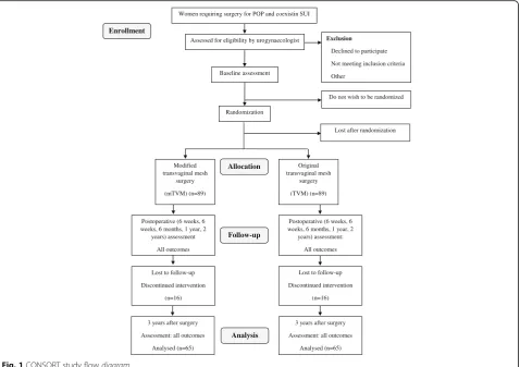
The present study is a single-center, prospective, double-blind (participant, investigator/surgeon, outcome assessor), randomized, controlled trial. The study will be conducted in accordance with the Declaration of Helsinki and has been approved by the local medical ethics committee at the University of Szeged under reference number 55/2016. The trial is registered under NCT 02935803, and patient recruitment started on 22 August 2016. The trial flow diagram is presented in Fig. 1 (CONSORT study flow dia-gram). The protocol follows the Standard Protocol Items: Recommendations for Interventional Trials (SPIRIT) checklist (see Additional file 1).
Patient recruitment and consent procedure
Consent to publish Written informed consent was obtained from the patients for publication of their indi-vidual details and accompanying images in this manu-script. The consent form is held by the authors in the patients’clinical notes and is available for review by the Editor-in-Chief.
Participants considered for trial
This will be a prospective longitudinal study involving all patients successively scheduled for operation for
symptomatic prolapse POP–Q grade II or III and
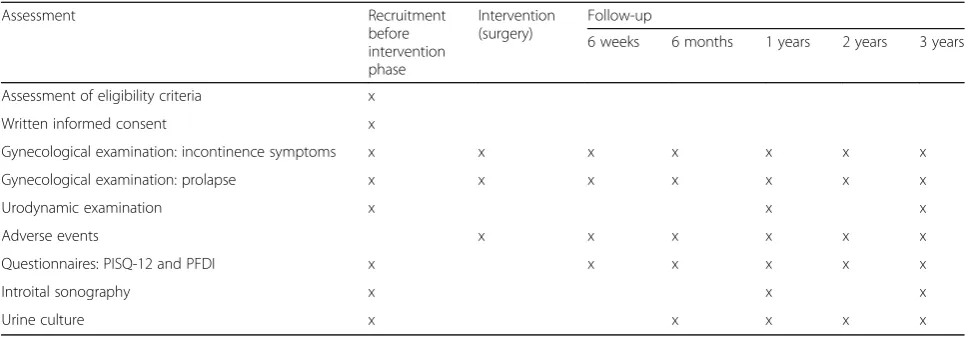
coexisting SUI, who will be included in the study after informed consent has been obtained. Patients will be recruited in the Department of Obstetrics and Gynaecol-ogy at the University of Szeged. The study will be con-ducted for an estimated maximum of 18 months (between August 2016 and December 2017). Table 1 provides an overview of the patient recruitment and evaluation plan.
The symptomatic POP–Q Stage II–III (determined by the gynecological examination using the International Continence Society quantification system) [18] anterior vaginal wall prolapse is defined as the maximum extent of the prolapsed anterior and middle compartments
being within 1 cm above and 6 cm below the hymen [19, 20]. According to the international POP guidelines (the EBU and NICE guidelines) [21, 22], if the condition disrupts the patient’s life and non-surgical treatment options have not helped, it should be treated surgically.
In all cases, SUI will be visualized after a complete physical examination is performed (verified by pad test/ Bonney test/two-dimensional [2D] introital sonography and urodynamic examination). The severity of SUI was assessed using the Ingelman–Sundberg classification [23]. Urodynamic examinations comprising uroflowme-try, cystometrography, the pressure-flow study, and the abdominal leak point pressure test will be performed be-fore surgery to objectively determine the coexisting symptomatic SUI based on the international guidelines (the EBU and NICE guidelines) [21, 22]. The abdominal leak point pressure test will be used as a standardized examination method for the evaluation of SUI with urine leakage as a sign. If the intra-abdominal pressure re-corded at the point of urine leakage was < 40 cmH2O,
the origin of the SUI was set as intrinsic sphincter defi-ciency (ISD) [24]. In the case of ISD, preoperative pelvic floor training (PFMT) will be recommended. If the pa-tient is unwilling to participate in PFMT or if the Enrollment
Women requiring surgery for POP and coexistin SUI
Assessed for eligibility by urogynaecologist Exclusion
Declined to participate
Not meeting inclusion criteria
Other Baseline assessment
Randomization
Do not wish to be randomized
Allocation
Lost after randomization
Modified transvaginal mesh
surgery
(mTVM) (n=89)
Original transvaginal mesh
surgery
(TVM) (n=89)
Postoperative (6 weeks, 6 weeks, 6 months, 1 year, 2
years) assessment
All outcomes
Postoperative (6 weeks, 6 weeks, 6 months, 1 year, 2
years) assessment:
All outcomes Follow-up
Lost to follow-up
Discontinued intervention
(n=16) Lost to follow-up
Discontinued intervention
(n=16)
3 years after surgery
Assessment: all outcomes
Analysed (n=65)
3 years after surgery
Assessment: all outcomes
Analysed (n=65) Analysis
[image:3.595.61.540.88.425.2]training was unsuccessful, we will recommend mesh surgery. This will also be the case for suspected urethral hypermobility, i.e. if the intra-abdominal pressure at the point of urine leakage was > 60 cmH2O.
Introital ultrasound findings
In all cases, introital ultrasound (GE Voluson 730) will be performed with a standardized bladder-filling volume of 300 mL. The vaginal probe (5–9 MHz) will be placed in the area of the vaginal introitus at the level of the ex-ternal urethral orifice, with the patient in a semi-sitting position. Ultrasound assessment of the bladder and urethra starts in the mid-sagittal plane.
During the sonography, we measure the longitudinal (L) distance between the bladder neck and the line through the lower edge of the pubic symphysis and the horizontal (H) distance between the bladder neck and the upper edge of the symphysis. The two distances are measured at rest (L1, H1), during contraction (L2, H2), on pressing or while coughing (L3, H3). Changes in these parameters during contraction of the levator muscle and on pressing serve to evaluate the reactivity of the pelvic floor muscles and the adequacy of the supportive structures of the urogenital organs [25]. The funneling of the proximal urethra during coughing as a typical stress urinary sign also will be examined [25].
Inclusion and exclusion criteria
Inclusion criteria
Female adults aged > 40 years with coexisting pelvic floor defects will be recruited, at least one year following de-livery, irrespective of parity and pre- or postmenopausal state, medically and physically fit for the measurement and therapeutic surgeries, and, in the case of systemic or local estrogen treatment, stable for the past three months before inclusion.
Exclusion criteria
Exclusion criteria are: urge, mixed incontinence or occult SUI; prolapse < grade II or > grade III POP–Q ([21, 22], https://uroweb.org/guideline/urinary-incontinence), apical or posterior compartment prolapse, dysuria (bladder tumor, neurogenic urinary bladder damage), a history of mesh use or anti-incontinence pelvic procedures; preg-nancy (urine test to accomplish); lactation period not yet finished; current urinary tract or vaginal infection, men-struation on the day of examination; contraindications for measurements or interventions, for example, acute inflam-matory or infectious disease, tumor or fracture; de novo systemic or local estrogen treatment (< 3 months); de novo drug treatment with anticholinergics or other bladder ac-tive substances (tricyclic antidepressants and selecac-tive sero-tonin reuptake inhibitors) and cancer of the pelvic organs.
Concealment of group allocation from participants
[image:4.595.56.539.98.267.2]After the screening phase, patients will be randomly assigned to one of the two therapy groups (the TVM group or modified TVM (mTVM) group). The alloca-tion sequence will be generated by the independent uro-gynecology secretariat using online randomization software (http://randomization.com); allocation ratio = 1:1 (TVM group:mTVM group). The allocation will be concealed in sealed, opaque, sequentially numbered en-velopes, which will be stored at the operating theatre. All the women recruited will be numbered consecutively corresponding to the numbered envelopes. The envelope will not be opened until the general narcosis of the study participant has been established. To increase the validity of the trial, the assigned procedure will be blinded for the study participants throughout the follow-up period. The participants will be informed of allocated treatment after completing the study forms 12 months after the procedures. If major complications occur, the study par-ticipants and the outcome assessor will be also informed Table 1Schedule of assessments/data collection
Assessment Recruitment
before intervention phase
Intervention (surgery)
Follow-up
6 weeks 6 months 1 years 2 years 3 years
Assessment of eligibility criteria x
Written informed consent x
Gynecological examination: incontinence symptoms x x x x x x x
Gynecological examination: prolapse x x x x x x x
Urodynamic examination x x x
Adverse events x x x x x x
Questionnaires: PISQ-12 and PFDI x x x x x x
Introital sonography x x x
of the allocated treatment at the time of any suspected complication(s).
Randomization
The patients will be randomized to one of the study groups using a computer-generated list. Allocation con-cealment will be ensured by enclosing assignments in sealed, opaque, sequentially numbered envelopes, which will only be opened when the general narcosis of the study patient has been established [26].
Postoperative outcomes and sequalae will be assessed by an outcome assessor who is a gynecologist in a sub-specialization program in urogynecology and also well trained in the transvaginal mesh operation. The outcome assessor will remain blinded to the type of intervention throughout the study.
Blinding
Participants will be blinded against the type of TVM sur-gery received (original TVM vs mTVM). The participant information document will not provide any information on the differences in surgical protocols such that the women could ascertain their group allocation. All inves-tigators involved in data acquisition, data analyses, and statistics will also be blinded against group allocation. The surgeons in charge of the therapy cannot be blinded against group allocation and therefore will not be in-volved in data acquisition, data reduction, data analyses, or statistics.
Measurement outcomes
Baseline (before intervention phase) and follow-up mea-surements (of primary, secondary, and tertiary out-comes) after six weeks to three years will be performed at the Division of Urogynaecology, Department of Ob-stetrics and Gynaecology, University of Szeged, Hungary, by an experienced urogynecologist who will be blinded to group allocation of participants and who will not operate on the patients (Table 1).
Primary endpoints
The primary outcome measures will be a significant im-provement in POP repair and objective cure of SUI after the surgery. The efficacy of POP repair will be under-stood as a significant (> 3 cm) improvement during follow-up at points Aa, Ba, C, and D using the POP–Q system (International Continence Society) [19, 20]. Anti-incontinence efficacy is classified as no further SUI, as diagnosed by cough tests and urodynamic examinations. Besides the gynecological and urodynamic examinations, sonographic findings from introital ultrasound examina-tions will be analyzed in terms of anatomical success both before and following surgery and during follow-up.
Secondary and tertiary endpoints
The secondary measurement outcome will comprise the intraoperative findings and postoperative factors. As con-cerns the long-term postoperative complications of the mesh procedures, we will determine the extrusion rate, the presence of de novo urge symptoms (DNUS) or urin-ary tract infection (UTI), and the need for reoperation. The diagnosis of DNUS will be set if detrusor pressure changes are detected in cystometrographic pressures after the surgeries. The postoperative complications that will lead to reoperation will be infection, recurrent descent or incontinence, implant extrusion, chronic pelvic pain, and total retention. Operative and perioperative complications (six weeks after the procedures) described after TVM vs mTVM will be collected; overall frequency within all the cases will be calculated and severity will be graded using the IUGA classification comprising all the follow-up periods [27].
The subjective cure for prolapse and incontinence will be measured with a significant enhancement of the Pel-vic Organ Prolapse/Urinary Incontinence Sexual Ques-tionnaire (PISQ-12) and Pelvic Floor Distress Inventory (PFDI) scores. The PISQ-12 and PFDI are validated to assess the impact of SUI symptoms on quality of life and sexuality and relate well to the prolapse symptoms. Pre-vious research has demonstrated that the questionnaires correspond well with grade of prolapse and urodynamic findings [5, 13, 17]. Our research group has assessed the validity of the questionnaires in screening for subjective genitourinary symptoms (unpublished data). To guaran-tee blinding of the tertiary outcome, the participant will complete the questionnaire without the outcome asses-sor present and seal it in an envelope, which will be given to research staff. The subjective outcome assess-ment is largely performed by participant-completed questionnaire, thus avoiding interviewer bias.
Trial interventions
Participants will receive the allocated intervention, either the original TVM operation or mTVM surgery. The sur-gical interventions will be delivered by two surgeons with expertise in the specific intervention and subspecia-lized in urogynecology. They will not assess the meas-urement outcomes. Further details on the interventions are provided below.
Original transobturator four-arm transvaginal mesh
be anchored to the anterior side of the cervix using two non-absorbable Prolene® 2-0 sutures (Ethicon, Issy-les-Moulineaux, France), while the mesh will then be spread by securing its anterior parts beneath the bladder neck using two or three Monocryl® 2-0 absorbable sutures (Ethicon, Issy-les-Moulineaux, France). Conventional in-struments will be employed for the original TVM procedure.
Modified intervention surgery
In the modified surgical technique, the prosthesis is placed between 1.5 cm below the urethral meatus and the cervix and the anterior part of the mesh is anchored with a stabilizing suture to the periurethral tissue at the level of the mid-urethra to elevate the middle part of the urethra, leading to potentially more effective anti-incontinence. An additional document file presents the intervention (detailed description of the mTVM) in detail (see Additional file 2).
Assessment of safety: postoperative complications/ reoperations
In the current study, there are no anticipated risks or in-conveniences, as the examinations and intervention employed are well-known and widely used in pelvic floor defect surgery. The modification to the TVM surgery does not carry a higher risk for patients than that of the original TVM in the setting of a fully equipped oper-ation theatre. This makes the immediate detection and treatment of adverse events possible. Also, after leaving the operation room, all patients will be closely moni-tored for the occurrence of potential (severe) adverse events (short-term postoperative complications) on the postoperative intensive ward. Moreover, the inclusion of each individual patient in the study is indicated in the electronic hospital information system and, hence, is visible to all physicians and nurses involved in the care of the patient. This facilitates the reporting of (severe) adverse events to the principal investigator. The princi-pal investigator will report suspected unexpected serious adverse reactions to the Institutional Review Board.
Statistical methods
Hypothesis
Alternative hypothesis for primary outcome: it is hy-pothesized that the group undergoing the modified transvaginal mesh operation will have a statistically
higher improvement of continence measured by
gynecological and urodynamic examination, and from the questionnaire administered before and after the intervention phase.
Sample size calculation
As we have newly developed the modification to the prosthesis surgery, an exploratory pilot study was de-signed to evaluate the feasibility of the modification to the mesh for the treatment of SUI. Twenty patients with SUI and POP were recruited for a mTVM operation by the same two senior surgeons who are conducting this randomization study. The sample size calculation study was designed based on preliminary data on the 20 patients. The newly developed technique yielded an ob-jective SUI cure rate of 92% as opposed to 72% for the original TVM published by Sergent et al. [13, 17]. Sample size calculations were performed with G*Power software [29], using the statistical model for an χ2 approach. Consequently, sample size was estimated the-oretically and an effect size of = 0.1, indicating a small effect, will be accepted. The sample size was calculated for the primary outcome of the SUI cure rate with the following assumptions:α= 0.05, power (1–βerror prob-ability) = 0.8, number of groups = 2. Based on these as-sumptions, a total sample size ofN= 130 was estimated. In anticipation of dropouts (10%: n= 16) or a violation of protocol (10%: n= 16), a final sample size of N= 162 (81 participants per group) results.
Statistical analyses
Analysis of the patients will follow the CONSORT flow diagram (Fig. 1) through the phases of the study (enroll-ment [assessed, excluded, randomized], allocation [con-trol group and experimental group with intervention received or not received], follow-up [lost to follow-up, discontinued intervention] and analysis) [30].
All statistical analyses will be conducted using SPSS software version 22 (IBM, Armonk, NY, USA). All tests will be two-sided and significance will be set atP< 0.05. Efficacy measurements were adjusted by intention-to-treat analysis. Missing values will be replaced using the last observation carried forward (LOCF) method. No subgroup analyses are planned. Standard deviations, 95% confidence intervals, and median will be used for the de-scriptive analyses. Primary and secondary outcome ana-lysis: the Chi-square test or Fisher’s exact test will be employed to identify any objective outcome differences among groups.
carried out to determine the effects of the modified operation on subjective cure rate for POP and SUI, and the Bonferroni post hoc test will be used to test the dif-ference between means. All statistical analyses will be completed after the final measurement of the last patient during the last clinical appointment after intervention. The repeated measure design with seven points in time allows us to monitor how patients change over time in both short-term (before/during intervention) and long-term situations (before/after intervention).
Discussion
Coexisting POP and SUI are increasingly recognized as a major health and financial concern affecting 63–80% of postmenopausal women [31]. TVM is the standard sur-gical method for the anatomical restoration of middle compartment Stage II–III prolapse; however, it should be supplemented with a mid-urethral sling to achieve better SUI treatment. Moreover, the combined mesh and sling operations yield unfavorably more frequent compli-cation rates and may provoke voiding dysfunction and recurrent UTI. Furthermore, following the original TVM, some residual SUI can develop because of the backward dislocation of the mesh.
To the best of our knowledge, the present study is the first to investigate a surgical modification to TVM for more effective anti-incontinence. Should this newly de-veloped modification be proved successful in treating SUI, it could be introduced in clinical practice due to its simplicity.
Additional files
Additional file 1:SPIRIT 2013 checklist: recommended items to address in a clinical trial protocol and related documents. (DOC 127 kb)
Additional file 2:Intervention surgery. (DOC 30 kb)
Abbreviations
ANOVA:Analysis of variance; LOCF: Last observation carried forward; mTVM: Modified transvaginal mesh operation; PFDI: Pelvic Floor Distress Inventory; PFMT: Pelvic floor training; PISQ-12: Pelvic Organ Prolapse/Urinary Incontinence Sexual Questionnaire; POP: Pelvic organ prolapse; POP-Q: Pelvic organ prolapse quantification system; SUI: Stress urinary incontinence; TVM: Transvaginal mesh operation; UTI: Urinary tract infection
Acknowledgements
None
Funding
No funding was received.
Authors’contributions
ZF and AS are responsible for this study. ZF and ZK conceived and developed the study design and drafted and revised the protocol. GN contributed to the study, especially with regard to the development of the therapy protocols, and is responsible for the physiotherapy interventions. AS and ZK are responsible for data acquisition and analyses. ZF and RR hold responsibility for patient recruitment. ZF prepared the manuscript and is the
corresponding author. All the authors read and approved the final manuscript.
Competing interests
The authors declare that they have no competing interests.
Publisher’s Note
Springer Nature remains neutral with regard to jurisdictional claims in published maps and institutional affiliations.
Author details
1Division of Urogynaecology, Department of Obstetrics and Gynaecology,
University of Szeged, Semmelweis u. 1., 6725 Szeged, Hungary.2Department of Obstetrics and Gynaecology, Blekinge Hospital, Karlskrona, Sweden.
Received: 16 April 2017 Accepted: 31 October 2017
References
1. Nygaard I, Barber MD, Burgio KL, Kenton K, Meikle S, Schaffer J, et al. Prevalence of symptomatic pelvic floor disorders in US women. JAMA. 2008;300:1311–6.
2. Dieter AA, Wilkins MF, Wu JM. Epidemiological trends and future care needs for pelvic floor disorders. Curr Opin Obstet Gynecol. 2015;27:380–4. 3. Markland AD, Richter HE, Fwu CW, Eggers P, Kusek JW. Prevalence and
trends of urinary incontinence in adults in the United States, 2001 to 2008. J Urol. 2011;186:589–93.
4. Wu JM, Matthews CA, Conover MM, Pate V, Jonsson FM. Lifetime risk of stress urinary incontinence or pelvic organ prolapse surgery. Obstet Gynecol. 2014;123:1201–6.
5. Takahashi S, Obinata D, Sakuma T, Matsui T, Takenobu Y, Igarashi T, et al. Transvaginal mesh (TVM) reconstruction with TVT/TOT sling for vaginal prolapse concurrent with stressurinary incontinence. Aktuelle Urol. 2010;41 Suppl 1:S20–3.
6. Baessler K, Maher CF. Mesh augmentation during pelvic-floor reconstructive surgery: risks and benefits. Curr Opin Obstet Gynecol. 2006;18:560–6. 7. Hiltunen R, Nieminen K, Takala T, Heiskanen E, Merikari M, Niemi K, et al.
Low-weight polypropylene mesh for anterior vaginal wall prolapse: a randomized controlled trial. Obstet Gynecol. 2007;110:455–62.
8. Nguyen JN, Burchette RJ. Outcome after anterior vaginal prolapse repair: a randomized controlled trial. Obstet Gynecol. 2008;111:891–8.
9. US Food and Drug Administration. FDA safety communication: update on serious complications associated with transvaginal placement of surgical mesh for pelvic organ prolapse. http://www.fda.gov/downloads/ medicaldevices/safety/alertsandnotices/ucm262760.pdf. Accessed 13 July 2011.
10. Tunitsky E, Abbott S, Barber MD. Interrater reliability of the International Continence Society and International Urogynecological Association (ICS/ IUGA) classification system for mesh-related complications. Am J Obstet Gynecol. 2012;206(442):e1–6.
11. Serati M, Sorice P, Bogani G, Braga A, Cantaluppi S, Uccella S, et al. TVT for the treatment of urodynamic stress incontinence: efficacy and adverse effects at 13-year follow-up. Neurourol Urodyn. 2015;36:192–7.
12. Nauth MA, Fünfgeld C. Correction of cystocele and stress incontinence with anterior trans obturator mesh. Eur J Obstet Gynecol Reprod Biol. 2007;136:249–53.
13. Sergent F, Sentilhes L, Resch B, Verspyck E, Medeiros R, Descamps P, et al. Treatment of concomitant prolapse and stress urinary incontinence via a transobturator subvesical mesh without independent suburethral tape. Acta Obstet Gynecol Scand. 2010;89:223–9.
14. Yonguc T, Bozkurt IH, Sen V, Aydogdu O, Yonguc GN, Gunlusoy B. Double-sling procedure for the surgical management of stress urinary incontinence with concomitant anterior vaginal wall prolapse. Int Urol Nephrol. 2015;47:1611–7.
15. van der Ploeg JM, Oude Rengerink K, van der Steen A, van Leeuwen JH, Stekelenburg J, Bongers MY, et al. Transvaginal prolapse repair with or without the addition of a midurethral sling in women with genital prolapse and stress urinary incontinence: a randomised trial. BJOG. 2015;122:1022–30. 16. Abbott S, Unger CA, Evans JM, Jallad K, Mishra K, Karram MM, et al.
pelvic reconstructive surgery: a multicenter study. Am J Obstet Gynecol. 2014;210:163. e1–8.
17. Sergent F, Gay-Crosier G, Bisson V, Resch B, Verspyck E, Marpeau L. Ineffectiveness of associating a suburethral tape to a transobturator mesh for cystocele correction on concomitant stress urinary incontinence. Urology. 2009;74:765–70.
18. Bump RC, Mattiasson A, Bø K, Brubaker LP, DeLancey JO, Klarskov P, et al. The standardization of terminology of female pelvic organ prolapse and pelvic floor dysfunction. Am J Obstet Gynecol. 1996;175:10–7. 19. Persu C, Chapple CR, Cauni V, Gutue S, Geavlete P. Pelvic Organ Prolapse
Quantification System (POP–Q)–a new era in pelvic prolapse staging. J Med Life. 2011;4:75–81.
20. Reid F. Assessment of pelvic organ prolapse: a practical guide to the pelvic organ prolapse quantification. Obstet Gynaecol Reprod Med. 2014;24:170–6. 21. Burkhard FC, Bosch JLHR, Cruz F, Lemack GE, et al. EBU Guideline Urinary
incontinence; 2016. http://uroweb.org/guideline/urinary-incontinence/. 22. National Institute for Health and Care Excellence. Urinary incontinence in
women: management 2015 NICE guidelines [CG171]. London: NICE; 2015. https://www.nice.org.uk/guidance/cg171.
23. Ingelman-Sundberg A. Urinary incontinence in women, excluding fistulas. Acta Obstet Gynecol Scand. 1952;31:266–91.
24. Majoros A, Hamvas A, Keszthelyi A, Romics I. Value of testing the abdominal leak point pressure in the differential diagnosis of urinary stress
incontinence. Orv Hetilap. 2003;144:2321–5. Article in Hungarian. 25. Tunn R, Petri E. Introital and transvaginal ultrasound as the main tool in the
assessment of urogenital and pelvic floor dysfunction: an imaging panel and practical approach. Ultrasound Obstet Gynecol. 2003;22:205–13. 26. Viera AJ, Bangdiwala SI. Eliminating bias in randomized controlled
trials: importance of allocation concealment and masking. Fam Med. 2007;39:132–7.
27. Haylen BT, Freeman RM, Swift SE, Cosson M, Davila GW, Deprest J, et al. Joint IUGA/ICS Working Group on Complications Terminology. An International Urogynecological Association (IUGA)/International Continence Society (ICS) joint terminology and classification of the complications related directly to the insertion of prostheses (meshes, implants, tapes) and grafts in female pelvic floor surgery. Neurourol Urodyn. 2011;30:2–12. 28. de Tayrac R, Brouziyne M, Priou G, Devoldère G, Marie G, Renaudie J.
Transvaginal repair of stage III-IV cystocele using a lightweight mesh: safety and 36-month outcome. Int Urogynecol J. 2015;26:1147–54.
29. Faul F, Erdfelder E, Lang AG, Buchner A. G*Power 3: A flexible statistical power analysis program for the social, behavioral, and biomedical sciences. Behav Res Methods. 2007;39:175–91.
30. Moher D, Hopewell S, Schulz KF, Montori V, Gotzsche PC, Devereaux PJ, et al. i CONSORT 2010 explanation and elaboration: updated guidelines for reporting parallel group randomised trials. BMJ. 2010;340:c869.
31. Molander U, Milsom I, Ekelund P, Mellström D. An epidemiological study of urinary incontinence and related urogenital symptoms in elderly women. Maturitas. 1990;12:51–60.
• We accept pre-submission inquiries
• Our selector tool helps you to find the most relevant journal • We provide round the clock customer support
• Convenient online submission • Thorough peer review
• Inclusion in PubMed and all major indexing services • Maximum visibility for your research
Submit your manuscript at www.biomedcentral.com/submit