INTRODUCTION
The role of the cephalic neural crest cells (NCCs) in the construction of craniofacial structures has been deciphered using a cell-marking technique applied in the avian embryo (for a review, see Le Douarin and Kalcheim, 1999). By constructing quail-chick chimeras in ovo, it was shown that NCCs, which exit from the diencephalon posterior half down to rhombomere 2, colonize the facial buds that are at the origin of the upper beak [derived from the nasofrontal and nasolateral together with the maxillary buds (Mx)], and the lower jaw [derived from the mandibular buds (Md)] (Couly et al., 1996; Köntges and Lumsden, 1996; Lee et al., 2004; Cerny et al., 2004).
It has been shown in our laboratory that duplication of the lower-jaw skeleton (composed of Meckel’s cartilage and associated membrane bones) can be induced by grafting a fragment of ventral foregut endoderm removed from a definite area of 6-somite stage (6 ss) quail embryos, at the presumptive level of the first branchial arch (BA1) in stage-matched chicken (Couly et al., 2002). Similar Meckel’s cartilage duplication was obtained before by grafting Bmp4-soaked beads into Md at embryonic day 3.5 (E3.5) (Barlow and Francis-West, 1997; Mina et al., 2002) and more recently by replacing Md ectoderm by the so-called ‘frontal ectodermal zone’ (FEZ) (Hu et al., 2003).
In a precedent work (Brito et al., 2006), we demonstrated that Sonic Hedgehog(Shh) expression in the ventral foregut endoderm, from the early somitic stages onwards, is crucial for the survival and further development of the cephalic NCCs that colonize BA1. Excision of the presumptive forehead area, including the precordal plate (PcP) and the
anterior-most Shh-expressing foregut endoderm, definitively impairs Shhexpression in BA1 foregut endoderm. Although mesencephalic NCCs migrate normally to BA1 under these conditions, their survival does not ensue and lower jaw does not develop. The forehead, which contains Shh-producing structures at that stage (PcP and anterior ventral endoderm), can be substituted for by exogenous Shh provided through a heparin bead applied to the section surface, hence in contact with the ventral foregut endoderm. In such a case, BA1 ventral endoderm starts to express Shhand a lower jaw develops. The crucial role of Shh on facial skeleton development is in line with the effect of Shh gene inactivation in the mouse, where holoprosencephaly, cyclopia and complete absence of facial skeleton (including lower jaw) were observed, in spite of the presence of BA1 at E9.5 (Chiang et al., 1996). Part of those phenotypes were also obtained by grafting cells producing antibodies against Shh in the presumptive facial area of 7-14 ss chick embryos (Ahlgren and Bronner-Fraser, 1999), and also by conditional knockout of the Smoothened(Smo) gene in the cephalic NCCs (Jeong et al., 2004). By contrast, overexpression of Shhin Md ectoderm of E2 chick embryos resulted in the formation of ectopic cartilage that branched off from the endogen Meckel’s cartilage (Haworth et al., 2007).
In order to document further the role of Shh in BA1 patterning and lower-jaw development, we have tested the effect of this morphogen administered to the presumptive territory of BA1 in 5-8 ss chick embryos, through grafting quail fibroblasts of the QT6-line engineered to secrete Shh [QT6-Shh cells (Duprez et al., 1998)]. Surprisingly, the presence of an exogenous source of Shh in BA1 mesenchyme, resulted in the induction of supernumerary Meckel’s cartilages, which develop in mirror-image orientations, caudolateral to the normal jaw. Development of these structures is preceded by specific heterotopic gene activities in BA1 caudal region. This process is strikingly similar to the molecular events associated with digit duplication resulting from the transfer of posterior limb bud mesenchyme (zone of polarizing activity or ZPA), or of Shh-producing cells, into the anterior region of the developing limb bud (Saunders and Gasseling, 1968; Riddle et al., 1993).
Induction of mirror-image supernumerary jaws in chicken
mandibular mesenchyme by Sonic Hedgehog-producing cells
José M. Brito1, Marie-Aimée Teillet2,3and Nicole M. Le Douarin1,*Previous studies have shown that Sonic Hedgehog (Shh) signaling is crucial for the development of the first branchial arch (BA1) into a lower-jaw in avian and mammalian embryos. We have already shown that if Shhexpression is precociously inhibited in pharyngeal endoderm, neural crest cells migrate to BA1 but fail to survive, and Meckel’s cartilage and associated structures do not develop. This phenotype can be rescued by addition of an exogenous source of Shh. To decipher the role of Shh, we explored the consequences of providing an extra source of Shh to the presumptive BA1 territory. Grafting quail fibroblasts engineered to produce Shh (QT6-Shh), at the 5- to 8-somite stage, resulted in the induction of mirror-image extra lower jaws, caudolateral to the normal one. It turns out that the oral opening epithelium, in which Shh, Fgf8and Bmp4are expressed in a definite pattern, functions as an organizing center for lower-jaw development. In our experimental design, the extra source of Shh activates Fgf8, Bmp4 andShh genes in caudal BA1 ectoderm in a spatial pattern similar to that of the oral epithelium, and regularly leads to the formation of two extra lower-jaw-organizing centers with opposite rostrocaudal polarities. These results emphasize the similarities between the developmental processes of the limb and mandibular buds, and show that in both cases Shh-producing cells create a zone of polarizing activity for the structures deriving from them.
KEY WORDS: Neural crest, Chick embryo, BA1, Meckel’s cartilage, Shh, Bmp4, Fgf8, Lower jaw
Development 135, 2311-2319 (2008) doi:10.1242/dev.019125
1CNRS, UPR 2197, Laboratoire de Développement, Evolution et Plasticité du Système nerveux, Institut de Neurobiologie Alfred Fessard, F-91198 Gif-sur-Yvette, France. 2CNRS, UMR 7622, Laboratoire de Biologie du Développement, Paris, France. 3UPMC Univ Paris 06, UMR 7622, Laboratoire de Biologie du Développement, Paris, France.
*Author for correspondence (e-mail: nicole.ledouarin@academie-sciences.fr)
Accepted 6 May 2008
D
E
V
E
LO
P
M
E
N
MATERIALS AND METHODS
Graft of QT6-Shh cells
Quail fibroblasts secreting Shh (QT6-Shh cells) (Duprez et al., 1998) were maintained in the standard culture medium (DMEM supplemented with 10% FBS and selecting antibiotic G418). Cells to be grafted were prepared as described before (Teillet et al., 1998). Twenty-four hours before the graft,
they were seeded (1⫻106cells/ml standard medium without G418) into (60
mm) bacteria plates not treated for cell adhesion. Cell aggregates were
transferred and grafted into chick embryos in ovo (Gallus gallus, JA57,
ISA-France). The fate maps of the head region constructed by Couly served as landmarks (Couly and Le Douarin, 1985; Couly and Le Douarin, 1990). Operated embryos were reincubated at 38°C under humidified and ventilated atmosphere until they reach E3-4 for whole-mount, E4-6 for immunochemistry or in situ hybridization on sections, or E10-12 for morphology and skeleton studies.
Immunohistological analysis
Embryos were fixed at E4-6 in ‘Formoy’ solution, dehydrated through ethanol, cleared in toluene and embedded in paraffin wax. Serial sections at
7 μm were immunostained with QCPN monoclonal antibody (mAb)
(Hybridoma Bank), which recognizes all quail cells, including QT6 cells and HNK1 mAb, which recognizes migrating NCCs and nerve fibers.
In situ hybridization of whole mounts and sections
Embryos at E3-6 were fixed in 4% formaldehyde in PBS. We used the whole-mount in situ hybridization procedure of Henrique et al. (Henrique et al., 1995). Antisense RNA probes were synthesized as described previously for Fgf8 (Crossley et al., 1996), Shh (Riddle et al., 1993), Bmp4(Francis et
al., 1994), Pitx1 (Henrique et al., 1995), Dlx5 (Pera et al., 1999), MyoD
(Pourquié et al., 1996), dHand(Howard et al., 1999)and Sox9 (Kordes et
al., 2005).
Skeletal staining
Embryos at E10-11 were fixed overnight in acetic alcohol containing 15% Alcian Blue. Bones were stained using Alizarin Red in alcohol and soft tissues were cleared with 1% KOH in 20% glycerol (Ojeda et al., 1970).
RESULTS
Shh-producing cells grafted in BA1 presumptive territory of 5-8 ss chick embryos induce
triplication of Meckel’s cartilage and mandibular bones
Shh-producing cells (QT6-Shh cells) (Duprez et al., 1998) were grafted in the right BA1 presumptive territory of 5-8 ss chick embryos in ovo (Fig. 1A, circle). Seven hours after the graft, at 10 ss, QT6-Shh cells, revealed by in situ hybridization with a Shh probe, were found in close contact with the host BA1 ectoderm and pharyngeal endoderm (Fig. 1B). At E7-11, either one (n=2/12) or two (n=7/12) hemi-lower-beaks had ectopically developed caudal to the normal jaw on the operated side (Fig. 1C, 1-3). Other embryos (n=3/12) looked normal. Generally, the supernumerary structures were similar in size and shape to the endogenous lower beak. Interestingly, they looked like a series of mirror images (Fig. 1C). Analysis of the skeleton by Alcian Blue and Alizarin Red staining revealed the presence of recognizable cartilage and bone elements derived from BA1. The supernumerary Meckel’s cartilages had their proximal part associated with the normal Meckel’s cartilage (Fig. 1D, arrow), thus forming a large structure from which two or three separate branches emerged (Fig. 1E, 1-3). The disposition of Md bones (dentary and splenial), present in all ectopic beaks, confirmed the mirror-image orientation (Fig. 1E,F, arrows). By contrast, the proximal (dorsal) cartilages of the lower jaw (quadrate and articular) were not duplicated and showed normal shape and size (Fig. 1D, arrowhead). In the non-grafted side, no supernumerary element developed (not shown).
Bilateral grafts of QT6-Shh cells were also performed (see Fig. S1 in the supplementary material). Out of 25 operated embryos, only one survived up to E10. In this only case, the supernumerary beaks on the right side were similar in size and shape to the normal one and presented mirror images as described for right unilateral grafts (see Fig. S1B-D in the supplementary material). By contrast, on the left side, a small bifurcated ectopic beak developed, showing some cartilage and bone elements (see Fig. S1D-F in the supplementary material).
Grafts of control QT6 cells not carrying the Shhconstruct, were not capable of inducing supernumerary skeletal structures (n=4) and did not affect the jaw morphology (see Fig. S2A-C in the supplementary material).
[image:2.612.318.558.58.334.2]These experiments show that an extra source of Shh applied in the BA1 presumptive territory before NCC migration can trigger supernumerary lower-jaw development without interfering with the growth and orientation of the normal jaw.
Fig. 1. Effect of QT6-Shh cell graft in BA1 presumptive territory on lower-jaw morphogenesis.(A) Graft of QT6-Shh cells (outlined), under the ectoderm, lateral to the posterior mesencephalon, in a 5 ss chick embryo, on its right side. (B) Cross-section at the level of the graft at 10 ss. In situ hybridization for Shhshows the QT6-Shh cells in contact with lateral ectoderm (Ect) and foregut endoderm (End). (C) Lateral right view of the head at E11 showing the normal lower beak (1) and two supernumerary hemi-lower-beaks (2,3) with mirror-image polarities (1/2 and 2/3). (D) Skeleton preparation of the embryo shown in C. The right lower jaw shows a large proximal Meckel’s cartilage (arrow) prolonged by three distinct branches of cartilage (1-3). The quadrate and articular (arrowhead) are not perturbed. (E) Higher magnification of D showing that each branch of Meckel’s cartilage (1-3) is associated with a dentary bone (white arrows) oriented in mirror image. (F) Higher magnification of structures 1 and 2 viewed through the left side (control side) allows the comparison of the normal left Meckel’s cartilage (nMc) and the larger right experimental one (eMc), and shows mirror-image duplication of splenial (black arrows) and dentary bones (white arrows). Scale bar: 50 μm in B.
D
E
V
E
LO
P
M
E
N
Grafted QT6-Shh cells remain in the proximal BA1 In order to obtain further information on the mechanism of action of Shh in lower-jaw development, we traced the grafted QT6-Shh cells in embryos operated unilaterally on the right side, both through their content of Shhtranscripts and by using the quail-specific QCPN mAb. Three days after the graft, at E4, BA1 was hypertrophied on the grafted side with an abnormal posterior Md extension (Fig. 2A,B, arrow). Frontal serial adjacent sections were alternatively immunostained with QCPN or hybridized with a Shh probe (Fig. 2B,C-E⬘). In the four cases analyzed, no QT6-Shh cells were found in the distal Md (Fig. 2C,C⬘), only very few of them, which were QCPN positive, remained in the medial part of Md prominence (Fig. 2D,D⬘, arrowheads) and most of the QCPN-positive cells, which also contained Shhtranscripts, were localized within Md proximal half (Fig. 2E,E⬘,E⬙). Thus, although devoid of QT6-Shh cells, the distal part of BA1 exhibited both hyperplasia and the onset of duplication process (Fig. 2C,C⬘; Fig. 5E), suggesting that Shh might act indirectly on BA1 outgrowth and development. Next observations showed that Shh action is primarily mediated through gene expression pattern modifications in the developing BA1 ectoderm.
Shh alters gene expression pattern in BA1 ectoderm and mesenchyme
[image:3.612.52.335.59.276.2]One possibility that could account for the response of BA1 distal mesenchyme to the extra source of Shh located in the proximal region is that the morphogen is primarily acting on the ectoderm with which the QT6-Shh cells were in close contact at E2 (see Fig. 1B). In turn, the ectoderm would be able to induce the growth and patterning of BA1 mesenchyme-derived structures. We thus analyzed the expression patterns of Fgf8and Bmp4 genes known to be crucial for BA1 development and normally distributed in a proximal (Fgf8) to distal (Bmp4) localization in BA1 anterior (rostral) ectoderm (Francis-West et al., 1994; Wall and Hogan, 1995; Trumpp et al., 1999; Creuzet et al., 2004; Haworth et al., 2004; Liu et al., 2005). In E3 and E4 chick embryos grafted with QT6-Shh cells, both Fgf8and Bmp4 transcripts were also present in the caudal region of BA1 ectoderm on the grafted side (Fig. 3A,C, arrows) where none of them is normally expressed, as seen on the contralateral non-grafted side. Therefore, Shh-secreting cells induced an ectopic caudal expression of Fgf8 and Bmp4, thus affecting BA1 gene expression pattern along its rostrocaudal axis.
[image:3.612.52.335.524.739.2]Fig. 2. Localization, at E4, of QT6-Shh cells grafted in BA1.(A) Facial view showing the oral cavity and facial buds of an embryo grafted with QT6-Shh cells on its right side (left on the figure). The experimental mandibular bud (Md) is hyperplasic and presents an outgrowth in its lateroposterior region (arrow). (B) Lateral right view of the same embryo showing the Md and maxillary bud (Mx) on the operated side and the positions of serial, distal-to-proximal, sections (C-E). (C,C⬘) Distal section showing no quail cells with the QCPN mAb. This part of the grafted Md presents a lateroposterior outgrowth (arrow in C⬘). (D,D⬘) Medial section: few QCPN+ quail cells are present in this Md region (arrowheads in D⬘). (E-E⬙) Proximal sections: most of the Shh+ (E⬙) and QCPN+ (E,E⬘) cells are found in the proximal region of BA1 (arrowheads) close to the boundary with BA2. Some cells are detected in BA2 proximal region (arrow). Scale bar: 100 μm in C-E⬙.
Fig. 3. Gene expression analysis at E3-4 in BA1 grafted with QT6-Shh cells.(A-C) Facial views of whole-mount in situ hybridized chick embryos grafted with QT6-Shh cells in BA1 territory on the right side (left on the figure). The left BA1 (right on the figure) is the control side. Fgf8(A) and Bmp4(C) expression is expanded, respectively, in the posterior proximal and distal BA1 ectoderm. Fgf8is uniformly expanded (one arrow) while Bmp4shows two extra zones of expression (two arrows). Shh(B) is also ectopically induced in two extra zones in the posterior ectoderm of BA1 (two arrows), while it continues to be normally expressed in the oral endoderm. (D) Facial view at E4 showing a duplicated right BA1 (on the left) (I,I⬘) while BA2 (II) is normal. Orientation of oblique sections in E-I (medial to lateral) are indicated. (E-I) In situ hybridization on adjacent sections evidenced a partial colocalization of Fgf8(G) and Bmp4(I) transcripts in the duplicated BA1 ectoderm and a strong Shhexpression (H) in the ectodermal fold separating I and I⬘(arrow). Grafted QT6-Shh cells are visible on this section. (F) Pitx1is expressed in three distinct zones of BA1. Mes, mesencephalon; Rb, rhombencephalon. Scale bar: 100 μm in E-I⬘.
D
E
V
E
LO
P
M
E
N
Moreover, Shh itself was induced ectopically in BA1 caudal ectoderm (Fig. 3B, arrows), whereas it is normally expressed only in BA1 endoderm (Fig. 3B) (Brito et al., 2006). These extra sites of Fgf8and Bmp4expression led us to focus our attention on Noggin, the expression of which can be induced by Bmp4 in Md mesenchyme (Stottmann et al., 2001). We observed, in whole-mount E4 embryos, an abnormal site of Nogginexpression (n=4) located mediodistally in the caudal part of the grafted BA1 (Fig. 4A, arrow), in a region where Bmp4expression is also abnormally present (Fig. 3C, arrows).
[image:4.612.323.548.58.370.2]To better appreciate the new distribution of Fgf8, Bmp4and Shh in BA1 ectoderm, we performed oblique (mediolateral) serial sections through the experimental BA1 carrying two Mds (I-I⬘) at E4 (Fig. 3D,E-I; see Fig. S3 in the supplementary material). As described previously, QT6-Shh grafted cells were found in the proximal part of BA1 mesenchyme (Fig. 3E,H). Ectopic Shh expression was observed in a fold of BA1 ectoderm separating the two lateral expansions of the Md (Fig. 3H, arrow). Interestingly, Bmp4expression was superimposed to that of Shhin the ectodermal fold, while Fgf8 was not (Fig. 3G,H,I; see Fig. S3 in the supplementary material). Moreover, expression of these genes varied according to the lateral to medial position of the sections, and Shhwas almost absent in the ectoderm of the more lateral ones (see Fig. S3C1 in the supplementary material). In more medial sections (see Fig. S3E,F in the supplementary material), where BA2 (II) could be visualized, none of these genes was detected in BA2 ectoderm. Furthermore, Pitx1, a specific marker of BA1 (Lanctôt et al., 1999), was not expressed in BA2 mesenchyme, but only in the ectoderm at the BA1/BA2 limit (see Fig. S3E4,F4, arrow in the supplementary material). By contrast, distinct domains of Pitx1 expression could be distinguished in experimental BA1 mesenchyme (Fig. 3F; see Fig. S3C4-F4 in the supplementary material). Pitx1whole-mount in situ hybridization at E4 confirmed this observation showing its caudolateral extension in BA1 (Fig. 4B, arrow). This showed that QT6-Shh grafts did not endow BA2 with BA1 properties. Notably, BA2 still normally expressed Hoxa2 in these embryos (data not shown). This indicated that the supernumerary structures were exclusively derived from BA1.
Thus, the presence of an extra Shh signal in BA1 mesenchyme was responsible for the ectopic expression of Shh, Bmp4 andFgf8 in caudal BA1 ectoderm, which exhibited a fold separating two Mds
(Fig. 3H arrow). As schematized in Fig. 8C,C⬘, the expression patterns of these genes, in relation with the ectodermal fold, probably determines the rostrocaudal orientation of the supernumerary lower jaws observed later and consequently their mirror-image polarity.
[image:4.612.51.270.60.203.2]This prompted us to examine Gli3expression, the loss of function of which was implicated in polydactylism (Hui and Joyner, 1993; te Welscher et al., 2002a; Littingtung et al., 2002). Gli3 can act as a transcriptional activator of hedgehog target genes, or as a repressor in absence of Shh signaling (for a review, see Ruiz i Altaba, 2006). Moreover, Gli3expression in the limb bud, has been shown to be controlled by dHandand reciprocally (te Welscher et al., 2002b), Fig. 4. Gene expression at E3-4 in BA1 mesenchyme after graft of
QT6-Shh cells.(A-D) Expression patterns of Noggin (A), Pitx1 (B), Gli3 (C) and dHand(D) in BA1 mesenchyme at E3 and E4. On the grafted side (left) Noggin, Pitx1and dHand expression is extended in a mediocaudal direction (arrows). (C) Gli3is downregulated in the region corresponding to dHandexpression (arrow).
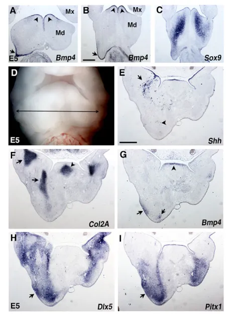
Fig. 5. Gene expression analysis at E5 in BA1 grafted with QT6-Shh cells.(A-C) Frontal sections from distal (A) to half-proximal (B,C) regions of E5 mandibular buds (Mds). (A,B) Bmp4is strongly expressed in the caudal-most ectoderm in the QT6-Shh grafted side (arrow) as in the rostral ectoderm of the Md, where it is normally expressed (arrowheads). (C) Sox9expression is caudally extended in the
mesenchyme of the grafted Md. Mx, maxillary bud. (D) Facial view of a grafted embryo at E5 with the location (double-headed arrow) of a series of transverse sections (E-I) indicated according to the proximodistal axis of the Mds. (E) Shhexpression shows the grafted QT6-Shh cells (arrow) in the proximal region close to the oral endoderm expressing Shh. Very few Shh-producing cells are found in the distal mesenchyme (arrowhead). (F) Col2a1expression shows expansion of Meckel’s cartilage (proximal part and induced branch) in the grafted side (arrows). The arrowhead indicates tongue cartilage. (G) Bmp4 transcripts are present in the tip of the tongue mesenchyme (arrowhead) and in the Md distal ectoderm and mesenchyme in the experimental side (arrows). (H,I) Dlx5and Pitx1expressions are extended distally and show a colocalization with Bmp4in the
mesenchyme in close contact with lateral ectoderm (arrows). Scale bar: 500 μm in A-C,E-I.
D
E
V
E
LO
P
M
E
N
and dHandmisexpression in the anterior limb bud resulted in ectopic Shhexpression and mirror-image digit duplication (Charité et al., 2000; Fernandez-Teran et al., 2000; McFadden et al., 2002). In our experiments, we observed that normal Gli3expression in the medial BA1 posterior half at E3 became considerably reduced after the graft of QT6-Shh cells (Fig. 4C, arrow). By contrast, dHandtranscripts, which are normally present only in the distal BA1 along its entire rostrocaudal axis, showed a lateral expansion in its caudal part, which coincides with absence of Gli3expression (Fig. 4C,D).
By contrast, the graft of control QT6 cells did not alter the expression pattern of the genes encoding either the signaling molecules or transcription factors involved in BA1 development (n=4) (see Fig. S2D-F in the supplementary material).
Thus, QT6-Shh grafts induce Shh, Fgf8and Bmp4expression in the caudal region of BA1 ectoderm leading to ‘rostralization’ of the ectoderm and mesenchyme of the caudal-most part of BA1.
Shh triggers the program of lower-jaw development
We observed that ectopic expression of Bmp4and Fgf8induced by QT6-Shh cells in BA1 caudal ectoderm at E3-4 resulted, at E5, in the growth of caudal expansions lined at their tip by a thickened ectodermal layer still expressing Bmp4 (Fig. 5A,B, arrows). This thickening recalls the apical ectodermal ridge of the limb bud as already described in other experiments (Richman and Tickle, 1992). Moreover, Sox9 (Fig. 5C) and collagen 2A1 (Col2a1) (Fig. 5F), expressed in precartilage nodules of NCC origin, were largely expanded caudally and distally in the grafted BA1. At the same time, Dlx5 and Pitx1 areas of expression were extended in BA1 overgrowth in contact with the Bmp4-expressing ectoderm (Fig. 5G-I, arrows). At E6, in the normal mandible, Cbfa1, a master gene for ossification, was expressed around Col2a1-expressing Meckel’s cartilage and in contact with the ectoderm (Fig. 6C,D). Similarly, in the ectopic mandible, Cbfa1mRNA was present around the induced Meckel’s cartilage (Fig. 6C,D, arrow) in the vicinity of the lateral and distal ectoderm where Bmp4transcripts (Fig. 6B,B⬘) and a faint expression of Shh(Fig. 6E, arrow) were also present.
We were then interested in investigating whether QT6-Shh cell transplants could trigger the development of the nervous and muscular tissues normally associated with the mandible. We looked at specific markers of nerve fibers (HNK1 immunostaining) and muscle cells (MyoD gene expression) in grafted E6 chick embryos. HNK1-positive nerve fibers were detected in the ectopic as well as the normal mandibles. HNK1 labeling was found between Meckel’s cartilage anlage (expressing Col2a1) and Bmp4-expressing ectoderm (Fig. 6F, arrows; Fig. 6B⬘,C⬘, outlined). MyoDtranscripts were also observed in the supernumerary Md, associated with HNK1-immunostained nerve fibers (Fig. 6G,G⬘, arrow).
These results indicate that, at early stages of chick development (5-8 ss), before NCC emigration, Shh can induce the triplication of lower jaws from the caudal part of BA1. The genes encoding the signaling molecules crucial for the onset of jaw development, such as Bmp4,Fgf8andShhfirst are activated in ectopic positions in the ectoderm. This is accompanied by the expression of transcription factors (such as Pitx1,Dlx5,dHand) in the mesenchyme. These processes involve the triggering of the developmental program for cartilage and bone differentiation from NC-derived mesenchyme and of muscle from somite-derived cells.
Shh does not induce ectopic BA1 skeletal structures in other cephalic regions than presumptive BA1 territory
[image:5.612.53.340.59.300.2]We have demonstrated so far that Shh can trigger the molecular program for supernumerary lower-jaw development in BA1 territory, knowing that Meckel’s cartilage could not develop in the absence of Shh, even though the different elements constituting BA1 (ectoderm, endoderm, mesoderm and NC-derived mesenchyme) were present (Brito et al., 2006; Melnick et al., 2005; Yamagishi et al., 2006). In a second step, we have addressed the issue of whether Shh could instruct other cephalic regions to acquire BA1 properties. Thus, we grafted QT6-Shh cells in the presumptive BA2 at 8-9 ss (Fig. 7A, circle). Three days later, at E4, QT6-Shh cells were found in the proximal part of BA2 (Fig. 7D, arrowhead). In contrast to the BA1 grafts, Bmp4 and Fgf8expression patterns were not modified (Fig. Fig. 6. Gene expression analysis at E6 in BA1 grafted with QT6-Shh cells: cell differentiation.(A) Facial view of a grafted E6 chick embryo showing caudal mandibular bud (Md) extension in the grafted side (arrow).
(B-G) Frontal serial sections proximodistally at the level of the Mds. (B,B⬘) Bmp4expression in the distal ectoderm and mesenchyme of the ectopic Md. (C,C⬘) Col2a1 expression indicates an extra Meckel’s cartilage (arrow) near the Bmp4 zone of expression. (D,D⬘) Cbfa1transcripts are present in the mesenchyme of normal and ectopic Mds close to the Bmp4domain and around Col2A1-expressing Meckel’s cartilage anlage. (E) Distal region of the extra Md with a faint expression of Shhin the ectoderm (arrow). (F) Nerve fibers immunostained with HNK1 mAb close to the ectopic Meckel’s cartilage (arrows; see outlines in B⬘and C⬘). (G) Frontal section in Md proximal half, in the grafted side, shows an extension of muscle (MyoDexpression) and HNK1+ nerves (square enlarged in G’). (G⬘) MyoD expression is associated with nerve (HNK1 immunostaining). Scale bar: 500 μm in B-G.
D
E
V
E
LO
P
M
E
N
7E,F). Moreover, no ectopic induction of Shhgene activity in BA2 ectoderm was detected. In fact, a downregulation of the endogenous Shh expression was observed in BA2 (n=4) (Fig. 7D, arrows). Moreover, Pitx1expression was not induced in BA2 mesenchyme (Fig. 7G). These experiments showed that, although QT6-Shh cells are capable of modifying Fgf8, Bmp4and Pitx1expression in BA1, no similar effect occurs in BA2. Later on, at E11, in most cases (n=4/5), no supernumerary skeletal structures were detected among BA2 derivatives (Fig. 7C). Only one embryo showed a small bifurcation of the basihyal (data not shown). The morphology of the face and neck was normal except for the larger middle and inner ear observed on the grafted side (Fig. 7B, arrow). This shows that the patterning effect of Shh on BA1 is not reproducible in BA2.
In another experiment, QT6-Shh cells were grafted more rostrally, lateral to the level of the prosencephalon/mesencephalon boundary (Fig. 7H, circle). At E4, the QT6-Shh cells were dispersed in the post-optic region and around the eye, but no ectopic expression of Shhand Fgf8was observed (data not shown). Curiously, Shh-producing cells were able to induce ectopic expression of Bmp4and Pitx1in the post-optic region (Fig. 7K,L, arrows). At E11 (n=4), while head morphology was normal with no supernumerary bones or cartilages in the post-optic region (Fig. 7I,J), the eye was hypertrophied on the grafted side when compared with the control side (Fig. 7K,K⬘,L,L⬘). Altogether, these data show that each rostrocaudal domain of the cephalic region responds differently to Shh treatment.
DISCUSSION
In the present work, we have evidenced the capacity of the morphogen Shh to induce lower-jaw triplication. Shh-producing cells (line: QT6-Shh) were applied to the presumptive BA1 territory of chick embryos, before migration of cephalic neural crest cells. Supplementary lower-jaw outgrowth is preceded by ectopic expression of a gene cascade in the caudal part of the developing branchial arch, similar to the one operating during normal development of Meckel’s cartilage and associated structures. It involves the expression of Shh, Bmp4and Fgf8in the ectoderm and, later on, Bmp4in the mesenchyme, in which transcription factors like Pitx1, Dlx5,dHandand genes specific for development of cartilage, bone (Sox9, Cbfa1and Col2A1) and muscle (MyoD) are sequentially activated.
The arrangement of the extra jaws that are induced by the QT6-Shh cells is strikingly reproducible: the proximal part of the Meckel’s cartilage is common and distal branches grow separately as mirror images.
[image:6.612.51.392.58.296.2]Shh signal is pivotal for the development of Meckel’s cartilage and associated structures It has previously been shown in our laboratory that a fragment of anterior ventral foregut endoderm is capable of inducing a supernumerary lower beak when grafted in BA1 presumptive area of 6 ss chick embryos. Moreover, excision of the same fragment of endoderm on one side is followed by the absence of the corresponding Meckel’s cartilage (Couly et al., 2002). In recent work, we showed that after excision of Shh-expressing anterior foregut endoderm along with the entire forehead at early neurula stages (5-6 ss), the cephalic NCCs which colonize BA1 exiting from the mesencephalon migrated normally but were further on subjected to massive apoptosis, whereas Shhwas not expressed in the pharyngeal endoderm. Fgf8and Bmp4transcripts were absent in BA1 ectoderm at E3-4 and the embryos were further deprived of Meckel’s cartilage and lower jaw (Brito et al., 2006; Le Douarin et al., 2007). These data suggested that the ability of the anterior pharyngeal endoderm to instruct BA1 to form an ectopic lower beak (Couly et al., 2002) might be related to its capacity to produce Shh. This prompted us to graft Shh-producing cells in the presumptive BA1 at 5-8 ss in order to see if these cells can fully mimic endoderm grafts in this process. It turned out that an ectopic source of Shh, placed on the right side of the embryo, leads to the triplication of the lower jaw with mirror-image polarities along the rostrocaudal axis. It has to be stressed that, in Couly’s experiments, the grafted ventral pharyngeal endoderm exerts a strong effect on the final shape and position of the ectopic lower jaw: with QT6-Shh cells, the endogenous and extra Meckel’s cartilages are fused on their proximal regions that distally diverge in three independent branches; by contrast, the endoderm graft leads to the formation of one ectopic Meckel’s cartilage parallel to the endogenous one. When the ventral pharyngeal endoderm was grafted bilaterally, a complete extra lower jaw was even induced (Couly et al., 2002).
Fig. 7. QT6-Shh cells grafted in presumptive BA2 or in the post-optic region do not induce BA1 extra-skeletal pieces.(A) Graft of QT6-Shh cells in the presumptive BA2 territory in a 9 ss chick embryo (dotted circle). (B,C) At E11, the grafted embryo shows a normal beak morphology but presents a larger otic vesicle (arrow). No ectopic skeletal structure is observed. (D-G) Facial views of E3-4 BA2-grafted embryos. (D) Shhexpression is reduced in BA2 grafted side (arrows), when compared with the control side. (E) Bmp4, (F)Fgf8and (G) Pitx1expression is not affected. (H) QT6-Shh cell graft in the presumptive post-optic region of a 5 ss chick embryo (dotted circle). (I,J) E11 operated chick embryo shows normal morphology and skeletal structures on the grafted side. (K,L) Induction of Bmp4and Pitx1expression in the post-optic region (arrows) where these genes are not expressed in control side (K⬘,L⬘).
D
E
V
E
LO
P
M
E
N
In none of the experiments reported here (Couly et al., 2002; Brito et al., 2006) (the present work) was the morphogenesis of the proximal (dorsal) part of the jaw perturbed. When the forehead was excised together with the PcP and the rostral Shh -expressing endoderm, no Meckel’s cartilages and associated bones were present but the quadrate and articular cartilages, as well as the squamosal, developed normally (Brito et al., 2006). In the experiments by Couly et al. (Couly et al., 2002), no duplication of these cartilages occurred. The same is true for the experiments that we describe here. Thus, Shh is likely to control the formation and extension of Meckel’s cartilage and associated structures, whereas the formation of the proximal (dorsal) skeleton (quadrate and articular) depends upon different mechanisms. In spite of the differences in the final shape of the supernumerary lower beaks obtained with QT6-Shh cells or with foregut endoderm grafts, Shh alone is sufficient to initiate the lower-jaw developmental program in the caudal presumptive BA1 territory. Induction of mirror-image duplication of Meckel’s cartilage by Shh-producing cells recalls the effect of the same type of graft in the anterior mesenchyme of the limb bud. The latter results in digit duplication with mirror-image orientation, owing to the induction of a new ZPA (Riddle et al., 1993). However, in the limb bud, Shhexpression was not induced in the ectoderm.
Shh induces the expression of a BA1-specific gene cascade along the BA1 rostrocaudal axis in both ectoderm and mesenchyme
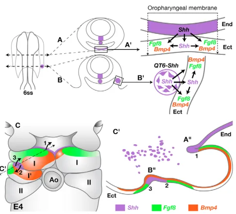
Events that characterize the patterning of BA1 ectoderm have been previously described in the chick embryo (Haworth et al., 2004; Haworth et al., 2007; Brito et al., 2006). As shown in our schematic model (Fig. 8), at 6 ss, the pharyngeal endoderm expressing Shhis in close contact with the ventral ectoderm, thus forming the oro-pharyngeal membrane (Fig. 8A⬘), which further on will form the epithelium lining the oral opening (Fig. 8A⬙). At this stage (E4), expressions of both Fgf8and Bmp4genes are activated in the oral epithelium, which corresponds to the rostral-most BA1 ectoderm. Moreover, Shhis expressed in the pharyngeal endoderm in close contact with Fgf8proximally and Bmp4distally (Fig. 8C, part I, on the right). In our experiments, 7 hours after the graft (at 10 ss), the QT6-Shh cells are located between the endoderm and ectoderm of the presumptive BA1 (Fig. 8B; see Fig. 1B). Later on (at E4), the quail fibroblasts will be found exclusively in the proximal BA1 area (Fig. 8C⬘, purple dots). Re-patterning of BA1 ectoderm by QT6-Shh cells involves a cascade of events similar to those taking place normally in BA1 ectodermal epithelium under the influence of the foregut endoderm. A new ‘oral-like epithelium’ is induced in which Fgf8, Bmp4and Shhare expressed in the caudal part of BA1 ectoderm (Fig. 8C, left; Fig. 8C⬘; see Fig. 3 and Fig. S3 in the supplementary material). This rostrocaudal re-patterning is also observed in the mesenchyme through upregulation of Pitx1,Nogginand dHand, and reduction or loss of Gli3. This suggests that, at 5-8 ss, the capacity to respond to Shh, far from being restricted to the region fated to become the oral epithelium (rostral BA1 ectoderm), is in fact present over the entire BA1 ectoderm.
The respective spatial disposition of the ectodermal areas expressing Shh, Bmp4and Fgf8is highly significant concerning the rostrocaudal orientation of the future extra jaws induced by QT6-Shh (Fig. 8C: 1-3).
Both Bmp4 and Fgf8 have previously been shown to be crucial for lower-jaw development by other groups. Conditional knockout of Bmp4in distal BA1 ectoderm completely hampered lower-jaw development (Liu et al., 2005). Other authors found that application
of Bmp2/4-soaked beads in chick BA1 mesenchyme, at E3, induced Meckel’s cartilage duplication (Barlow and Francis-West, 1997; Mina et al., 2002). In our experimental design, prior to lower-jaw duplication, Pitx1 (Lanctôt et al., 1999), Dlx5(for a review, see Depew et al., 2005) and dHand(Yanagisawa et al., 2003) were associated with Bmp4expression and followed by the activation of genes involved in chondrocytes (Sox9and Col2a1) (Mori-Akiyama et al., 2003), osteoblasts (Cbfa1) (Ducy et al., 1997) and muscle (MyoD) (Kablar et al., 1998) differentiation.
The control of Fgf8expression by Bmp4 was demonstrated in Chordinand Nogginknockout mice (Stottmann et al., 2001). In these cases, Bmp activities were enhanced while the proximal part of BA1 was devoid of Fgf8expression, resulting in the absence of lower jaw. The same phenotype was obtained by downregulation of Bmp4expression in BA1 ectoderm, leading to an increase of Fgf8 territory, which extended distally (Liu et al., 2005). Head infection with RCAS-Fgf8 also resulted in shorter lower-jaw formation (Abzhanov and Tabin, 2004).
[image:7.612.319.562.56.277.2]It appears therefore that a crucial equilibrium between the production of Bmp4 and Fgf8 is necessary for the extension of Meckel’s cartilage. Our experiments, which consist of the administration of exogenous Shh prior to BA1 development, produce extra sources of Bmp4 and Shh without downregulating Fgf8 production. This disposition favors the development of extra jaws. In these experiments, the presence of exogenous Shh can lead Fig. 8. A model for oral epithelium induction by the foregut endoderm or by the graft of QT6-Shh cells in the chick BA1 territory.(A,B) A 6 ss chick embryo cross-sectioned at the normal oropharyngeal level (A), and at the level of grafted QT6-Shh cells (purple spot) (B). Gene inductions elicited by normal (A⬘) and ectopic (B⬘) sources of Shh (purple) are indicated. (C) Expression of Fgf8(green), Bmp4(red) and Shh(purple) in BA1 ectoderm of QT6-Shh grafted (on the left) and control (on the right) sides at E4. Curved arrows indicate growth orientation of the further normal (1) and induced (2 and 3) Meckel’s branches. (C⬘) Virtual section of the experimental BA1 (broken line) indicates three distinct lower jaw organizing centers (1-3) presenting Fgf8/Shh/Bmp4alternations in the normal (1) and induced (2,3) jaw anlagen. I, first branchial arch; I⬘, induced part of I; II, second branchial arch; Ect, ectoderm; End, endoderm; Ao, aortic arch. A⬙and
B⬙, normal oral epithelium and induced ‘oral-like epithelium’.
D
E
V
E
LO
P
M
E
N
to an increase of NCC number, and NCCs are known to be able to produce Bmp antagonists such as Noggin (Stottmann et al., 2001; Smith and Graham, 2001). We could demonstrate that graft of QT6-Shh cells in BA1 resulted in induction of ectopic Nogginexpression, corresponding to Bmp4areas (see Fig. 3C, Fig. 4A). Under these conditions, the level of Bmp4 could be restricted to a rate compatible with the production of Fgf8 in a quantity appropriate for the formation of the supernumerary jaws. The fact that infection of chicken Md with RCAS-Nogginprevents the formation of cartilage and bones supports this view (Foppiano et al., 2007).
It is interesting that the graft of QT6-Shh cells results in reduction or loss of Gli3expression in BA1. This is in line with the fact that inactivation of Gli3in mice leads to upregulation of Fgf8transcripts in several sites, such as in the apical ectodermal ridge of the limb bud (Aoto et al., 2002), and results in polydactylism (Litingtung et al., 2002; te Welscher et al., 2002a). A hypothesis to account for the loss of Gli3after QT6-Shh cell grafts in BA1 is an indirect action through dHand, which has already been shown to control Gli3 expression in the posterior limb bud of mouse embryo (Fernandez-Teran et al., 2000; te Welscher et al., 2002b). Moreover, it is known that Shh does not act directly on Gli3gene expression but rather is involved in preventing the cleavage of full-length Gli3 (activator form 190 kDa) to its repressor form (83 kDa) (Wang et al., 2000).
In our experiments, the striking effect of QT6-Shh cell grafts on BA1 development is the induction of ectodermal foci similar to the normal one acting as organizer of lower-jaw extensions. This process leads to the development of two extra lower jaws owing to the maintenance of a balance between Fgf8and Bmp4expression levels.
BA1 ectoderm has a specific capacity to respond to Shh signal
The last aspect of this work was to investigate whether Shh was able of instructing other cephalic areas to acquire BA1 properties. We demonstrated that QT6-Shh cell grafts in the presumptive area of BA2 neither induced ectopic skeletal elements nor triggered Fgf8, Bmp4 and Shh expression in the ectoderm or Pitx1 in the mesenchyme. This is in agreement with previous studies in which graft of foregut endoderm or FEZ ectoderm into BA2 did not trigger the development of BA1 structures (Couly et al., 2002; Hu et al., 2003). Moreover, infection of BA2 ectoderm by RCAS-Shhdid not result in ectopic skeleton development (Haworth et al., 2007). Indeed, Shhexpression was disturbed in the experimental side of BA2 and no hyperplasia was seen comparable with the one observed in BA1. We also showed that graft of QT6-Shh cells in the presumptive post-optic region at 4 ss induced ectopic expression of Pitx1and Bmp4, but not of Shhand Fgf8, and no ectopic cartilages and bones were found (Fig. 7J). Similarly, implanting Shh-soaked beads in chick embryo Mx at HH15 did produced no phenotype (Lee et al., 2001). By contrast, graft of fibroblasts infected with RCAS-Shh, into the naso-frontal prominence of a chick embryo at HH20-21 induced a supernumerary egg tooth and a cartilage rod derived from the nasal septum (Hu and Helms, 1999). Although these experiments and the ones described here were carried out at different stages of development, altogether they show that each cephalic region has its own properties to respond to Shh.
In conclusion, Shh is a pivotal signal for the development of the lower jaw, including Meckel’s cartilage and associated skeletal, muscular and neural structures. These morphological events are preceded by Shh, Fgf8and Bmp4 induction in BA1 ectodermal epithelium, giving rising to a ‘new oral-like epithelium’. It is notable that this molecular response to Shh is specific to BA1 and cannot be induced in other cephalic regions. Moreover, Shh-producing
endodermal cells of the oral epithelium act in lower-jaw development as a zone of polarizing activity (ZPA) comparable with the ZPA in limb patterning. An extra source of Shh applied at an early developmental stage to the BA1 presumptive territory is able to induce two extra zones of polarizing activity caudal to the normal one.
We thank Sophie Gournet for illustrations and Christine Vincent for technical assistance. We also thank T. Jaffredo for Cbfa1/Runx2probe. QCPN antibody was obtained from the Developmental Studies Hybridoma Bank developed under the auspices of the NICHD and maintained by The University of Iowa, Department of Biological Sciences, Iowa City, IA 52242. We thank E. Dupin for reading the manuscript. J.B. received fellowships from the Coordenação de Aperfeiçoamento de Pessoal de Nivel Superior (CAPES) and Fondation pour la Recherche Médicale (FRM). This work was supported by the Centre National de la Recherche Scientifique (CNRS), Université Paris-Sud, and by grants from the Association pour la Recherche contre le Cancer [ARC-3929, ARC-4815 (M.-A.T.)] and the Fondation L. Bettencourt Shuller to N.L.D.
Supplementary material
Supplementary material for this article is available at http://dev.biologists.org/cgi/content/full/135/13/2311/DC1
References
Abzhanov, A. and Tabin, C. J.(2004). Shh and Fgf8 act synergistically to drive cartilage outgrowth during cranial development. Dev. Biol. 273,134-148. Ahlgren, S. C. and Bronner-Fraser, M.(1999). Inhibition of sonic hedgehog
signaling in vivo results in craniofacial neural crest cell death. Curr. Biol. 9, 1304-1314.
Aoto, K., Nishimura, T., Eto, K. and Motoyama, J.(2002). Mouse GLI3 regulates Fgf8 expression and apoptosis in the developing neural tube, face, and limb bud. Dev. Biol. 251, 320-332.
Barlow, A. J. and Francis-West, P. H.(1997). Ectopic application of recombinant BMP-2 and BMP-4 can change patterning of developing chick facial primordia.
Development124, 391-398.
Brito, J. M., Teillet, M. A. and Le Douarin, N. M.(2006). An early role for sonic hedgehog from foregut endoderm in jaw development: ensuring neural crest cell survival. Proc. Natl. Acad. Sci. USA103, 11607-11612.
Cerny, R., Lwigale, P., Ericsson, R., Meulemans, D., Epperlein, H. H. and Bronner-Fraser, M.(2004). Developmental origins and evolution of jaws: new interpretation of “maxillary” and “mandibular”. Dev. Biol. 276, 225-236. Charité, J., McFadden, D. G. and Olson, E. N.(2000). The bHLH transcription
factor dHAND controls Sonic hedgehog expression and establishment of the zone of polarizing activity during limb development. Development127, 2461-2470.
Chiang, C., Litingtung, Y., Lee, E., Young, K. E., Corden, J. L., Westphal, H. and Beachy, P. A.(1996). Cyclopia and defective axial patterning in mice lacking Sonic hedgehog gene function. Nature383, 407-413.
Couly, G. and Le Douarin, N. M.(1985). Mapping of the early neural primordium in quail-chick chimeras. Developmental relationships between placodes, facial ectoderm, and prosencephalon. Dev. Biol. 110, 422-439.
Couly, G. and Le Douarin, N. M.(1990). Head morphogenesis in embryonic avian chimeras: evidence for a segmental pattern in the ectoderm corresponding to the neuromeres. Development108, 543-558.
Couly, G., Grapin-Botton, A., Coltey, P. and Le Douarin, N. M.(1996). The regeneration of the cephalic neural crest, a problem revisited: the regenerating cells originate from the contralateral or from the anterior and posterior neural fold. Development122, 3393-3407.
Couly, G., Creuzet, S., Bennaceur, S., Vincent, C. and Le Douarin, N. M. (2002). Interactions between Hox-negative cephalic neural crest cells and the foregut endoderm in patterning the facial skeleton in the vertebrate head.
Development 129, 1061-1073.
Creuzet, S., Schuler, B., Couly, G. and Le Douarin, N. M.(2004). Reciprocal relationships between Fgf8 and neural crest cells in facial and forebrain development. Proc. Natl. Acad. Sci. USA 101, 4843-4847.
Crossley, P. H., Minowada, G., MacArthur, C. A. and Martin, G. R.(1996). Roles for FGF8 in the induction, initiation, and maintenance of chick limb development. Cell84, 127-136.
Depew, M. J., Simpson, C. A., Morasso, M. and Rubenstein, J. L.(2005). Reassessing the Dlx code: the genetic regulation of branchial arch skeletal pattern and development. J. Anat. 207, 501-561.
Ducy, P., Zhang, R., Geoffroy, V., Ridall, A. L. and Karsenty, G.(1997). Osf2/Cbfa1: a transcriptional activator of osteoblast differentiation. Cell89, 747-754.
Duprez, D., Fournier-Thibault, C. and Le Douarin, N.(1998). Sonic Hedgehog induces proliferation of committed skeletal muscle cells in the chick limb.
Development125, 495-505.
D
E
V
E
LO
P
M
E
N
Fernandez-Teran, M., Piedra, M. E., Kathiriya, I. S., Srivastava, D., Rodriguez-Rey, J. C. and Ros, M. A.(2000). Role of dHAND in the anterior-posterior polarization of the limb bud: implications for the Sonic hedgehog pathway. Development127, 2133-2142.
Foppiano, S., Hu, D. and Marcucio, R. S.(2007). Signaling by bone
morphogenetic proteins directs formation of an ectodermal signaling center that regulates craniofacial development. Dev. Biol. 312, 103-114.
Francis, P. H., Richardson, M. K., Brickell, P. M. and Tickle, C.(1994). Bone morphogenetic proteins and a signalling pathway that controls patterning in the developing chick limb. Development120, 209-218.
Francis-West, P. H., Tatla, T. and Brickell. P. M.(1994). Expression patterns of the bone morphogenetic protein genes Bmp-4 and Bmp-2 in the developing chick face suggest a role in outgrowth of the primordial. Dev. Dyn. 201, 391-398.
Haworth, K. E., Healy, C., Morgan, P. and Sharpe, P. T.(2004). Regionalisation of early head ectoderm is regulated by endoderm and prepatterns the orofacial epithelium. Development131, 4797-4806.
Haworth, K. E., Wilson, J. M., Grevellec, A., Cobourne, M. T., Healy, C., Helms, J. A., Sharpe, P. T. and Tucker, A. S.(2007). Sonic hedgehog in the pharyngeal endoderm controls arch pattern via regulation of Fgf8 in head ectoderm. Dev. Biol. 303, 244-258.
Henrique, D., Adam, J., Myat, A., Chitnis, A., Lewis, J. and Ish-Horowicz, D. (1995). Expression of a Delta homologue in prospective neurons in the chick.
Nature375, 787-790.
Howard, M., Foster, D. N. and Cserjesi, P.(1999). Expression of HAND gene products may be sufficient for the differentiation of avian neural crest-derived cells into catecholaminergic neurons in culture. Dev. Biol. 215, 62-77. Hu, D. and Helms, J. A.(1999). The role of sonic hedgehog in normal and
abnormal craniofacial morphogenesis. Development126, 4873-4884. Hu, D., Marcucio, R. S. and Helms, J. A.(2003). A zone of frontonasal ectoderm
regulates patterning and growth in the face. Development 190,1749-1758. Hui, C. C. and Joyner, A. L.(1993). A mouse model of greig
cephalopolysyndactyly syndrome: the extra-toesJ mutation contains an intragenic deletion of the Gli3 gene.Nat. Genet. 3, 241-246.
Jeong, J., Mao, J., Tenzen, T., Kottmann, A. H. and McMahon, A. P.(2004). Hedgehog signaling in the neural crest cells regulates the patterning and growth of facial primordia. Genes Dev. 18, 937-951.
Kablar, B., Asakura, A., Krastel, K., Ying, C., May, L. L., Goldhamer, D. J. and Rudnicki, M. A.(1998). MyoD and Myf-5 define the specification of musculature of distinct embryonic origin. Biochem. Cell Biol. 76, 1079-1091. Köntges, G. and Lumsden, A.(1996). Rhombencephalic neural crest
segmentation is preserved throughout craniofacial ontogeny. Development122, 3229-3242.
Kordes, U., Cheng, Y. C. and Scotting, P. J.(2005). Sox E gene expression distinguishes different types and maturation stages of glial cells in developing chick and mouse. Brain Res. Dev. Brain Res. 157, 209-213.
Lanctôt, C., Moreau, A., Chamberland, M., Tremblay, M. L. and Drouin, J. (1999). Hindlimb patterning and mandible development require the Ptx1 gene.
Development126, 1805-1810.
Le Douarin, N. M. and Kalcheim, C.(1999). The Neural Crest(2nd edition). New York: Cambridge University Press.
Le Douarin, N. M., Brito, J. M. and Creuzet, S.(2007). Role of the neural crest in face and brain development. Brain Res. Rev. 55, 237-247.
Lee, S. H., Fu, K. K., Hui, J. N. and Richman, J. M.(2001). Noggin and retinoic acid transform the identity of avian facial prominences. Nature414, 909-912. Lee, S. H., Bédard, O., Buchtová, M., Fu, K. and Richman, J. M.(2004). A new
origin for the maxillary jaw. Dev. Biol. 276, 207-224.
Litingtung, Y., Dahn, R. D., Li, Y., Fallon, J. F. and Chiang, C.(2002). Shh and Gli3 are dispensable for limb skeleton formation but regulate digit number and identity. Nature418, 979-983.
Liu, W., Selever, J., Murali, D., Sun, X., Brugger, S. M., Ma, L., Schwartz, R. J., Maxson, R., Furuta, Y. and Martin, J. F.(2005). Threshold-specific
requirements for Bmp4 in mandibular development. Dev. Biol. 283, 282-293.
McFadden, D. G., McAnally, J., Richardson, J. A., Charité, J. and Olson, E. N. (2002). Misexpression of dHAND induces ectopic digits in the developing limb bud in the absence of direct DNA binding. Development129, 3077-3088. Melnick, M., Witcher, D., Bringas, P., Jr, Carlsson, P. and Jaskoll, T.(2005).
Meckel’s cartilage differentiation is dependent on hedgehog signaling. Cells
Tissues Organs179, 146-157.
Mina, M., Wang, Y. H., Ivanisevic, A. M., Upholt, W. B. and Rodgers, B. (2002). Region- and stage-specific effects of FGFs and BMPs in chick mandibular morphogenesis. Dev. Dyn. 223, 333-352.
Mori-Akiyama, Y., Akiyama, H., Rowitch, D. H. and de Crombrugghe, B. (2003). Sox9 is required for determination of the chondrogenic cell lineage in the cranial neural crest. Proc. Natl. Acad. Sci. USA100, 9360-9365. Ojeda, J. L., Barbosa, E. and Bosque, P. G.(1970). Selective skeletal staining in
whole chicken embryos; a rapid Alcian blue technique. Stain Technol. 45, 137-138.
Pera, E., Stein, S. and Kessel, M.(1999). Ectodermal patterning in the avian embryo: epidermis versus neural plate. Development126, 63-73.
Pourquie, O., Fan, C. M., Coltey, M., Hirsinger, E., Watanabe, Y., Breant, C., Francis-West, P., Brickell, P., Tessier-Lavigne, M. and Le Douarin, N. M. (1996). Lateral and axial signals involved in avian somite patterning: a role for BMP4. Cell84, 461-471.
Richman, J. M. and Tickle, C.(1992). Epithelial-mesenchymal interactions in the outgrowth of limb buds and facial primordia in chick embryos. Dev. Biol. 154, 299-308.
Riddle, R. D., Johnson, R. L., Laufer, E. and Tabin, C.(1993). Sonic hedgehog mediates the polarizing activity of the ZPA. Cell75, 1401-1416.
Ruiz i Altaba, A.(2006). Hedgehog-Gli Signaling in Human Disease(1st edn). Georgetown: Landes Bioscience.
Saunders, J. W. and Gasseling, M.(1968). Ectodermal-mesenchymal interaction in the origin of the limb symmetry. In Epithelial-Mesenchymal Interaction(ed. R. Fleischmayer and R. E. Billingham), pp. 78-79. Baltimore: Williams and Wilkins. Smith, A. and Graham, A.(2001). Restricting Bmp-4 mediated apoptosis in
hindbrain neural crest. Dev. Dyn. 220, 276-283.
Stottmann, R. W., Anderson, R. M. and Klingensmith, J.(2001). The BMP antagonists Chordin and Noggin have essential but redundant roles in mouse mandibular outgrowth. Dev. Biol. 240, 457-473.
te Welscher, P., Zuniga, A., Kuijper, S., Drenth, T., Goedemans, H. J., Meijlink, F. and Zeller, R.(2002a). Progression of vertebrate limb development through SHH-mediated counteraction of GLI3. Science298, 827-830. te Welscher, P., Fernandez-Teran, M., Ros, M. A. and Zeller, R.(2002b).
Mutual genetic antagonism involving GLI3 and dHAND prepatterns the vertebrate limb bud mesenchyme prior to SHH signaling. Genes Dev. 16, 421-426.
Teillet, M., Watanabe, Y., Jeffs, P., Duprez, D., Lapointe, F. and Le Douarin, N. M.(1998). Sonic hedgehog is required for survival of both myogenic and chondrogenic somitic lineages. Development125, 2019-2030.
Trumpp, A., Depew, M. J., Rubenstein, J. L., Bishop, J. M. and Martin, G. R. (1999). Cre-mediated gene inactivation demonstrates that FGF8 is required for cell survival and patterning of the first branchial arch. Genes Dev. 13, 3136-3148.
Wall, N. A. and Hogan, B. L.(1995). Expression of bone morphogenetic protein-4 (BMP-protein-4), bone morphogenetic protein-7 (BMP-7), fibroblast growth factor-8 (FGF-8) and sonic hedgehog (SHH) during branchial arch development in the chick. Mech. Dev. 53, 383-392.
Wang, B., Fallon, J. F. and Beachy, P. A.(2000). Hedgehog-regulated processing of Gli3 produces an anterior/posterior repressor gradient in the developing vertebrate limb. Cell100, 423-434.
Yamagishi, C., Yamagishi, H., Maeda, J., Tsuchihashi, T., Ivey, K., Hu, T. and Srivastava, D.(2006). Sonic hedgehog is essential for first pharyngeal arch development. Pediatr. Res. 59, 349-354.
Yanagisawa, H., Clouthier, D. E., Richardson, J. A., Charite, J. and Olson, E. N.(2003). Targeted deletion of a branchial arch-specific enhancer reveals a role of dHAND in craniofacial development. Development130, 1069-1078.