1
EVALUATION OF THE USEFULNESS OF LAMINATED LAYER
ANTIGENS IN THE SEROLOGICAL FOLLOW UP OF CYSTIC
ECHINOCOCCOSIS IN HUMANS
OGHENEKARO E. OKITI
SCHOOL OF ENVIRONMENT AND LIFE SCIENCES, UNIVERSITY OF
SALFORD, SALFORD, UK.
Submitted in Partial Fulfillment for the Degree of Master of
Philosophy.
2 DEDICATION
3 TABLE OF CONTENTS
Dedication………..2
Table of Contents ………...3
List of abbreviation ………..9
Acknowledgment………..10
Abstract……… 11
Chapter 1 1.0 Introduction……… 13
1.1 Echinococcosis ...13
1.2 Distribution /Epedemiology...15
1.3 Public health complications ... .18
1.4 Life cycle of Echinococcus granulosus………. 19
1.5 Echinococcus metacestode……… 20
1.6 The Laminated layer... .23
1.7 Development in the intermediate host………. 25
1.8 Human Hydatidosis...28
1.9 Pathology...29
1.10 Diagnosis ... 32
1.11 Treatment... ..39
1.11.1 Surgery ...40
1.11.2 Pair ...41
1.11.3 Chemotherapy ...42
1.12 Changes in cyst morphology and follow up after treatment... 43
1.13 Cyst Classification ... 44
4
1.15 The role of cytokine in echinococcosis... 51
1.16 Post- treatment follow-up……….. 55
Rationale of Study………..56
2 Materials and Methods ...59
2.1 Antigenic materials ...59
2.1.1 Hydatitid cyst fluid (HCF) ...59
2.1.2 Laminated Layer (LL)... …….59
2.2 Sera ...60
2.3 Enzyme Immunosorbent Assay (ELISA)………..63
2.4 SDS-PAGE...64
2.5 Western Blot / immunoblot ...65
2.6 Lectin Assay ... ..66
2.7 Affinity chromatography ...66
Chapter 3 3.0 Characterisation of Laminated layer extracts ...68
3.1 Introduction ………....68
3.2 Material and Method……….75
3.3 Results………....76
3.3.1 SDS-PAGE……… ….76
3.3.2 Reactivity of Laminated layer in ELISA……….76
3.3.3 Immunoblots………..77
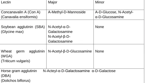
3.4 Lectin Binding Analysis………...79
5 CHAPTER 4 – Lectin Affinity purification
4.1 Introduction ………..85
4.2 Aim……….87
4.3 Results………...88
4.4 Discussion……….91
CHAPTER 5 The Use of laminated layer in follow-up after treatment . 5.1 Introduction... ………..94
5.2 Results... ………. 98
5.3 Discussion...119
Chapter 6 General discussion... ..122
References... 129
Appendix... 157
List of Tables: Table 2.1: Summary of patients information………..61
Table 3.1 Carbohydrate specifications and Specificities……… 80
Table 5.1: Summary of patients infromation………...96
Table 5.2: Antibody response (untreated patients)………98
6 List Of Figures.
Figure 1.1 Life cycle of Echinococcus……….20
Figure 1.2 Structure of hydatid cyst ……….23
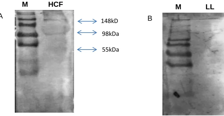
Figure 1.3 Classifiction of cyst ultrasound images ...48 Figure 3.1 An SDS Page Image showing the dissociation of the LL proteins as compared to SHF ………76
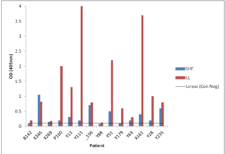
Figure 3.2: Initial ELISA for fourteen confirmed hydated patients screemed against cyst fluid (SHF) and laminated layer (LL) antigens ………77
Figure 3.3 Immunoblot images of the reactivity of HCF and LL with total IgG with pooled positive sera ………77
Figure 3.4 Blot images for negative controls for the reactivity of HCF and LL with total IgG……….78
Figure 3.5 Blot Images the reactivity of HCF and LL with IgG1 with pooled positive sera ……… .78
Figure 3.6 Blot images for the negative controls for the reaction of HCF and LL to IgG1……….79
Figure 3.7 Blot imagines for showing positive results of the reactivity of HCF and LL with IgG4……… …81
Figure 4.1 The chart recorder showing run through and elution peaks of crude LL extract. ……….89
7
Figure5.2 Graphs showing response of HCF and LL with P160 to IgG, IgG1 and IgG4...106 Figure 5.3 Graphs showing response of HCF and LL with X269 to IgG, IgG1 and IgG4...107 Figure 5.4 Graphs showing response of HCF and LL with X345 to IgG, IgG1 and IgG4...108 Figure 5.5 Graphs showing response of HCF and LL with X161 to IgG, IgG1 and IgG4...109 Figure 5.6 Graphs showing response of HCF and LL with Y13 to IgG, IgG1 and IgG4 ………..110
Figure 5.7 Graphs showing response of HCF and LL with Y28 to IgG, IgG1 and IgG4 ………..111
Figure 5.8 Graphs showing response of HCF and LL with Y51 to IgG, IgG1 and IgG4 ………..112
Figure 5.9 Graphs showing response of HCF and LL with Y63 to IgG, IgG1 and IgG4 ………...113
Figure 5.10 Graphs showing response of HCF and LL with Y88 to IgG, IgG1 and IgG4………..114
Figure 5.11 Graphs showing response of HCF and LL with Y111 to IgG, IgG1 and IgG4……… .. 115
Figure 5.12 Graphs showing response of HCF and LL with Y179 to IgG, IgG1 and IgG4……… ..116
8
9
List of Abbreviations
HCF Hydatid Cyst Fluid
SDS Sodium dodecyl sulphate
PAGE Polyacrylamide gel electrophoresis WHO World Health Organisation
PNPP p-nitrophenylphosphate PBS Phosphate buffered saline
PAIR Puncture, Aspiration, Injection and Re-aspiration ELISA Enzyme-Linked Immunosorbent Assay.
LL Laminated layer GL Germinal layer
CE Cystic Echinococcosis AE Alveolar Echinococcosis TEMED Tetramethylethylenediamine TRIS Trishydroxymethylaminomethane BCB Bicarbonate carbonate buffer BCIP Bromochloroindolyl phosphate KDa Kilodalton
NCP Nitrocellulose paper AgB Antigen B
Ag5 Antigen 5
10
ACKNOWLEDGEMENTS
I wish to express my gratitutde to my supervisor, Prof. Micheal T. Rogan, for his guidance, continuous encouragement and great help during the course of my study. Sincere thanks also goes to Dr Eberhard Zeyhel (AMREF) for the supply of sera samples.
I would also like to thank my parents for their wonderful help, making funds available and for always being there.
Many thanks to Dr Anthony Bodell who have being of immense help throughout the course of my study.
To my colleague, Judy Mwangi, I say thank you for being there, thank you for your show of friendship.
11
ABSTRACT
Cystic Echinococcosis is a zoonotic infection of humans caused by the metacestode (larval) stages of the cestode Echinococcus granulosus (family Taeniidae). Diagnosis of the infection often involves immunodiagnostic approaches using cyst fluid antigens and these have also been used in serological follow up of patients after surgical treatment or chemotherapy. However the usefulness of other metacestode antigenic extracts for these purposes has not been fully investigated.
12
13
CHAPTER 1
INTRODUCTION
1.1 Echinococcosis
The genus Echinococcus of cestode parasites belonging to the family Taeniidae
includes also the well-known tapeworms such as Taeniasaginata. Echinococcus
species have life cycles that always involve a definitive host harbouring an adult worm, and an intermediate host carrying the metacestode or the larva. The definitive host (mostly carnivore) become infected by the ingestion of protoscolices that are contained in the bladder-like metacestode lodged in intermediate host viscera (Diaz et.al, 2011) Protoscolices later develop into 3mm long gut –dwelling adult tapeworms, which produce eggs. The intermediate host is infected by accidentally ingesting eggs passed out with faeces from definitive host. Hydatid disease is the term associated with infection by larval Ehinococcus (Diaz et. al, 2011). Traditionally, four species are recognised in the genus. They are Echinococcus granulosus,
Echinococcus multilocularis, Echinococcus oligarthrus and Echinococcus vogeli. Canids are definitive hosts for all species in the genus with the lion strain of E. granulosus being an exception (Diaz et. al, 2011).Domestic ungulates mainly act as intermediate hosts for E. granulosus, while wild rodents and
lagomorphs carry out this role for the other three species.
14
hydatid disease caused by E. multilocularis (Brunetti, et. al, 2010). Cystic hydatid disease can remain unobserved for many years, as the slow growing hydatid cyst by and large causes pathology only through the compression, contracting and putting pressure on the host organ, which is most commonly liver or lung, or any other organ. Consequently, symptoms are rather non-specific and are dependent on the precise location. Complications not associated with organ contraction can be experienced and are also important; they include bacterial superinfection and cyst rupture, which can bring about the risk of anaphylactic shock and or the initiation of a secondary infection. The disease has a worldwide distribution. It is a highly prevalent disease, especially where pastoralism is an important activity and regular dosage of domestic dogs with the drug praziquantel have not been allowed due to politics, institutional conditions. Hence, cystic hydatid disease is important in areas of Central Asia, South America, China, and Africa (Jenkins et. al, 2005).
15
1.2 Distribution/Epidemiology
The genus Echinococcus is an important one because it consists of a number of zoonotic species that can cause serious ill health in man. The genus is consisted of at least 4 species, but evidence gathered from recent molecular studies suggests there should be a taxonomic revision to at least 5 species or even possibly 6 (Le et al, 2002; McManus, 2002; Thompson & McManus, 2002). With the species E. granulosus, there is also a significant strain variation. The definitive host with each species is a carnivore, whilst the intermediate host could be any of a large number of mammalian species. The parasite is pathogenically and economically significant in intermediate and unusual intermediate hosts, where the larval parasite develops into a hydatid cyst. The genus is distributed worldwide, although geographical distribution of a number of species is limited (Torgerson and Budke, 2003).
E.granulosus is distributed globally, it is found on all continents, with highest prevalence in parts of Eurasia (especially the Mediterranean countries, the Russian Federation and adjacent independent states, and China), East and North Africa, Australia and South America (Eckert et al., 2001). According to Eckert et al (2001) and Ito et al (2003), there is also clear evidence for the emergence and re-emergence of human cystic echinococcosis in parts of China, central Asia, Eastern Europe, and Israel. Communities that are involved in sheep farming are known to harbour the highest rates of infection, emphasising the public health importance of the sheep-dog cycle and the sheep strain of E. granulosus in transmission to people. (Thompson and McManus, 2001; McManus, 2002).
16
guarding and or herding animals. Transmission of E. granulosus is predominantly in a cycle between dog definitive hosts harbouring the small intestinal tapeworm, and livestock (especially sheep). The distribution of this parasite in the United Kingdom is restricted, being found mainly in mid and southern Wales. The zoonotic strains of E.granulosus are present in every country in Europe, except Ireland, Iceland and Denmark (Torgerson and Budke, 2003). It is most extremely endemic in large parts of China and a significant re-emerging zoonosis in the former Soviet Republics in central Asia (Torgerson et al, 2002a,b).
The parasite is also present throughout the Indian subcontinent and the Middle East.
E.granulosus is widely circulated or diffused in Africa, and of a major problem in Northern African countries such as Tunisia, Morrocco, Libya and Algeria. There are specific concerns for the parasite in the South of the Sahara in certain locations like the Turkana in Kenya. The parasite is present in Canada and Alaska of the North America region, and appears to put on mainly a sylvatic cycle. In the continental USA, the parasite occurs at irregular intervals with just a few communities, foci such as certain communities in Utah and California (Torgerson and Budke, 2003).
It is also widespread in South America, especially in Argentina, Uruguay and Peruvian Andes. In Australia a sylvatic cycle between dingoes and wallabies can also occur with over 25% of dingoes and up to 65% of macropod marsupials infected (Jenkins and Morris, 1995; Jenkins, 2002).
17
usually involved. The latter transmission cycle is the commonest and presents the greater risk, threat to human health. The highest incidence rates in man have
often being from, or noticed in areas where there are close relationship with man and domestic livestock, with man often using dogs as working dogs. The common source of infection for dogs is by feeding on offal from infected sheep, which most times harbour G1 zoonotic strain that is in many cases responsible for human CE.
18
Emerging also in other former communist countries like Bulgaria is a similar pattern (Todorov and Boeva, 1999).However, providing resources were made available, a decrease, reduction in prevalence, even eradication would have been a possibility. This mainly is due to the factors that affect dynamics of transmission.
1.3
PublicHealth Implications
Worldwide currently, over three million people are afflicted with echinococcosis, and the extent of the morbidity associated with both AE and CE is estimated to result in more than 1.5million disability adjusted life years (DALY‘S) lost ( Budke et. al, 2006; Togerson et al, 2010). AE is of significant public health concern, especially in parts of Central and Eastern Europe and notably, Northwest China (Vuitton et. al, 2003). Even though, in many endemic areas, the annual incidence of AE may appear low (0.03-1.2 per 100 000 inhabitants) (Vuitton et. al, 2003), the estimation is that there are many cases remaining undiagnosed (Brunetti et al, 2010).
19
1.4 Life Cycle of Echinococcus granulosus
Two hosts are required for the life cycle of the parasite, a definitive and an intermediate host. Dogs are the main definitive hosts for E. granulosus while foxes are for E. multilocularis. The major intermediate hosts for E. granulosus are ungulates and wild rodents for E. multilocualris (See review by, Ammann and Eckert, 1996). The intestine of the definitive host harbours the adult worm and eggs passed out in the faeces are ingested by the intermediate host. Gastroenteric enzymes digest the external coating of the eggs, following its oral ingestion and the oncosphere larva is freed. The embryos possess hooklets with which they attach to the mucosa of the intestine (Tüzün et al, 2002). They penetrate the wall and enter the portal venules and lymphatics, from where they are transported to the liver, lungs, organs and tissues of the systemic circulation. Ultimately, the oncosphere develops into a cyst within which the protoscoleces are produced.
20
Figure 1.1: Life cycle of Echinococcus granulosus
(Source:
http://images.google.co.uk/images?imgurl=http://www.biochemj.org/bj/362/0297/bj3620297f0 1.gif)
1.5 Echinococcus Metacestodes
21
through an increase in diameter. E. oligarthrus and E. vogeli develop in similar way but tend to form multi-chambered cysts. Growth by E. multilocularis is different and contrasting to what is obtained with the other species in that it grows by
outward budding, thereby giving rise to a labyrinth of chambers and tubules (Diaz et. al, 2011).
22 cyst rupture (Brunetti et. al, 2010).
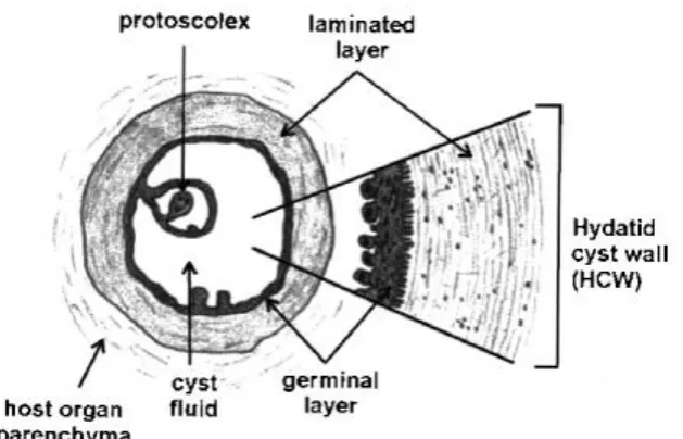
Budding off from the germinal membrane are the brood capsules and protoscolices (PSC) (See review by, Zhang et al, 2003). ―A mature fertile cyst is frequently unilocular (having a single cavity) and visualized as a clear (anechoic), fluid-filled lesion, usually with a cystwall visible‖ (Rogan et. al, 2006). The cyst wall is made up of a syncitial germinal layer that gives rise to brood capsules and protoscolices, and a non- living laminated layer adjoining the host tissue. The living germinal layer is not distinct or differentiated within the cyst wall in ultrasound. However, cysts vary in both size (1-20cm or more) and internal structure (Rogan et al, 2006).
Host tissues usually enclose or surround the parasitic cyst or endocyst to form a pericyst. The endocyst is largely consisted of a thick (0.2-2mm) acellular laminated layer. A thin (10-20µm) germinal layer may be present in healthy cysts, which lines the inside of the laminated layer, which may give rise to brood capsules containing protoscolices. The central cavity of a healthy cyst is filled with clear fluid, and varying sizes of daughter cysts which are formed by internal growth; may also be present. In degenerating, degenerated or dead cysts, a viable germinal layer or protoscolices may no longer be contained in the endocyst, there is the possibility of infiltration, it may have occurred and pericyst often shows signs of partial or complete calcification. (Wang et al, 2003).
23
[image:23.595.77.392.208.410.2]Richard and Lightowlers, 1986; McManus and Bryant, 1995). The cyst wall is consisted of an inner thin multinucleated germinal layer and an outer thick acellualr laminated layer.
Figure 1.2: Structure of the Hydatid cyst.
(Source:http://images.google.co.uk/images?imgurl=http://pathmicro.med.sc.edu/parasitology /EchinococcusLifeCycle)
1.6 The Laminated Layer
24
immune responses. (Coltorti and Varela-Diaz, 1974; Bortoletti and Ferreti, 1978; Richards et al, 1983 ; Harris et al, 1989 ; Rogan and Richards, 1989; Holcman et al, 1994; Gottstein and Felleisen, 1995). It consists of a protein-polysaccharide complex: the carbohydrate component appears to be built up of glucose, galactose, glucosamine and galactosamine (Kilejian and Schwabe, 1971; McManus and Bryant, 1986; Leducq and Gabrion, 1982). Protection of the parasite from the hosts immune response could stem from the carbohydrate (Coltorti and Varela-Diaz, 1974; Leducq and Gabrion, 1982; Rogan and Richards, 1986), presumably inhibiting complement activation (Smyth and Mcmanus, 1989). The laminated layer is parasitic in origin, secreted by the germinal layer (Bortoletti and Feretti, 1978; Harris et al, 1989; Holcman et al, 1994). Both layers, laminated and germinal layers are joined together by cystoplasmic connections.
The germinal layer is made of undifferentiated, proliferative totipotential cells, which produce brood capsules that project into the lumen of the mother cyst or daughter cysts. Formation of the protoscolex occur from budding of the germinal layer of the brood capsules which eventually may break away from their attachment to the germinal membrane and form ‖hydatid sand‖ in the cyst fluid. When ingested by the
25
stained by periodic acid, Schiffs reagent (PAS) and in histological studies provides a useful marker (Kilejian et al, 1962 ; Craig et al, 1995). This layer is developed
from or given rise to by the germinal (inner) layer (Bortoletti and Ferretti, 1978; Holcman et al, 1989), and its structure may also be contributed to by host material (Kilejian and Schwabe, 1971; Pezzella et al, 1984). It has been shown that more galactosamine than glucosamine is contained in the laminated layer (LL) of E.
granulosus. Nevertheless, there are more glucosamine than galactosamine in protoscoleces (Px) and hydatid cyst fluid (HCF). Also presented in this layer is acid muco-polysaccharide (Richards, 1984).
1.7 Development In The Intermediate Host
Hatched parasitic embryos migrate through the intestinal mucosa and enter venules and lymphatics. Between 60-70% of the embryos are filtered by the liver, and 15-25% by the lungs, 10-15% reaches other organs via the systemic circulation (Sinner
et. al, 1991). Undestroyed embryos are transformed into small cysts that will grow 2-3cm each year. The parasitic cyst wall is consisted of a germinal layer (endocyst) and a laminated proteinaceous membrane (ectocyst). The host forms a dense fibrous capsule (pericyst) which is in reaction against the cyst, and this contains blood vessels that provide nutrients to the parasite (Sinner et. al, 1991).
26
cyst tegument may reflect the parasites immunoprotectivemechanism (Holcman et. al, 1994). Mature eggs of E. granulosus possess a thick embryophore and its
ultrastructure shows it consists of thick elongated blocks that are united by electron-lucid cement (Holcman et. al, 1997), and surrounding the oncosphere is a thin cytoplasmic oncosphere membrane. The most eminent granules of penetration glands occupy the region of the nuclei in the hatched oncosphere. Various different functions are ascribed to the secretion of the penetration glands. They are involved in penetration and are totally pushed out during this process (Holcman et. al, 1997). The secretion causes lysis of host tissue in the surrounding of the invading oncosphere, enabling the oncosphere to resist the host cellular attack by maintaining a zone of necrosis of surrounding cells during the development of laminated layer (Heath, 1971).
The penetration glands support and assist in adhesion and protection against digestive enzymes or immune response of the host ( Lethbridge, 1980; Fairweather and Threadgold, 1981), they may also contribute to the formation of microvilli (Harris
et. al, 1989; Holcman et al, 1994). There are three recognised types of penetration glands (Swiderski, 1983), displaying a range of electron densities (Holcman et. al, 1994) different functions could be ascribed to different secretions. Harris et. al, (1989), suggested that much of the membrane needed for the extension of the microvilli from the epithelium could come from the fusion of penetration granule membranes with the outer plasma membrane. The three pairs of hooks located in the region opposite to the nuclei are equipped with a complex muscle system.
27
to cut tissue for penetration, in contrast to the hooks found on protoscolex or scolex which are lacking in independent musculature and only function to provide anchorage (Antoniou and Tselentis, 1993). The hook region, seen as the smaller lobe at light microscopy is incorporated in the metacestodes eventually (Heath and Lawrence, 1981), by day 2-3 after activation. The post-oncospheral development is a complex process of acquisition of biochemical and morphological properties difficult to be observed in vivo. ―There is a great number of studies on in vitro culture of protoscolex from fertile hydatid cysts to adult strobilate stage and a significant number on the ultrastructure of E. granulosus protoscolex tegument and of the germinal and laminated layers of the hydatid cyst‖ (Morseth, 1967; Bortoletti and Ferretti, 1973, 1978; Lascano et al, 1975; Conder et al, 1983, Rogan and Richards, 1986; Casado et al, 1992).
However, studies on in vitro culture of oncospheres are few, this, perhaps is due to the risk associated with handling of E. granulosus eggs (Heath and Smyth, 1970; Heath and Lawrence, 1976, 1981) and only two studies deal with ultrastructural development of the oncosphere to early metacestode (Harris et. al, 1989, Holcman
et. al, 1994). In the obvious absence of cell multiplication, cellular reorganization of an early metacestode takes place. There is a rapid increase in number and size of the microvilli by day 1 and up to day 2. In the epithelium of the metacestodes, electron-lucid vesicles start to appear increasing in size and number continuously.
―In a 3days old metacestode, long microvilli are substituted by old short microtriches
28
microvilli‖. (Holcman& Heath, 1997). This layer is the first of a series that stems from
the germinal membrane, and eventually appears to be a series of adjacent laminations. The large microvilli are completely by day 5 substituted by short and microfilamentous microtriches that project into the laminated layer. The appearance of the second laminated layer is between day 6-8. Its more electron-dense than the first lamination and is represented on its outer and inner surfaces by particulate material.
―Some microtriches appear to open into or be covered by the particulate material of the second lamination‖ (Holcman and Heath, 1997,). The laminated layer that
surrounds the metacestode of the E. granulosus is involved in protection of the parasite from the host immune response. Lamination first appears very early in post- oncospheral development. The organization of the laminated layer in the early metacestode of E. granulosus suggests that the outer sheet of the laminated layer is likely to be constantly replaced. The cyclical production possibly, is an intrinsic characteristic of the laminated cover essential in the creation of layers that could eventually be depressed as the cyst grows, and serve to divert host cellular response to the parasite. Before a naive host would be expected to mount an antibody-mediated immune response, the full development of the first lamination is completed (Holcman and Heath, 1997).
1.8 Human Hydatidosis
29
the abdominal cavity, heart, bone, muscle, nervous system, and or other locations can be affected (Khuroo, 2002; McManus et al, 2003). The cystic larvae grow slowly, and its growth is well tolerated by the host, leading occasionally to large parasitic masses (Moro, et al, 1999; McManus et al, 2003). Human echinococcosis occurs when eggs that have been shed in the faeces of definitive hosts are ingested by man.
Usually, the initial phase of CE is asymptomatic with small, well encapsulated cysts, which after an undefined period of several months to years; the infection may become symptomatic as a space-occupying lesion.
However, according to Pawlowski et. al, (2001), 60% of infections will remain asymptomatic. The commonest organ involved is the liver, with over two third of cysts usually. Infection in the lungs accounts for 20% of cases, with involvement of other organs accounting for less than 10% of cases (Torgerson and Burdke, 2003).
1.9 Pathology
30
unavailable (Brunetti et. al, 2010). CE cases remain asymptomatic most times until the cyst compresses or ruptures and there is spillage of its contents into neighbouring tissues and organs, by which time the disease is well advanced already (Brunetti et. al, 2010).
The pathological damage or dysfunction caused by cysts is mainly by the gradual process of space-occupying repression or the displacement of vital host tissue, vessels or organs. Clinical manifestations are consequently determined primarily by the site and number of cysts and these are quite variable. A massive release of cyst fluid and dissemination of protoscolices is what can follow and in most cases is what follows accidental rupture of cysts, resulting, occasionally in anaphylactic reactions and or multiple secondary cystic echinococcosis, since protoscolices have the
potential of developing into cysts within the intermediate host (Schantz and Gottstein, 1986).
There have been reports of cystic echinococcosis presenting for medical attention in people that are aged from younger than 1year to older than 75years, with fairly similar rates in both sexes. Following surgery on primary cysts, recurrence may occur. About 60% of all cases of cystic echinococcosis may be asymptomatic, although an unknown proportion may become symptomatic. ―About 0.2 per
31
seldomly in the heart, brain, ovaries, vertebral column (1% or less each) (Menghebat
et al., 1993). Symptoms presented by cystic echinococcosis can be highly variable, and can be dependent not only on the organ involved, but also on size of cysts and their position within the organ, the mass effect within the organ and upon surrounding structures, and related complications to cyst rupture and secondary infection. In response to cyst leakage or rupture, manifestations of systemic immunological responses may be evident. Common complications involving cystic echinococcosis include rupture into the biliary tree with secondary cholangitis, obstruction of the biliary by daughter cysts or extrinsic compression, rupture into the bronchial tree, intracystic or subphrenic abscess formation, development of a bronchobiliary fistula and intraperitoneal rupture (with or without anaphylaxis).
32
release of many thousands of larvae (protoscolices), with each having the capability to differentiate into another hydatid cyst (Rogan et al, 2006).
1.10 Diagnosis
The detection of the space occupying cysts or lesions caused by metacestode(s) of
33
1997 ; Gharbi et al, 1997). Diagnosing CE early can bring about significant improvements in the quality of the management and treatment of the disease. Early stages of the infection are asymptomatic in most cases, so, cheap and relatively easy to use methods are required for large-scale screening of populations at high risk. Providing such an approach is immunodiagnosis, which can also confirm clinical findings (Zhang et al, 2003).
Immunodiagnosis is vital in that it plays an important as well as a complementary role. Its usefulness is not only for primary diagnosis but also for follow-up of patients after surgical or pharmacological treatment. Detection of antibody in sera is more sensitive than the detection of circulating antigen, and remains the method of choice (Zhang et al, 2003). Serological testing of cystic hydatid disease (CE) has a very long history, and almost all serological tests that have been developed have been used in the diagnosis of human cases. Among the various tests, there are considerable differences in sensitivity and specificity. Non-specific and insensitive tests, like the Cassoni Intradermal test, the complement fixation test, the latex agglutination test, the indirect haemagglutination test have been replaced by the enzyme-linked immunosorbent assay (ELISA), the indirect immunofluorescence antibody test, immunoelectrophoresis (IEP), and immunoblotting (IB) in routine laboratory procedures, applications (Lightowlers and Gottstein, 1995).
―The lipoproteins antigen B (AgB) and antigen 5 (Ag5) (Oriol and Oriol, 1975), the
34
immunoblotting and or by immunoprecipitation of radiolabelled antigen and SDS-PAGE‖ (Shepherd and Mcmanus, 1987; see review by, al-Yaman and Knobloch, 1989; Lightowlers et al, 1989; Shapiro et al, 1992).
―Antigen B, with a molecular mass of 120KDa is a polymeric lipoprotein that can be
measured as a circulating antigen in patients blood (Kamiya and Sato, 1990; Liu et al,1993) has been suggested to play an important role in the biology of the parasite-host relationship (Shepherd et al, 1991; see review by, Rigano et al,2001). Antigen B is a highly immunogenic molecule, appearing ladder like under reduced condition on SDS-PAGE, with three bands with molecular sizes of approximately 8 or 12, 16, and 24KDa (Chordi and Kagan, 1965 ; Oriol et al, 1971 ; Shepherd and Mcmanus, 1987 ; Lightowlers et al, 1989 ; Leggatt et al, 1992), suggesting that it comprises polymers of 8KDa subunits. The smallest subunit has proved the most useful target in diagnostic studies‖ (Ortona etal, 2000; Rott et al, 2000).
Ag5 is a lipoprotein with a very high molecular mass complex composed of 57 and 67KDa components that dissociates into 38 and 22 to 24KDa subunits under reducing conditions (Lightowlers et al, 1989). ―According to history, the demonstration of serum antibodies precipitating antigen 5 (arc5) by immunoelectrophoresis or similar techniques has been one of the most widely used immunodiagnostic procedures for CE‖ (Shepherd and McManus, 1987).
35
Craig, 1994; Eckert and Deplazes, 2004). Studies on the hydatid cyst of E. granulosus have indicated the occurrence of high levels of host IgG heavy chain in the germinal layer of non-fertile cysts and suggests the host immune response could be destructive of protoscolex production by bringing about or causing apoptosis of the germinal membrane, possibly opening up an avenue for vaccination against established cyst (Blanton et al,1991 ; Lawn et al, 2004).
Following the knowledge and understanding of the smallest size of the lipoprotein of antigen B to be 8KDa and believed to be Echinococcus specific with diagnostic potentials, Barbieri et al (1993) went on to prepare a mixture of lipoproteins antigens that contained the relevant diagnostic AgB and Ag5 from bovine hydatid cyst fluid by heparin- affinity chromatography. A standardized antigen mixture of high sensitivity and specificity for human hydatid serology has been provided by this heparin- binding lipoprotein fraction (HBLF) (Barbieri, et al, 1993; Barbieri, et al,1994).
36
The cDNA library prepared with E. granulosus protoscolices obtained from hydatid cysts of Uruguayan sheep were immunoscreened and allowed the isolation of a reactive phage clone (ʎ3C3) which was characterised further. Their results showed
affinity- purified monospecific polyclonal antibodies against lambda 3C3 reacted by western blotting with HBLF bands of 8, 16, 24 and 32 KDa apparent molecular mass. Consequently, the pattern described as corresponding to AgB subunits (Lightowlers, et al, 1989) was reproduced with anti- ʎ3C3 antibodies.
Recent research has demonstrated that AgB, encoded by a gene family constituted of member genes, exhibits variation to a very high degree (Frosch, et al, 1994; Chemale, et al, 2001; Arend, et al, 2004; Muzulin, et al, 2008). Five 8KDa subunit genes from E. granulosus so far have already being identified. They are named as EgB8/1, EgB8/2, EgB8/3, EgB8/4 and EgB8/5 (Haag, et al, 2004). EmB8/1-EmB8/5 have also being identified in E. multilocularis (Mamuti, et al, 2006, 2007). AgB
recombinant subunits were found to self assemble by Monteiro et al, (2007) into high molecular mass homo-oligomers with structural features that are similar to those of the parasite- produced AgB while they studied the recombinant subunits of AgB1, AgB2 and AgB3.
37
genes of AgB antigen family, to investigate their serological reactivity and differences in the recognition of specific antibodies, to identify potential subunit antigens for immunodiagnostic tests and to proving a basis for standardization of AgB antigen.
They (Jiang Li et al, 2012) analysed the reactivity of a panel of 243 serum samples from CE, AE, CC patients and NH with 8 recombinant subunit antigens by ELISA. They also made comparison of three paralogous subunits from E. granulosus (EgAgB1- EgAgB3) and E. multilocularis (EmAgB1-EmAgB3), respectively for their reactivities in CE and AE sera detection. Their results showed that all of the three orthologous subunits (EgAgB1 vs EmAgB1, EgAgB2 vs EmAgB2 and EgAgB3 vs EmAgB3) were not different statistically when detecting CE or AE sera and therefore suggested that there may be a similarity in their epitopes
The diagnosis of lung hydatid disease is based on chest imaging using X-rays or computed tomography (CT). Serological tools are used only to confirm the diagnosis because of low sensitivity and incomplete specificity (Santivanez and Garcia, 2010). The assay performance is dependent mainly on the format of the test and nature of antigen used but can also vary according to the characteristics of the disease such as organs involved, number of cysts and presence of any cyst complications (Zhang et al, 2003; Zhang and McManus, 2006). Previously, the use of synthetic peptides or recombinant antigens derived from sequences of the two major components of cystic fluid antigen B (AgB) and Ag5 have been proposed for use as reproducible antigens to improve test reliability and allow better standardization (Ortona et al, 2000;
Virginio et al, 2003; Carmena et al, 2006).
38
data on serological diagnosis of lung CHD and how published p176 studies do not allow estimations of its sensitivity or provide further details for pulmonary cases. Santivanez et al (2012) did a study and applied p176 ELISA in a series of known cases and responses of those patients were compared to the responses of noninfected controls to provide further information on the test performance of the assay for the diagnosis of lung CHD as well as its performance in relation to disease characteristics. The use of p176 was to counter the variability, high variability in results obtained with the use of cyst fluid as the antigenic source/ material.
Results obtained for the sensitivity of the p176 ELISA for the diagnosis of lung CHD cases was almost 80%, despite the fact that the restricted numbers of samples with isolated pulmonary CHD prevented a more precise assessment of sensitivity. In the long run, the simpler, cheaper, semiquantitative ELISA format and the potential for better reproducibility make this ELISA a good alternative for the diagnosis and posttreatment follow-up of lung CHD. Diagnosis of infection in human is based on the identification of infiltrative or cystic lesions by imaging techniques such as ultrasonography or computed tomography (Brunetti et al, 2010).
The diagnosis of AE is strengthened by immunodiagnostic tests such as ELISAs especially using native protoscolex or metacestode antigens, purified fractions (Em2 antigen), or recombinant antigens (ІІ/3-10‾, Em10- or Em18- antigen) with variable sensitivities and specificities (Gottstein, et al, 1993; Brunetti, et al, 2010; Schweiger, et al, 2011). The study undertaken by Barth et al (2012) was to validate the
39
type carbohydrate antigen called Em2 (Hulsmeier et al, 2002) which is a major antigen of the laminated layer of the E. multilocularis metacestode that is also present in the cyst fluid (Deplazes and Gottstein, 1991; Gottstein et al, 1992) is recognised by the monoclonal antibody mAb Em2 G11.
Barth et al (2012) were able to show that the mAb Em2 G11 is strongly positive in the laminated layer of E. multilocularis lesions in various human tissues in all
samples studied. According to them, no protoscolices were found in all investigated material of 49 AE patients which confirmed protoscolices are a very inconstant diagnostic feature (Marty et al, 2000), and therefore submitted that the mAb Em2 G11- positive laminated layer is the crucial immunohistological hallmark for diagnosis of AE. The mAb Em2 G11 is also said to be species specific as no positive results were recorded at all for CE neither in the laminated layer, germinal layer, calcareous corpuscles nor in the protoscolices when stained with mAb Em2 G11 (Barth et al, 2012).
1.11 Treatment
In endemic regions, asymptomatic hepatic cystic echinococcosis are common and up to 75% of infected people may remain free of symptoms for more than 10years (Frider et al,1999). ―Cysts may be seen to expand, become septate, or calcify when
patients are monitored with serial ultrasound‖. A greater occurrence of this condition
40
percutaneous aspiration (Filice et al, 1990; Wang et al, 1994 ; Akhan, et al, 1996 ; WHO/OIE, 2001).
The possibility of cyst recurrence however remains the main problem with this form of treatment. Recurrence rates of cyst after surgery have been reported as being between 2% and 20% (WHO/OIE, 2001). However, these rates may be subjected to some level of inaccuracy since occurrence of cysts in patients after surgery could also be because it had been missed in the initial examination or because of subsequent reinfection after exposure to eggs. In establishing and validating whether cysts are truly recurrent after surgery or have by other means arisen, the use of ultrasound based cyst morphology could also be of benefit (Wang et al, 2003).
Surgical removal of the lesions is included in the treatment options for CE, and CE in most parts of the world is the most common reason for abdominal surgery. Ninety percent success rate has been attributed to surgery (Pawlowski et al, 2001). The PAIR (Puncture-Aspiration-Injection- Reaspiration) technique is an alternative to surgery (WHO/OIE, 1996). Chemotherapy, with drugs such as benzimidazoles, have also been used with some success. An indication for a wait and see approach to treatment is employed in calcified cysts (Torgerson and Budke, 2003).
1.11.1 Surgery
The principal and mainstay therapy for large cysts, infected cysts, those that are superficial and likely to rupture, and those in vital and anatomical sites or exerting considerable and substantial mass effect has always been surgery. ―Surgical options
41
without omentoplasty), or (palliative) tube drainage of infected cysts. Cyst extrusion (Barrett‘s technique) is also a surgical option for pulmonary disease‖. More radical
surgery is associated with a higher complication rate but also a lower relapse rate. Recurrence usually is due to either insufficient cyst removal or previously undetected cysts. Percentage of reported recurrence rates range from 2-25% (Ammann and Eckert, 1996).
1.11.2 Pair
The puncture, aspiration, injection, reaspiration (PAIR) technique was introduced in the mid-190‘s (Gargouri et al,1990; Filice and Brunetti, 1997). Under ultrasound guidance, the cyst is punctured, as much cyst fluid is aspirated as possible, followed by the injection of a protoscolicide (e.g, 95% ethanol), and cyst contents reaspirated between 15-20mins later. This technique should only be undertaken by skilled practitioners, with intensive- care support on ground in the event of anaphylaxis. Assessment should be made of cyst aspirates for the presence of protoscolices or bilirubin. The use of PAIR should only be in, or, with patients with chemotherapeutic cover so as to minimise the risk of secondary cystic echinococcosis. This technique has not been performed or experienced with children and or pregnant women.
42
PAIR is inadvisable (Anonymous, 1996). Percentage rates of complication ranges from 28% in the absence of albendazole (Men et al, 1999), to 5-10% with concomitant chemotherapy (Pelaez et al, 2000 ; Aygun et al,2001).
―A multicentre survey on PAIR carried out by the WHO informal working group on
echinococcosis (Filice et al, 2000), reported a 1% major complication (anaphylaxis or spillage) rate and a 13.7% minor (fever, rash, cyst infection, or haemorrhage)‖.
―The usage of PAIR with albendazole chemotherapy has been shown to be as
effective as pericystectomy for hepatic cystic echinococcosis in one randomised propective trial (Khuroo et al., 1997) with lower post-procedure morbidity and shorter hospital stay.
1.11.3 Chemotherapy
Albendazole and mebendazole, the benzimidazole compounds have been the bedrock, the fundamental chemotherapy for cystic echinococcosis. ―Treatment with
albendazole (10mg/kg in divided doses – usually 400mg – twice daily) results in the disappearance of up to 48% of cysts and a substantial reduction in size of a further 24%‖ (Horton, 1997). Mebendazole (40-50mg/kg per day in three divided doses) is
less capable of producing desired effect than albendazole. (Horton, 1997).
43
―Non-viability of cyst increases with duration of treatment – from 72% of cysts non-viable after 1 month to 94% of cysts non-non-viable after 3 months of treatment‖
(Gil-Grande et al, 1993), with the usual adverse effects including nausea, hepatotoxicity, neutropenia (which may not be reversible), and alopecia (occasionally).
All patients are advised to have regular monitoring of leucocyte counts and liver function tests. The protoscolicidal metabolite of albendazole is albendazole sulphide. Praziquantel (25mg/kg per day) has been used concurrently with albendazole for concomitant treatment of cystic echinococcosis, and early trial in man has shown improved efficacy over albendazole alone (Mohamed et al,1998). ―Albendazole and
mebendazole are listed as category C drugs in pregnancy in the USA (Gilbert et al, 2001), and category D and B£ respectively in Australia (Anonymous, 2000). Neither drug is definitely inadvised in pregnancy. Specialist advice should be sought if treatment during pregnancy is likely‖.
1.12 Changes in Cyst Morphology and Follow Up After Treatment.
44
Every cyst begins as a typical small unilocular cyst with clear cyst fluid. In some cases, the cyst is termed to be sterile as no further development takes place. While in most other cases, there is subsequent development of protoscolices within the brood capsules attached to the germinal layer. ―For many cysts, change from this
form is not experienced, and ultrasound examination can show growth of the cyst but no change in internal structure over several years‖ (Rogan et al, 2006). Other cysts show a significant level of variation in internal structure, such as calcification, collapsed cyst walls and the presence of additional daughter cysts internally. Exhibition of such heterogeneity in cyst structure has for clinicians been an issue in terms of how to treat CE. The presence of daughter cysts, could be an indication that PAIR is a less favourable option; a distinct, small, unilocular cyst might give a good response to chemotherapy, while a small, calcified cyst might signify poor parasite viability and a good prognosis and would thus be a candidate for long term observation only (Pawlowski, 1997).
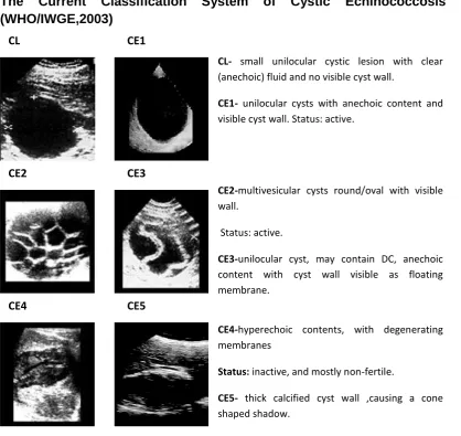
1.13 Cyst classification
45
Type 2, fluid collection with a split wall, Type 3, fluid collection with septa, Type 4, heterogenous echo patterns, and Type 5, presence of reflecting thick walls indicative of calcification‖ (Gharbi et al, 1981 ; Wang et al, 2003).
Gharbi‘s classification was the basis, foundation for all subsequent ultrasound
classifications, which included largely minor modifications and or additional categories. Fifty nine clinical CE cases formed the basis of Lewall and McCorkell‘s CE classification and 3 categories were proposed by them. Type 1, simple fluid-filled cysts; Type IR, lesions containing wavy membranes, representing detached endocyst secondary to rupture; Type 2, lesions contained of daughter cysts and/ or a formed echogenic material; and Type 3, dead, densely calcified lesions (Lewall and McCorkell, 1985).
46
With the use of ultrasound morphology to monitor cyst development or regression on the increase, a more universal classification was considered necessary. With this aim, the WHO Informal Working Group on Echinococcosis (WHO-IWGE) proposed recently a standardized classification to bring about the unification and simplification of the various ultrasound CE classifications (WHO/OIE, 2001).Six categories were proposed by WHO-IWGE: ―Type CL, unilocular cystic lesion(s) with uniform anechoic content, with pathognomic signs that is inclusive of visible cyst wall and ―snowflake‖
signs; Type CE2, is a multivesicular, and multiseptated cysts; Type CE3, anechoic content with a detached laminated membrane from the cyst wall visible as a floating membrane or as a ―water lily‖; Type CE4, hyperechoicheterogenous, or hyperechoic
degenerative contents, daughter cysts not present; and Type CE5, are cysts that are distinctive by thick calcified wall that is arch-shaped, giving a cone-shaped shadow, variation in the degree of calcification may be from partial to complete. Also included in the WHO-IWGE standardized ultrasound classification of CE are the size and biological status of the hydatid cyst(s). In summary, cyst size ‹5cm is classified as small (s), medium/middle (m) size is from 5-10cm, and ›10cm is large (L). Biological status also should be classified as ―active‖ (which comprises of group 1: Types CE1 and CE2), ―transitional‖ (these are group 2: Type CE3), or ―inactive‖ (comprising of
group 3: Types CE4 and CE5) (WHO/OIE, 2001). This classification for CE by WHO is yet to be applied in many clinical situations or mass screening programmes (Wang
47
ectocyst, presence of daughter cysts and cyst calcification have all been highlighted by all of the proposed classifications, these may be considered as regressive signs though not consequently indicating total cyst non-viablity.
48
The Current Classification System of Cystic Echinococcosis Cysts (WHO/IWGE,2003)
Figure 1.3: Classification of ultrasound images.
CE1-CE2 cyst types are considered fully viable, CE3 cyst type is considered and classified a transitional stage, and may remain viable or regress to a non-viable stage; CE4-CE5 are degenerative , non-viable and usually inactive stages.
1.14 Natural History of Hydatid Cyst
A change in morphology might or might not be noticed in cysts over a relatively long period of time in treated or untreated individuals. Distinctly, slow degenerative changes are shown by some hydatid cyst and have a natural history or evolution. The details of changes are less well defined and have nonetheless been addressed by several authors (Pawlowski, 1997; Daeki et al, 2000; Teggi and Di Vico, 2001).
CE4-hyperechoic contents, with degenerating membranes
Status: inactive, and mostly non-fertile.
CE5- thick calcified cyst wall ,causing a cone shaped shadow.
Status: inactive.
CL CE1
CE2 CE3
CE4 CE5
CL- small unilocular cystic lesion with clear (anechoic) fluid and no visible cyst wall.
CE1- unilocular cysts with anechoic content and visible cyst wall. Status: active.
CE2-multivesicular cysts round/oval with visible wall.
Status: active.
CE3-unilocular cyst, may contain DC, anechoic content with cyst wall visible as floating membrane.
[image:48.595.72.491.78.473.2]49
―The most recent interpretation is based on the current WHO classification as a
progressive natural history of cyst development from CE1 to CE5‖ (Macpherson et al,2004). It is enormously important to understand the possible developmental fate of a cyst, to make possible the monitoring and prediction of disease progression, regression and recurrence (Rogan et al, 2006).
However, there is some debate and controversy as to whether the WHO progression is too simplified, and alternative classifications have been suggested by some authors that would also relate to size of cysts and indicate a different order of events (Wang et al, 2003).Follow up of cases can be for as long as 8-10years after treatment, but there is huge variation between studies in respect to monitoring points, making comparisons difficult. Therefore, it might not be completely as to what stages a cyst has passed through between scans (Rogan et al, 2006). Based on several literature descriptions, it appears that several developmental pathways can come from a typical unilocular CE1 cyst (Wang et al, 2003; Teggi and Di Vico, 2001). The viability of the parasite tissues within the cyst is the important feature; the cyst has the ability to regenerate in some form if viable portions of germinal layer, brood capsule wall or protoscolices are present. Usually, CE1 cysts are viable and fertile (possessing protoscolices), while CE5 cysts are dead and calcified (Rogan et al, 2006). The remaining types and their viability are more questionable.
50
contained, as demonstrated by some cases that had reverted to type CE1 (Larrieu et al,2004).CE2 cyst types are classified as active and are not accepted generally as showing degeneration. Viability of the parasitic material of the daughter cyst is not questionable, but the viability of the primary cyst is, and this is important when the natural history is being considered (Rogan et al, 2006).
The lack of understanding of the origin of daughter cysts is a main problem, as there is no histological support for the assumption that these arise from the germinal layer. This interpretation has come under arguments based on ultrasound images that seem to show a series of progressively larger daughter cysts forming around the periphery of the primary cyst. It is however impossible to say that the cysts have arisen from the intact germinal layer from these images given that the germinal layer is less than 1mm thick (Rogan et al, 2006). In addition, the internal production of a laminated layer would not be allowed due to the polarity and orientation of the tissues within the germinal layer (Bortoletti and Ferretti, 1978). The relevance of this is particularly with those ultrasound studies that conspicuously show formation of daughter cysts by internal growth, endogenous proliferation of the germinal layer (Czermark et al., 2001). A vesicle, in these cases seen forming from the mother cyst wall would have the laminated layer on the inside and the germinal layer on the outside, which according to Rogan et al., (2006), is incorrect.
51
hydatid cysts is possessed by protoscolices and is directly involved with causing secondary hydatidosis, and altered physiological conditions can trigger cystic differentiation of protoscolices in vitro (Smyth and Barrett, 1980). Studies have also shown direct evidence from cysts in livestock that protoscolices show cystic development within degenerate primary cysts (Rogan, 1988). Although, the appearance of the fluid within daughter cysts is usually anechoic (clear), the fluid within the primary cyst is often more hyperechoic (dense), which signifies the presence of some sort of debris or infiltrate (Rogan et al, 2006). This material was referred to as ―matrix‖ by Lewall and McCorkell (Lewall and McCorkell, 1985) and
cysts such as this are often full of pus or leucocyte infiltrate at surgery and debris from a degenerate primary cyst (Abu-Eshy, 1998; Teggi and Di Vico, 2001). Bacterial infection, although not always, is sometimes present (Schipper et al, 2002). It is clear that the germinal layer and the cyst wall cannot be intact if cellular infiltrate or bacterial cells are present in the cyst cavity, therefore, the formation of daughter cysts is involved with damage or primary cyst degeneration (Rogan, 1988; Teggi and Di Vico, 2001; Wang et al, 2003).
1.15 The Role of Cytokines in Echinococcosis
In regards to the investigation of cytokine production in AE and CE patients, several studies have been undertaken to determine the underlying immunological responses to infection and disease. Much of the current understanding of Echinococcus
52
induction of a Th1 response, recruiting Th1 cytokines such as IFN-γ (Mourglia-Ettlin
et. al, 2011), which then switches to a Th2 response, predominantly inducing IL-4, IL-5, IL-10, and IL-13, in chronic and progressive disease stages, bringing about the hallmark response characteristics of most helminthic infections (Rogan, 1998; Mourglia-Ettlin et. al, 2011). Degenerating cysts in murine models are by contrast associated with Th1 cell activity and the production of IFN-γ (Rogan, 1998) which
shows the protective effect of Th1 cytokines during infection and disease. Even though immunological studies of early infections are more difficult in human populations, according to data available from epidemiology studies by Yang et. al, (2009); Vuitton, (2003) have shown that different cytokine profiles are displayed by natural courses of human AE and CE at different stages of disease progression.
53
However, following successful drug treatment with albendazole, the polarized Th2 response in advanced disease tend to revert back to Th1 (Rigano et. al, 1995; Rigano et. al, 1999). It is important to note that both Th1 and Th2 cytokines are produced in patients with active and/ or progressive disease; nonetheless, it is only those patients who are able to elicit a Th1 response that have been shown to respond well to chemotherapy, where patients maintaining higher levels of IL-4 and IL-10 do not (Rigano et. al, 1995; Rigano et. al, 1995; Rigano et. al, 1999; Rigano et. al, 1999).A Th1 profile also correlates well with good prognosis in patients following the removal of cysts surgically and in those with inactive, late-stage of CE5 cysts (Rigano et. al, 2004; Rigano et. al, 2004). An Algerian study of 177 patients with CE showed that the Th1/Th2 skew is correlated and related to clinical stage, disease progression and prognosis, with Th1 cytokine being associated with protection and susceptibility to disease associated with Th2 (Mezioug and Touil-Boukoffa, 2009). The ability of the patient to maintain a Th1 response or yield to a Th2 response eventually decides whether he/she is vulnerable or resistant to disease and respond successfully to treatment (YuRong, et. al, 2012).
54
with inactive or regressive disease (Craig, 1986; Daeki et. al, 2000; Khabiri et. al, 2006).Contrastingly, levels of IgG2 and IgG3 become elevated when cysts become infiltrated and/ or destroyed by the host (Daeki et. al, 2006), during which time clearly noticeable decreased levels of IgG1 and IgG4 are observed (Bayraktar, et. al, 2005). In murine studies of both AE and CE, antibody titers were found to be comparatively consistent in affected mice, notwithstanding the susceptibility of the host strains (Vuitton et. al, 2006), but changes according to the severity of the infection were observed (Vuitton et. al, 2006). This is in line, in consistency with the natural growth of cysts of both E. multilocularis and E. granulosus where protection against the immune response is provided by the intact cyst wall.
55
cytokines, mostly IFN-γ, are well correlated with IgG2 levels and disease
progression. Consequently, useful markers or indicators of disease activity and of the natural course of disease/cyst development can be provided by measuring cytokine and antibody profiles, particularly the IgG subclasses (Rigano et. al, 1995; Vuitton, 1997; Daeki et. al, 2000).
An indication to whether a patient would respond to treatment or not may also is offered by these immune profiles. Regardless of this, there remains a substantial variance in the immunological response between patients that may be affected by the parasite strain and/ or antigen type produced, which influences the development of T helper subsets. Antigen dose and the genetic background of the host are other factors that may also contribute (Emery et al, 1997; Eiermann et. al, 1998). Contributing significantly also is the general well- being and health of an individual to disease susceptibility and this is especially influenced by conditions underlying such as malnutrition and/ or coinfections with tuberculosis (Vuitton, 2003) or HIV (Sailer et al, 1997; Wellinghausen et. al, 1999; Zingg et. al, 2004).
1.16 POST-TREATMENT FOLLOW-UP
56
monitoring of CE patients after surgery and during chemotherapy has been emphasized. Despite limitations encountered with serological tests, due to their cost effectiveness and improvement facilities, they are probably best choice for follow-up assessment of CE after either surgery and / or chemotherapy (Rafiei et. al, 2008).
Serological diagnosis in a routine laboratory depends mainly on the detection of immunoglobulin class G (IgG) antibodies directed against different antigens of E. granulosus or E. multilocularis (Grimm et. al,1998). Sensitivity and specificity of the serological tests depend on the stage of the disease, the localization of the parasites, the antigens, and the techniques used (Gottstein, 1992; Craig, 1993). One of the most widely used antigens is the cyst fluid (CF) of E. granulosus cysts of sheep or cattle origin, and the enzyme-linked immunosorbent assay (ELISA) is one of the most commonly used techniques in serodiagnostic laboratories.In cases of CE of the liver, antibodies against CF antigens can be detected with a high diagnostic sensitivity by this method (Grimm, et. al., 1998).
Rationale of study
57
antigen B (AgB). To bring about the development of a means of measuring the immunogenic activity in sera of CE patients using antigenic markers, which, may potentially create antigen/ antibody profiles signalling the progression or regression of disease in relation to/ with particular categories of cyst.
The use of these markers in association with IgG subclasses may bring out and reveal information distinctively, with emphasis particularly on the development of a more specific means of monitoring success or failure of therapy during post-treatment, follow-up and surveillance. Studies by Doiz et. al, (2001) showed that antibodies from CE patients with specific proteins of molecular weights 39KDa and 42KDa from the antigen B/5 rich fraction of HCF by western blot analysis may be useful in the status of the disease, as these bands were present in patients with progressive disease, but absent in cured patients.
58
The aim of this study is to investigate the recognition of crude antigens by sera of patients of confirmed hydatid disease of different cyst types and stages in relation to their reactivity with whole immunoglobulin (IgG) and IgG subclasses 1 and 4 in an attempt to identify, classify and determine disease categorisation using immunological markers.
59
CHAPTER 2
MATERIALS and METHODS
2.1 Antigenic Materials
2.1.1 Hydatid Cyst Fluid (HCF)
Liver and lung of infected sheep were collected from a local UK abattoir brought back to the laboratory at University of Salford. Carefully, cysts were removed, aspirated and antigen or hydatid cyst fluids were collected according to the method described by Rogan et. al, (1991). A brief description, hydatid cysts were aseptically aspirated using sterile 5ml syringe and needle, the fluid was centrifuged and the supernatant obtained was used as the crude hydatid cyst fluid and this was put in sterile 50ml bottle and stored at -20℃ until use.
2.1.2 Laminated Layer (LL)
60
2.2 Sera.
Serum samples used for the purpose of this current study were collected as part of the African Medical and Research Foundation (AMREF) Hydatid Control Programme, based in Lokichoggio, Kenya under the management of Dr Eberhard Zeyhle. Collection of many of the samples was made over a long period of time, but all samples were stored at -20°C without repetitive freeze thaw. Each patients diagnosis was confirmed at AMREF by the using ultrasound examination. Each patient was monitored at regular intervals by ultrasound and details of cyst size and morphology recorded by photographs and notes. The classification of cyst types observed was based on matching the ultrasonographers‘ notes to the WHO 2003
classification system under the guidance of Dr Zeyhle. Serum samples were taken at each observation point. Most patients were put on a schedule to take one or more courses of albendazole at 20mg/kg/day (see Table 2.1). Some patients experienced a delay before their first course of albendazole was taken and these could be regarded as ―untreated‖ for the preliminary part of their observation. Treatment was
refused by some other patients and these also were regarded as untreated.
61
[image:61.595.68.534.293.761.2]For optimisation pourposes of the crude antigen ELISA assays and analyses of CE patients sera, negative controls comprised a pool of normal sera from a CE endemic area (Turkana, Kenya). For Western blot analysis and ELISA detection of total serum IgG and IgG subclass antibodies from the sera of infected individuals probed, normal human sera was used for negative controls (Sigma-Aldrich, UK).
Table 2.1 Summary of Patient Information
Patient ID Gender/ Age Cyst series Organ Involvement ALB Chemotherapy (mg/kg body weight)/
surgery
Sera Timeline
B142 Female 6
CE1, CE4
R/ liver Untreated
0-42.7months P160 Female
16
CE2, CE3
R/ liver Untreated
0-33.9months X345 Female
20
CE2, CE3
Omentum/ mesentery
Untreated 0-3.6months
X161 Female 17
CE3, CE4
R/ liver 27.6mths ALB
(20mg).
0-31.6months
Y88 Female
5
CE1 R/ liver 4.8mths 2
courses ALB (20mg);
17.8mths ALB (20mg).
0-8.4months
Y13 Female
20
CE1, CE3, CE4, CE5
R/ liver, lower abdomen
3.8mths 3
courses ALB (20mg);
22.2mths ALB (20mg);
58.7mths ALB (20mg); 62.3mths endocystectomy . 0-209.8month s