Chauviac, Francois-Xavier and Robertson, Giles and Quay, Doris H.X.
and Bagneris, Claire and Dumas, C. and Henderson, B. and Ward, J.
and Keep, Nicholas H. and Cohen-Gonsaud, Martin (2014) RpfC (Rv1884)
atomic structure shows high structural conservation within the resuscitation
promoting factor catalytic domain.
Acta Crystallographica Section F:
Structural Biology Communications F70 (8), pp. 1022-1026. ISSN
1744-3091.
Downloaded from:
Usage Guidelines:
Please refer to usage guidelines at
or alternatively
Acta Crystallographica Section F
Structural Biology Communications
ISSN 2053-230X
The RpfC (Rv1884) atomic structure shows high
structural conservation within the
resuscitation-promoting factor catalytic domain
Francois-Xavier Chauviac,a‡ Giles Robertson,aDoris H. X. Quay,aClaire Bagne´ris,a Christian Dumas,bBrian Henderson,cJohn Ward,d Nicholas H. Keepa* and Martin Cohen-Gonsauda,b*
a
Crystallography, Institute for Structural and Molecular Biology, Department of Biological Sciences, Birkbeck, University of London, Malet Street, London WC1E 7HX, England,bCentre de Biochimie Structurale, CNRS UMR 5048, 29 Rue de Navacelles, 34090 Montpellier, France; INSERM U1054, Universite´ Montpellier I, Montpellier, France,cDepartment of Microbial
Diseases, UCL–Eastman Dental Institute, University College London, 256 Gray’s Inn Road, London WC1X 8LD, England, anddThe
Advanced Centre for Biochemical Engineering, Department of Biochemical Engineering, University College London, Torrington Place, London WC1E 7JE, England
‡ Current address: Virology Department, Structural Virology Unit, Institut Pasteur, 28 Rue du Docteur Roux, 75724 Paris CEDEX 15, France.
Correspondence e-mail: [email protected], [email protected]
Received 13 March 2014 Accepted 5 June 2014
PDB reference:RpfC, 4ow1
The first structure of the catalytic domain of RpfC (Rv1884), one of the resuscitation-promoting factors (RPFs) from Mycobacterium tuberculosis, is reported. The structure was solved using molecular replacement once the space group had been correctly identified as twinned P21 rather than the apparent C2221by searching for anomalous scattering sites inP1. The structure displays a
very high degree of structural conservation with the previously published structures of the catalytic domains of RpfB (Rv1009) and RpfE (Rv2450). This structural conservation highlights the importance of the versatile domain composition of the RPF family.
1. Introduction
Resuscitation-promoting factors (RPFs) have attracted much interest since their discovery in the late 1990s. These proteins resuscitate bacteria that have entered a dormant state, allowing them to prolif-erate normally. Despite some key advances since the first protein identification and characterization, their precise mechanism of action
remains elusive. The protein was first isolated inMicrococcus luteus,
where a heat-labile, non-dialysable and trypsin-sensitive factor present in culture supernatants was able to resuscitate non-growing
cells (Mukamolovaet al., 1998). The factor was identified as a protein
and named resuscitation-promoting factor. In this same seminal study, corresponding genes in other GC-rich Gram-positive bacteria,
most notably inMycobacterium tuberculosis, were also identified. The
resuscitating function was later confirmed in M. tuberculosis
(Mukamolovaet al., 2002). This is an important finding, as one third
of the human population is latently infected withM. tuberculosisin a
dormant form. This represents a large population reservoir for reactivation of tuberculosis and also a potential novel therapeutic avenue for treating tuberculosis.
Sequence analysis coupled with homology modelling led to the hypothesis that the conserved RPF catalytic domain could be a transglycosidase belonging to the family of c-type lysozymes
(Cohen-Gonsaud, Keepet al., 2004). The prediction was confirmed by the first
solution structure of the RpfB catalytic domain fromM. tuberculosis,
which showed that the domain is a short version of the c-type
lyso-zyme lacking the first helix (Cohen-Gonsaud et al., 2005). Later,
various experiments unambiguously demonstrated that the RPF
domain is a peptidoglycan hydrolase (Mukamolova et al., 2006;
Telkovet al., 2006).
Five RPF paralogues are present inM. tuberculosis(rpfA–E). They
contain a conserved catalytic domain, but the domain composition
shows variability also found in other species (Ravagnaniet al., 2005).
binding (Ruggiero et al., 2009) and a prokaryotic membrane lipo-protein lipid-attachment site that may confer it with a juxtacrine function, while RpfA (407 amino acids) possesses a low composi-tional complexity domain that may confer an autocrine function
(Mukamolovaet al., 1998).
Initial studies showed that deletion of individualrpfgenes had no
significant phenotypic consequences (Downinget al., 2004; Tufariello
et al., 2004). This suggests that the mycobacterial RPF proteins are
functionally redundant. The deletion of the entire mycobacterialrpf
gene family is also dispensable for growth (Kana et al., 2008).
However, phenotypic alterations appear with the deletion of three or
morerpfgenes and reveal a functional hierarchy of the mycobacterial
Rpf proteins that has been reviewed elsewhere (Kana & Mizrahi, 2010).
The question arises as to the functional specificity of the various RPF paralogues. Is specificity based on small changes within the RPF catalytic domain structure itself or on the domain organization? The
solution structure (Cohen-Gonsaud et al., 2005) and various X-ray
structures of RpfB (Ruggieroet al., 2009, 2013; Squegliaet al., 2013)
and, very recently, the structure of RpfE have been published
(Mavriciet al., 2014). In this paper, we describe the X-ray structure of
the RpfC catalytic domain. Despite the presence of multiple copies in the asymmetric unit, twinning and strong noncrystallographic trans-lation, we succeeded in solving the structure using molecular repla-cement. The structure highlights the high degree of structural conservation within the RPF domains, which could explain why the mycobacterial paralogues are functionally redundant.
2. Methods
2.1. Protein preparation and crystallogenesis
The sequence coding for the catalytic domain of RpfC (residues
Gly68–Lys159 of UniProt RPFC_MYCTU) was cloned into theNdeI
andNheI sites of the pET15-TEV plasmid to generate a recombinant
protein containing a six-histidine tag at the N-terminus cleavable by
Tobacco Etch Virus(TEV) protease (Cohen-Gonsaud, Bartheet al., 2004). The N-terminus after cleavage corresponds to the first amino acid of the mature RpfC after predicted cleavage of the signal peptide. The experimentally determined start codon is residue 34 of
the UniProt entry (RPFC_MYCTU; Ramanet al., 2004) and the first
34 residues (34–67 of the UniProt entry) are the signal peptide. Therefore, we number the protein structure from residue Gly1, which is Gly68 in the UniProt entry. The last 17 residues of the protein were predicted to be disordered from the RpfB structure and were excluded from this construct.
Protein expression was carried out in Escherichia coli Rosetta2
(DE3) strain grown in ZYM5052 auto-induction medium at 25C for
36 h (Studier, 2005). Cells were harvested and lysed by sonication in
100 mM Tris pH 7.5, 2 mM-mercaptoethanol (BME) (buffer A).
The lysate was cleared by centrifugation at 48 000gfor 1 h at 4C. The
supernatant was loaded onto a nickel–NTA column (GE Healthcare)
equilibrated with buffer A and was eluted with bufferA
supple-mented with 300 mMimidazole (bufferB). The eluted protein
frac-tion was dialysed (3 kDa cutoff) against 20 mMTris pH 7.5, 2 mM
BME (bufferC) overnight at 4C in the presence of TEV protease.
The cleaved protein was further purified by gel filtration on a HiLoad Superdex 75 column (GE Healthcare, Amersham, England)
equili-brated in bufferCbefore being concentrated for crystallization trials.
Crystals grew readily in 22 of the 96 conditions of The Classics Suite (Qiagen, Hilden, Germany), but all belonged to the same space
group, with the condition 0.1Msodium citrate pH 5, 20%(w/v) PEG
6000 giving the best crystals. Some optimization of this condition was
carried out and a slight improvement was achieved using 0.1M
sodium citrate pH 5, 22%(w/v) PEG 6000. The crystals were
cryo-protected in the crystallization condition with 20% ethylene glycol.
2.2. Data collection, processing and phasing
Default processing of data sets using eitherXDS(Kabsch, 2010) or
iMosflm(Powellet al., 2013) always gave space group C2221. Data
sets were reprocessed inP21(Table 1) with care taken to use anRfree
selection that meant that all pseudoequivalent reflections were in the
refined or the free data set. A thin-shellRfreefile was obtained using
SFTOOLSfrom CCP4 (Winn et al., 2011) from an RpfC data set
indexed inC2221with unit-cell parametersa= 65.12,b= 142.88,c=
88.93 A˚ ,=== 90. The initial file was expanded to the lowest
symmetry space groupP1. From there, the file was modified to match
the unit-cell parameters to the integrated P21 data. The first
[image:3.610.312.565.103.302.2]re-Acta Cryst.(2014). F70, 1022–1026 Chauviacet al. RpfC
1023
Table 1
Data collection and processing.
Values in parentheses are for the outer shell.
Diffraction source ESRF beamline ID23-1 Wavelength (A˚ ) 1.0723
Temperature (C) 173
Detector ADSC Quantum 315r CCD Crystal-to-detector distance (mm) 216.5
Rotation range per image () 0.35
Total rotation range () 210
Space group P21
a,b,c(A˚ ) 66.23, 89.93, 78.09 ,,() 90, 115.08, 90
Mosaicity () 0.259
Resolution range (A˚ ) 44.97–1.90 (1.94–1.90) Total No. of reflections 266668 (17421) No. of unique reflections 64809 (4200) Completeness (%) 99.6 (100) Multiplicity 4.1 (4.1)
hI/(I)i 8.3 (2.6)
Rr.i.m.† 0.087 (0.439)
OverallBfactor from Wilson plot (A˚2) 18.1
† EstimatedRr.i.m.=Rmerge[N/(N1)]1/2, whereNis the data multiplicity.
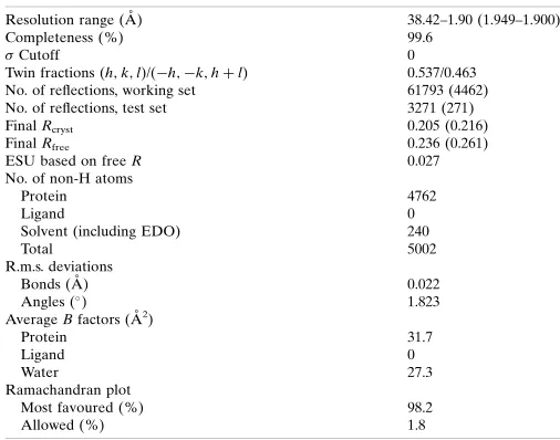
Table 2
Structure solution and refinement.
Values in parentheses are for the outer shell.
Resolution range (A˚ ) 38.42–1.90 (1.949–1.900) Completeness (%) 99.6
Cutoff 0 Twin fractions (h;k;l)/(h;k;hþl) 0.537/0.463 No. of reflections, working set 61793 (4462) No. of reflections, test set 3271 (271) FinalRcryst 0.205 (0.216)
FinalRfree 0.236 (0.261)
ESU based on freeR 0.027 No. of non-H atoms
Protein 4762 Ligand 0 Solvent (including EDO) 240 Total 5002 R.m.s. deviations
Bonds (A˚ ) 0.022 Angles () 1.823
AverageBfactors (A˚2)
Protein 31.7 Ligand 0 Water 27.3 Ramachandran plot
[image:3.610.46.296.104.262.2]indexing was carried out to set the angle to = 114 using the
transformation matrix (100, 001,110) with unit-cell parametersa=
65.12,b= 88.93,c= 157.02 A˚ . Finally, the softwareREINDEXfrom
CCP4 was used with settingsh=h,k=k,l=l/2 to give the correct
unit-cell lengthsa = 65.12,b= 88.93,c= 78.51 A˚ ,== 90,=
114.50. The free set was then reduced to theP2
1asymmetric unit and
used as the source of free reflection flags for all other data sets.
Initial phasing was carried out by MrBUMP(Keegan & Winn,
2008) using the crystal structure of the catalytic domain of RpfB
(PDB entry 3e05; Ruggieroet al., 2009). A solution with four copies in
the asymmetric unit was found inC2221but would not refine below
an Rfree of 0.500 using MOLREP (Vagin & Teplyakov, 2010).
However, two copies of this model were found in theP21unit cell and
refined with the use of twinning to a final Rfree of 0.236 using
REFMAC5 (Murshudov et al., 2011; see Table 2). There is a noncrystallographic translation of (0.554, 0.0, 0.109) in fractional coordinates of 50% of the origin peak. With the improvements in
molecular replacement including noncrystallographic translation
since this work was originally carried out, current versions ofPhaser
(McCoy et al., 2007) and MOLREP can solve this structure more
routinely from a single RpfB chain.
3. Results and discussion
3.1. Structure-solution problems
Many data sets were collected from crystals of RpfC or the point mutations RpfC_E13A or RpfC_E13M with and without potential substrates and including selenomethionine-substituted RpfC_E13M at the ESRF, SLS, SOLEIL and Diamond synchrotrons. The auto-matic space-group assignment for all data sets gave the space group
asC2221, with unit-cell parameters of arounda= 66,b= 141,c= 90 A˚ ,
=== 90. The resolutions of the data sets ranged from 3.0 to
1.9 A˚ . This would predict four copies of the RpfC chain in the
asymmetric unit. We failed to obtain a molecular-replacement
solu-tion using our NMR structure (PDB entry 1xsf; Cohen-Gonsaudet
al., 2005). Slightly better solutions were found using the crystal
structure of the RpfB catalytic domain, withRandRfreeof around
0.45 and 0.50, respectively, but these would not refine further. Attempts at Se or S SAD also did not give solutions. However, anomalous site searching using charge flipping (Dumas & van der
Lee, 2008), which works inP1, indicated that the data were probably
in space groupP21, as eight sites could be found using the SeMet
RpfC_E13M data in this space group. This data set did not yield a useable map, probably owing to the data set being twinned (0.41 from a Britton plot) and the presence of only weak anomalous signal that
only extended to around 3.8 A˚ as assessed byphenix.xtriage(Zwartet
al., 2005) and CTRUNCATE from CCP4. However, molecular
replacement with the C2221solution from the crystal structure of
RpfB (Ruggieroet al., 2009) in space groupP21(unit-cell parameters
a= 65,b= 88,c= 78 A˚ ,== 90,= 114.50) to give eight copies in
the asymmetric unit and refining with twin operators h;k;l and
h;k;hþlallowed refinement to acceptableRandRfreevalues on
carefully selecting the free set (see Table 2). Twinning was not
apparent from theL-test (Yeates, 1988) or the moments ofE, but was
[image:4.610.45.565.268.689.2]estimated for the final data set as 0.41 from theH-test (Padilla &
Figure 1
Yeates, 2003) and 0.45 in a Britton plot (Fisher & Sweet, 1980) as
tested byCTRUNCATE. Other data sets gave similar twinning. The
final refined twinning fraction in REFMAC5 for the deposited
structure was 0.463 forh;k;hþl. Despite soaking and
co-crys-tallizing with a range of substrates and substrate fragments, for
exampleN-acetylglucosamine (NAG), polymers of up to five repeats
of N-acetylglucosamine and NAG-N-acetylmuramic acid, and
peptidoglycan fragments that are generated by a number of enzymes, we never obtained clear density for substrates in the active site. We have therefore deposited the structure of the wild-type RpfC catalytic domain (PDB entry 4ow1).
3.2. Structure analysis
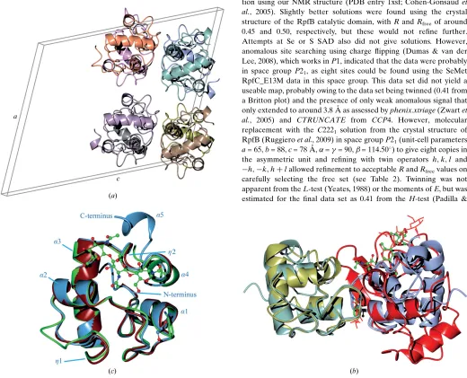
The asymmetric unit consists of eight copies of the RpfC chain. A set of four copies is generated by two twofold axes perpendicular to the crystallographic twofold; a single translation of (0.554, 0.0, 0.109)
then generates the second set of four copies (Fig. 1a). Coupled with
twinning the two folds give rise to the pseudo-C2221symmetry.
ChainsA,EandShave the most residues modelled into electron
density (Gly1–Lys86) with an extra helix beyond the end of the
conserved domain (Gly78). ChainBhas the least modelled residues
(Pro4–Gly78); the other chains are between these limits. We have modelled an ethylene glycol (the cryoprotectant) where a benzami-dine molecule is present in the RpfB structures with PDB codes 4kpm
(Squegliaet al., 2013) and 4emn (Ruggieroet al., 2013). As for the
benzamidine in 4kpm, this is only seen in one of the similar interfaces. Benzamidine and ethylene glycol are not all that similar, but this observation indicates that this region in RPFs prefers binding small organic molecules to water. This region is part of the predicted binding site of a hexasaccharide based on superposition of the
lyso-zyme-cleaved hexasaccharide complex with PDB code 1lzs (Songet
al., 1994). The crystal packing of the two adjacent chains close to the
benzamidine/ethylene glycol site is almost perfectly conserved in our RpfC structure and in the RpfB structures, despite there being no evidence of this contact being physiological. The two pairs of chain
superimpose with an r.m.s.d. of 1.1 A˚ over 149 residues using SSM
(Krissinel & Henrick, 2004), which is not much larger than that for the single chains (see below). The RPF domains are sufficiently close to clash with the superposed disaccharide in this region. The trisac-charide in 4kpm coincides with the other part of the cleaved
saccharide in 1lzs (Fig. 1b).
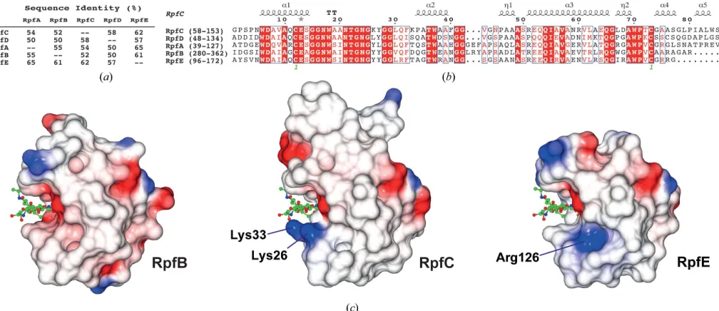
As expected, the structural conservation between the new RpfC catalytic domain structure that we have determined in this study and
the extensively studied RpfB domain is high. The calculated C
r.m.s.d. between the two structures (our structureversusPDB entry
4kl7; Squegliaet al., 2013) is only 0.90 A˚ for 76 residues aligned by
SSMwith 52% sequence identity over the domain (Figs. 2aand 2b).
Compared with the recent RpfE structure (PDB entry 4cge; Mavrici
et al., 2014), the calculated Cr.m.s.d. is even lower at 0.82 A˚ for 77
residues with 62% sequence identity (Figs. 1cand 2a). Most of the
backbone geometry is conserved, including the connecting loops between the helices. This is in accordance with the first NMR struc-ture that we determined, where the 30 calculated strucstruc-tures shared a
low r.m.s.d. of 0.57 A˚ , low thermal motion as shown by NOE (Nuclear
Overhauser Effect) ratios (Cohen-Gonsaud, Bartheet al., 2004) and a
well ordered fold for the RPF domain. The only difference observed is located within a short sequence insertion that is present in the RpfB
RPF domain compared with the other four M. tuberculosis RPF
proteins (Figs. 1cand 2b). In RpfC two residues display an elongated
conformation (42GVGN45), very similar to RpfE (137GSGS140), to
connect -helices 2 and 3, while a 310-helix (
321
GLRYAPR327) is
present in RpfB. This small change within the secondary-structure
composition does not change the relative orientation of-helices 2
and 3 within the RPF fold (Fig. 1c). The variation in surface charge
between RpfB and RpfE has previously been noted (Mavriciet al.,
2014). RpfC has two lysines, Lys26 and Lys33, on one side of the sugar-binding cleft, which are tyrosines in RpfA, RpfB and RpfD or a leucine in RpfE and serine or threonine in RpfA, RpfD and RpfE or
an aspartate in RpfB (Fig. 2b), respectively. This leads to a different
charge distribution around the ligand-binding pocket, which may
have a role in specificity (Fig. 2c). Mavriciet al.(2014) suggested that
[image:5.610.55.559.489.707.2]Acta Cryst.(2014). F70, 1022–1026 Chauviacet al. RpfC
1025
Figure 2
Arg126 may play a role in binding the peptide part of the pepti-doglycan, conferring specificity on RpfE.
4. Conclusion
The RpfC structure catalytic domain displays a high degree of structural conservation with the other members of the mycobacterial resuscitation-promoting factor family. Based on the structure that we
have solved, we propose that the five RPFs fromM. tuberculosishave
similar substrates, although variation in charge around the active site may give rise to small variations in the specificity for different peptidoglycan modifications. The high degree of conservation of the RPF domain explains why the protein is functionally redundant, but most importantly shows that the auxiliary domain composition is mainly responsible for the functional variability.
This work was supported by grants from the French Infrastructure for Integrated Structural Biology (FRISBI) ANR-10-INSB-05-01 (MC-G), an X-TB Struct EU grant (MC-G and NHK), MRC grant G0401038 (GR, NHK, BH and JW), a Bloomsbury studentship to F-XC and a Commonwealth Studies Commission studentship MYCS-2011-252 to DHXQ.
References
Cohen-Gonsaud, M., Barthe, P., Bagne´ris, C., Henderson, B., Ward, J., Roumestand, C. & Keep, N. H. (2005).Nature Struct. Mol. Biol.12, 270–273. Cohen-Gonsaud, M., Barthe, P., Pommier, F., Harris, R., Driscoll, P. C., Keep,
N. H. & Roumestand, C. (2004).J. Biomol. NMR,30, 373–374.
Cohen-Gonsaud, M., Keep, N. H., Davies, A. P., Ward, J., Henderson, B. & Labesse, G. (2004).Trends Biochem. Sci.29, 7–10.
Downing, K. J., Betts, J. C., Young, D. I., McAdam, R. A., Kelly, F., Young, M. & Mizrahi, V. (2004).Tuberculosis,84, 167–179.
Dumas, C. & van der Lee, A. (2008).Acta Cryst.D64, 864–873. Edgar, R. C. (2004).Nucleic Acids Res.32, 1792–1797. Fisher, R. G. & Sweet, R. M. (1980).Acta Cryst.A36, 755–760. Kabsch, W. (2010).Acta Cryst.D66, 125–132.
Kana, B. D., Gordhan, B. G., Downing, K. J., Sung, N., Vostroktunova, G., Machowski, E. E., Tsenova, L., Young, M., Kaprelyants, A., Kaplan, G. & Mizrahi, V. (2008).Mol. Microbiol.67, 672–684.
Kana, B. D. & Mizrahi, V. (2010). FEMS Immunol. Med. Microbiol. 58, 39–50.
Keegan, R. M. & Winn, M. D. (2008).Acta Cryst.D64, 119–124. Krissinel, E. & Henrick, K. (2004).Acta Cryst.D60, 2256–2268.
Mavrici, D., Prigozhin, D. M. & Alber, T. (2014). Protein Sci. 23, 481– 487.
McCoy, A. J., Grosse-Kunstleve, R. W., Adams, P. D., Winn, M. D., Storoni, L. C. & Read, R. J. (2007).J. Appl. Cryst.40, 658–674.
McNicholas, S., Potterton, E., Wilson, K. S. & Noble, M. E. M. (2011).Acta Cryst.D67, 386–394.
Mukamolova, G. V., Kaprelyants, A. S., Young, D. I., Young, M. & Kell, D. B. (1998).Proc. Natl Acad. Sci. USA,95, 8916–8921.
Mukamolova, G. V., Murzin, A. G., Salina, E. G., Demina, G. R., Kell, D. B., Kaprelyants, A. S. & Young, M. (2006).Mol. Microbiol.59, 84–98. Mukamolova, G. V., Turapov, O. A., Young, D. I., Kaprelyants, A. S., Kell, D. B.
& Young, M. (2002).Mol. Microbiol.46, 623–635.
Murshudov, G. N., Skuba´k, P., Lebedev, A. A., Pannu, N. S., Steiner, R. A., Nicholls, R. A., Winn, M. D., Long, F. & Vagin, A. A. (2011).Acta Cryst. D67, 355–367.
Padilla, J. E. & Yeates, T. O. (2003).Acta Cryst.D59, 1124–1130.
Powell, H. R., Johnson, O. & Leslie, A. G. W. (2013).Acta Cryst.D69, 1195– 1203.
Raman, S., Hazra, R., Dascher, C. C. & Husson, R. N. (2004).J. Bacteriol.186, 6605–6616.
Ravagnani, A., Finan, C. L. & Young, M. (2005).BMC Genomics,6, 39. Ruggiero, A., Marchant, J., Squeglia, F., Makarov, V., De Simone, A. & Berisio,
R. (2013).J. Biomol. Struct. Dyn.31, 195–205.
Ruggiero, A., Tizzano, B., Pedone, E., Pedone, C., Wilmanns, M. & Berisio, R. (2009).J. Mol. Biol.385, 153–162.
Song, H., Inaka, K., Maenaka, K. & Matsushima, M. (1994).J. Mol. Biol.244, 522–540.
Squeglia, F., Romano, M., Ruggiero, A., Vitagliano, L., De Simone, A. & Berisio, R. (2013).Biophys. J.104, 2530–2539.
Studier, F. W. (2005).Protein Expr. Purif.41, 207–234.
Telkov, M. V., Demina, G. R., Voloshin, S. A., Salina, E. G., Dudik, T. V., Stekhanova, T. N., Mukamolova, G. V., Kazaryan, K. A., Goncharenko, A. V., Young, M. & Kaprelyants, A. S. (2006).Biochemistry (Mosc.),71, 414– 422.
Tufariello, J. M., Jacobs, W. R. Jr & Chan, J. (2004).Infect. Immun.72, 515–526. Vagin, A. & Teplyakov, A. (2010).Acta Cryst.D66, 22–25.
Winn, M. D.et al.(2011).Acta Cryst.D67, 235–242. Yeates, T. O. (1988).Acta Cryst.A44, 142–144.