Why development matters in neurodevelopmental disorders
30
0
0
Full text
(2) 1 Why development matters in neurodevelopmental disorders George Ball and Annette Karmiloff-Smith Centre for Brain and Cognitive Development, Birkbeck, University of London. Abstract The importance of taking a truly developmental perspective to the study of neurodevelopmental disorders can be underappreciated. With illustrations from Williams, Down and Fragile X syndromes, this chapter makes a strong theoretical plea for a developmental approach from multiple levels. Section 1 argues that adult neuropsychological models are inappropriate for understanding neurodevelopmental disorders. Developmental change is then highlighted in the context of mapping: genotype to phenotype (Section 2), brain to cognition (Section 3), early basic-level underpinnings to cognitive-level outcomes (Section 4), and of evaluating environmental factors (Section 5). Practical tips for research into neurodevelopmental disorders are offered in Section 6, before general conclusions are drawn.. Author details George W. Ball, Research Assistant and Masters student Annette Karmiloff-Smith, Professorial Research Fellow Centre for Brain and Cognitive Development, Department of Psychological Sciences, School of Science, Birkbeck, University of London, Malet Street, London, WC1E 7HX..
(3) 2 1. Why is the adult neuropsychological model inappropriate for understanding neurodevelopmental disorders? Paradoxically, numerous studies of infants and children are not developmental at all, because they take static snapshots, targeting a specific age group. A truly developmental perspective embraces a developmental way of thinking, regardless of the age of the population studied (Karmiloff-Smith, 1992, 1998). Even research on infants can be non-developmental, simply examining performance in, for example, 5-month-olds, whereas some studies of adults are developmental, because they focus on change over time (Cornish et al., 2008; Tyler et al., 2009). In other words, neuro-cognitive development is dynamic across the entire lifespan, and there is no static end state. Indeed, to understand ontogenetic development in atypical or typical individuals, it is vital to trace developmental trajectories across time (Karmiloff-Smith, 1998; Cornish, Scerif, & Karmiloff-Smith, 2007), to assess progressive change from infancy onwards at the genetic, neural, cognitive, and behavioural levels, including the role of the environment, and to pinpoint how parts of the developing system may interact with other parts differently at different times across ontogenesis (KarmiloffSmith, 1998; Steele, Brown, & Scerif, 2012). A process that is vital, say, at Time 2 may no longer play a role at Time 5. Yet, its delay at Time 2 may have been crucial to a healthy developmental trajectory and outcome, because of its interactions with other parts of the developing system (Karmiloff-Smith, 1998). Indeed, developmental timing is among the most important of factors that must be taken into account when trying to understand human development, particularly in the case of neurodevelopmental disorders..
(4) 3 This is why, in our view, the adult neuropsychological model is inappropriate for explaining neurodevelopmental disorders. Adult models tend to be static, whereas neurodevelopmental disorders require a dynamic approach that focuses on trajectories and how they change over time (Thomas et al., 2009). Thus, we need to distinguish between the developed brain and the developing brain (Karmiloff-Smith, 2010, 2012a). Take the case of Williams syndrome (WS), a neurogenetic syndrome involving the hemizygous deletion of some 28 genes on chromosome 7. The fusiform face area (FFA) is particularly large in volume compared to the rest of the WS brain, and adolescents and adults with WS are very proficient at face processing tasks (Karmiloff-Smith et al., 2004). There are two possible explanations, one drawn from the adult neuropsychological approach, the other from a developmental perspective. The first would argue that the large FFA in WS causes the face processing proficiency – a brain-tobehaviour explanation. The second, by contrast, would argue that the unusual infant focus on faces in WS influences the enlargement of the FFA over time – a bidirectional brain-to-behaviour-to-brain interaction. Only a truly developmental approach can address such alternatives. But where does the WS infant fascination with faces stem from? Could this be “caused” by an early difference in the FFA? Or, is the following developmental scenario more likely? In the last three months of intra-uterine life the foetus does a great deal of auditory processing and recognises the intonation patterns of its mother’s voice at birth (Hepper, Scott, & Shahidullah, 1993). In the early years, infants with WS tend to pay particular attention to auditory input (Mervis, Morris, Bertrand, & Robinson, 1999). What is the first sound that the baby hears at birth? Usually it is its mother’s voice. Locating the voice would orient the baby to the mother’s.
(5) 4 face, which would display smiles and other encouraging stimuli. So, it is possible that the WS infant’s fascination with faces – a visual stimulus – stems from an earlier fascination with voices – an auditory stimulus – in combination with problems with visual disengagement and a heightened social drive (Frigerio et al., 2006; Riby & Hancock, 2008). Thus, the visual fixation on faces could initially derive from attention to the auditory modality and thence drive the progressive enlargement of the FFA. While this hypothesis may turn out to be inaccurate, we believe that it is an illustration of a vital developmental way of thinking when trying to explain neurodevelopmental disorders.. Later in Section 4, we provide a second example of how similar data across two syndromes (Williams syndrome and Down syndrome) can seem to replicate the double dissociation approach of adult neuropsychology, isolating two numerical systems. However, the explanations for these differing patterns in the two neurodevelopmental disorders are not due to impaired versus intact numerical modules. Rather, they are rooted in differences in earlier, more basic-level processes, such as saccadic eye movement planning and attention, which affect number as well as other cognitive domains over developmental time differently in each syndrome (see Section 4 below for more detail). Indeed, we advocate that any time a seeming dissociation is found in adolescents or adults with neurodevelopmental disorders, it is crucial to trace such cognitive-level outcomes back to their basic-level processes in early development and to consider their cascading effects over developmental time..
(6) 5 2. Developmental change counts in mapping genotype to phenotype. Many studies map specific genes to specific behavioural phenotypes, but rare are those that take account of changing gene expression over time. Yet, if a gene is expressed widely initially and becomes increasingly confined to certain brain regions, then the mapping from gene to phenotype will change (Karmiloff-Smith et al., 2012b). Developmental time – or what Elman and collaborators called ‘chronotopic constraints’ (Elman et al., 1996) – plays a crucial role, even in monogenic disorders. Take, for example, Fragile X syndrome, which is associated with the silencing of a single gene, whose protein product, FMRP, is involved in the regulation of multiple cascading processes leading to activity-dependent changes in dendritic spine morphology and synaptic regulation across cortex. FMRP is highly expressed in both foetal and adult brain tissues, but it interacts with multiple other proteins, a process which changes over developmental time. In adult cerebellum and cerebral cortex, FMRP and two of these proteins are colocalized. In the foetus, as in the adult, FMRP is located in cytoplasm, but in the foetus only one of the collaborating proteins is strongly expressed in nucleus (Scerif & Karmiloff-Smith, 2005). Thus, FMRP is likely to collaborate with sets of proteins in undifferentiated foetal neurones, which are different from those with which it interacts in differentiated adult neurones. These complex interactions suggest that a single gene dysfunction can initiate multiple cascading effects on cellular, neural, and cognitive phenotypes that vary across developmental time.. The importance of considering developmental change in mapping genotype to neural phenotype may also be seen in the phenotypic trajectories of neurogenetic disorders such as Down Syndrome (DS). DS, or trisomy 21, is.
(7) 6 caused in most cases by a non-disjunction after conception, resulting in an extra copy of chromosome 21. It has been argued that the DS genotype particularly impairs the development of late developing neural systems such as the hippocampus and prefrontal cortex (Edgin, 2013; Nadel, 1986). For example, while the density of myelinated fibres in hippocampal regions of neonates is somewhat reduced relative to typically developing controls, this hippocampal difference in density increases progressively throughout childhood and into early adulthood, resulting in much larger relative differences further down the developmental trajectory (Ábrahám et al., 2012). Such developmental findings might explain why, relative to typical controls, no hippocampal volume reduction is reported in the first year of life (Pennington, Moon, Edgin, Stedron, & Nadel, 2003) but is routinely identified in older children (Carducci et al., 2013; Jernigan, Bellugi, Sowell, Doherty & Hesselink, 1993; Pinter et al., 2001) and in adults (Aylward et al., 1999; Kesslak, Nagata, Lott, & Nalcioglu, 1994; Raz, Torres, Briggs, & Spencer, 1995). Thus, when mapping the DS genotype to the DS brain, consideration must be given to changes that occur in the neural profile across developmental time.. Development is also important when mapping genotype to cognitive phenotypic outcomes. Continuing with the above example from DS that reveals progressive delay in hippocampal development, the described neural trajectory also fits with findings in the cognitive domain. Interestingly, hippocampal-dependent processes, such as episodic memory and spatial learning, are severely impaired in adolescents and young adults with DS relative to implicit and working memory processes (Pennington et al., 2003; Vicari, Bellucci, & Carlesimo, 2000)..
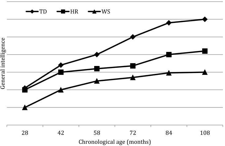
(8) 7 Crucially, however, a developmental trajectory approach does not automatically assume that this dissociation at the cognitive level is present in the same form earlier in the developmental trajectory. Indeed, Rast and Meltzoff (1995) found that two-year-olds with DS showed strength in memory for episodically-defined events relative to their acquisition of object permanence, for instance (see also, Paterson, Brown, Gsödl, Johnson, & Karmiloff-Smith, 1999, for similar differences between the start and end states for language and number in DS and WS).. Clearly, more in-depth research on the early cognitive profile of each neurodevelopmental disorder would better inform our understanding of their full developmental trajectories, but any account of genotype to phenotype mappings should be sensitive to possible changes in gene expression and cognitive phenotype over developmental time. Snapshots of a cognitive phenotype at Time 6, for instance, may tell the researcher nothing about the state of that cognitive phenotype at Time 1, unless considered within the context of development. Figure 1 illustrates this point. It plots the longitudinal trajectory of a child (HR) with a partial deletion of some 23 of the 28 genes in the WS critical region (Karmiloff-Smith et al., 2003, 2012b; Tassabehji et al., 1999, 2005) with respect to her general intelligence scores from chronological age 28 months (Time 1) to 108 months (Time 6), together with the average general intelligence trajectories of typically developing (TD) children and those with the full WS deletion for comparison. It is clear that HR’s general intelligence scores were at the same level as typically developing children at 28 months but became progressively impaired over developmental time, such that by 108 months, HR’s general intelligence score was markedly below TD children, although still higher.
(9) 8 than the average child with WS of the same chronological age. Had one taken a snapshot and drawn conclusions about genotype/phenotype relations when HR was 28 months, the conclusion would have been that her deleted genes had no impact on general intelligence. By contrast, examining the graph seven years later at 108 months, it is clear that she is seriously impaired, although not as severely as children with the full WS deletion, highlighting the importance of taking account of full developmental trajectories over time when mapping genotype to phenotype.. HR. WS. General intelligence. TD. 28. 42. 58 72 Chronological age (months). 84. 108. Figure 1. General intelligence scores of HR, a child with partial deletion WS, average general intelligence scores of typically developing children and of children with WS (full deletion), from chronological ages 28 to 108 months. (unpublished figure, reproduced from conference presentations by KarmiloffSmith).
(10) 9. 3. Developmental change counts in mapping brain to cognition. The cognitive phenotype of WS is characterized by relatively skilled language, a strong drive for social interaction, and proficient face-processing abilities, with scores falling within the normal range on standardized face processing tasks, like the Rivermead Face Memory Task (Wilson et al., 2008) and the Benton Facial Recognition Test (Benton, Sivan, Hamsher, Varney, & Spreen, 1994), alongside very impaired spatial and numerical cognition (Karmiloff-Smith et al., 2004). The initial excitement about the syndrome stemmed from the identification of this uneven cognitive profile and the possibility that it might represent “selective deficits of an otherwise normal modular system” (Clahsen & Temple, 2003, p. 347), with the modules for language and face processing considered intact and those for number and spatial cognition selectively impaired.. Electrophysiological studies of the WS brain have pointed to possible developmental arrest compared with the TD brain. Even in domains of relatively proficient behaviour, e.g., in face and language processing, the WS brain fails to become progressively specialized (Karmiloff-Smith, 2009), in contrast to the TD brain. Indeed, the TD brain initially processes incoming input bilaterally but, with development, a progressive specialisation and localisation of brain function emerges, giving rise to a shift to increasingly specialised hemispheric processing (predominantly right hemisphere for faces, predominantly left hemisphere for the morphosyntax of language) (Casey, Giedd, & Thomas, 2000; Choudhury & Benaisch, 2011; Durston et al., 2006; Johnson, 2001; Mills, Coffey-Corins, & Neville, 1997; Minagawa-Kawai, Mori, Naoi, & Kojima, 2007; de Haan, Pascalis, &.
(11) 10 Johnson, 2002). By contrast, several studies have now shown that the WS brain tends to continue processing faces and language bilaterally, even in adulthood (Grice et al., 2001, 2003; Karmiloff-Smith et al., 2004; Mills et al., 2000; Neville et al., 1994). In other words, whereas the TD brain presents with a gradual specialisation and hemispheric localisation of function over developmental time, this does not seem to occur in the development of the WS brain (Karmiloff-Smith, 2009). Moreover, in TD brains the temporal signature for faces becomes increasingly specific over developmental time, whereas WS brains tend to process faces, cars and other objects with the same temporal signature (Grice et al., 2001; Karmiloff-Smith, 2009; see also Golarai et al., 2010, for similar differences between TD and WS neural processing in fMRI studies).. Therefore, even when individuals with WS score in the normal range, such as on standardized face processing tasks, this does not mean a selective “sparing”, because it has been shown that the cognitive and neural processes underlying the proficient behaviour are different from those of TD controls (Karmiloff-Smith et al., 2004).. 4. The need to trace higher-level cognitive outcomes back to basic-level processes in infancy. Let us now take a concrete example of a cognitive domain – exact and approximate number – and the demonstration of a possible double dissociation in infancy, typical of the type found in studies of adult neuropsychology. Small exact number discrimination involves the computation of precise differences between 1, 2, 3 or 4 items. Large approximate number discrimination is the.
(12) 11 capacity to estimate whether two quantities (e.g., 8 versus 16 items) are different, without being able to count them, based on magnitude judgments. For example, studies of adult neuropsychological patients have yielded a double dissociation between numerical abilities, affecting two intra-parietal circuits one for computing exact number, the other for computing approximate numerical quantities (Butterworth, 2010; Dehaene, 1997; Demeyere, Lestor, & Humphreys, 2010).. Research on TD infants also yields two numerical sub-systems that develop at different rates (Brannon, 2006; Izard, Dehaene-Lambertz & Dehaene, 2008; Lipton & Spelke, 2003; Xu 2003). Indeed, TD infants are able to discriminate small numbers (one, two, three) as early as 3 months, but it is several months later that they can discriminate large approximate quantities: at 6 months they differentiate 8 from 16 dots, i.e., a ratio of 1:2, and by 9-10 months they distinguish 8 from 12 dots, a ratio of 2:3 (Brannon, Abbott, & Lutz, 2004; Xu & Arriaga, 2007). In a series of studies examining small exact number discrimination and large approximate number discrimination in infants with DS and infants with WS (Karmiloff-Smith et al., 2012a; Paterson, Brown, Gsödl, Johnson, & Karmiloff-Smith, 1999; van Herwegen, Ansari, Xu, & Karmiloff-Smith, 2008), we found that those with WS performed like TD controls on small number discrimination tasks, but showed significantly weaker discrimination of large approximate quantities. By contrast, infants with DS displayed the opposite pattern: they failed at small number discrimination but showed significant discrimination of large approximate quantities..
(13) 12 So, for the first time in research on infants with different neurogenetic syndromes, our studies yielded a double dissociation between small exact number and large approximate number, typical of the findings in adult neuropsychological patients. The results might therefore suggest that for WS, one or more of the 28 genes deleted on one copy of chromosome 7q11.23 contribute to a domain-specific deficit in large approximate number discrimination while leaving intact the small number system, and for DS, one or more of the extra genes on chromosome 21 contribute to a domain-specific deficit in small exact number discrimination while sparing the large number system. However, notions like “double dissociation”, “intact” and “sparing” are drawn from the adult neuropsychological literature, describing a system in its mature state. Is this an appropriate model for a system in the process of gradual development? Or, as we believe, do these sub-systems emerge over time, through early cross-domain/cross-modality interactions, as different brain circuits progressively specialize for different numerical functions (Karmiloff-Smith, 1998, 2009; Simon, 1997)?. It is therefore imperative to assess how other processes might interact over developmental time with infant sensitivity to numerical displays. We thus drew on earlier data on infant/toddler attention and saccadic eye movement planning in the two syndromes (Brown et al., 2003) and related these to the WS and DS numerical findings (Karmiloff-Smith et al., 2012a). The attention studies identified deficits in both syndromes – deficits in attention shifting in WS, deficits in sustained attention in DS. In the saccadic eye movement study, infants/toddlers with DS showed similar patterns to TD infants, i.e., efficient.
(14) 13 albeit slower saccadic eye movement planning. By contrast, infants/toddlers with WS displayed severe eye movement planning deficits (Brown et al., 2003). We thus predicted that infants/toddlers with WS would be impaired in their scanning of large numerical displays. To examine this further, after the experiment proper, we collected eye-tracking data (Tobii, 2003) from infants with WS and DS while they were viewing numerical displays on the computer screen. A preliminary examination of scanning patterns of the infants who provided useful data indicated that, like in our studies of eye movement planning and attention (Brown et al., 2003), those with DS tended to scan the overall array, whereas those with WS tended to remain fixated on a few individual items (see illustrations of scanning patterns in Karmiloff-Smith et al., 2012a).. Thus, for WS, a serious deficit in rapid saccade planning (Brown et al., 2003; Karmiloff-Smith et al., 2012a) may cause problems in visually disengaging from individual objects in displays. This likely explains why they succeed at small exact number discrimination, yet have difficulty discriminating large approximate quantities because they do not scan them adequately. By contrast, the opposite holds for infants with DS, because they have difficulties with sustained attention (Krakow & Kopp, 1982; Casey & Riddle, 2012), which interferes with their precise individuation of objects in small displays but enhances their rapid global scanning of large displays.. Thus, rather than identifying a double dissociation of spared and impaired modules between the two syndromes, it turns out that a single but different basic-level problem for each syndrome – attention shifting in WS, sustained.
(15) 14 attention in DS - contributes to the explanation of both the numerical deficits and the numerical proficiencies in each syndrome. The findings therefore do not require an explanation solely in terms of selective, domain-specific number abilities, as might be the case in adult neuropsychological patients whose brains had by then become specialized in terms of two independently functioning numerical sub-systems. Rather, the syndrome-specific infant differences are likely to be traceable to basic-level deficits and proficiencies in the visual and attention systems early in development, which have cascading effects on cognitive-level outcomes over ontogenetic time. Furthermore, identification of these different basic-level problems in early development may be particularly useful in informing the planning of early syndrome-specific intervention.. 5. Developmental change counts in understanding the impact of environmental factors. A question to emerge from studies of families with children with neurodevelopmental disorders is why the effects of positive environments are not greater. Indeed, unlike children from low SES environments, many children with neurogenetic syndromes who participate in academic research are well nourished, grow up in a caring environment, receive considerable cognitive stimulation, and do not suffer the physical and mental abuses reported in some contexts of early social adversity (Meaney & Szyf, 2005; Liston, McEwen, & Casey, 2009; Tomalski et al., 2013; Tottenham et al., 2010, 2011). For instance, we have recently shown that low SES has detrimental effects on the frontal cortex as early as 6-9 months of age (Tomalski et al., 2013). So, why do positive environments not compensate for genetic vulnerabilities? Is it just the severity of.
(16) 15 the genetic mutations that constrains environmental effects? Or is this not only due to mutated genes, but also because early environments for such children differ in more subtle ways than is commonly realised? Indeed, having a neurodevelopmental disorder not only involves genetic mutations; it also changes the environment in which the atypical infant develops in multiple subtle ways. In our view, the moment a parent is informed that their child has a genetic disorder, the parent’s behaviour necessarily subtly adapts. As a result, the baby’s responses within the dyadic interaction will also be subtly modified, and these interactional effects may increase over time. This is not only true of children with neurodevelopmental disorders. Even for well-stimulated typically developing children, cognitive development can be subtly fostered or hindered by differences in environmental conditions, e.g., in mother/infant interaction styles (Karmiloff-Smith et al., 2010, 2012a).. A couple of examples serve to illustrate the environmental changes that may obtain for children growing up with genetic disorders. The first comes from a study of the process of vocabulary learning in toddlers with Down syndrome (John & Mervis, 2010). When TD toddlers start to label things, their parents usually allow them temporarily to overgeneralize (e.g., call all animals “cat”). By contrast, in the case of toddlers with DS, parents tend to veto and correct overgeneralizations immediately (John & Mervis, 2010). In our view, this is because of the parents’ natural fear that their child with lower intelligence will never learn the right label if allowed to overgeneralize. Yet, initial overgeneralization in the TD child encourages category formation (e.g., by calling different animals “cat”, the child creates an implicit “animal” category), and.
(17) 16 categorization is known to be impaired in many neurodevelopmental disorders, including DS. The second example comes from motor development. Observational data from our lab reveal that parents of infants/toddlers with genetic syndromes often find it difficult (compared to parents of TD infants) to let their atypically developing offspring use the sensitive nerve endings in their mouth to explore object properties, or to crawl/walk uninhibited around the lab in order to fully discover their environment. This reticence is likely to be due to an understandable greater fear of accidents with respect to vulnerable children. These unconscious assumptions about what atypical children can and cannot learn may unwittingly lead their parents to provide less variation in their linguistic input to their child and in general offer a more cautious, less varied environment - subtle differences that are likely to compound over developmental time.. Thus, a richer notion of “environment” is required, i.e., how having a neurodevelopmental disorder may subtly change the social, cognitive, linguistic, emotional and physical environments to which the atypically developing child is progressively exposed.. 6. Practical tips In this section, we provide a few practical tips/recommendations for research in neurodevelopmental disorders that build on our developmental neuroconstructivist approach (Karmiloff-Smith, 1998; Farran & Karmiloff-Smith, 2012)..
(18) 17 a. We strongly recommend the tracing of full developmental trajectories from infancy onwards. This does not necessarily restrict the researcher solely to longitudinal studies, although the latter represent an important approach. Indeed, an optimal design for studying developmental disorders is to combine case studies with initial cross-sectional designs and then longitudinal group follow-up (Thomas et al., 2009). An understanding of underlying mechanisms will be furthered by the richer descriptive vocabulary provided by the trajectories approach (e.g., in distinguishing different types of delay and deviance).. b. We highlight the need for longitudinal studies. However, these should be not only at the cognitive level, but also at the neural level (Karmiloff-Smith, 2010). Understanding the extent to which the brains of children with neurodevelopmental disorders change over time is crucial. And it is just as necessary to identify regressive events as progressive changes, including the extent to which the atypically developing brain shows any indices of circuit specialisation.. c. We recommend examining microdevelopmental change in neurodevelopmental disorders. While this has been a relatively lively area of TD research (Karmiloff-Smith, 1984, 2013; Siegler & Svetina, 2002), it has rarely if ever been used for the study of neurodevelopmental disorders. Yet, understanding the processes of change over the very short term is just as important as identifying macro-developmental change..
(19) 18 d. In our view, researchers are still far from understanding how genetic mutations interact with the subtle changes that occur in the environments of children with neurodevelopmental disorders. We thus need a much more indepth account of environment changes. One avenue of promising research would be the study of twins in which one twin has a neurodevelopmental disorder and the other is typically developing. Subtle differences in the socio-linguistic, cognitive, and physical environments of each twin of equivalent chronological age and seemingly equivalent environment could be very informative.. 7. Development itself is the key to understanding neurodevelopmental disorders.. We started this chapter by highlighting an important difference between adult neuropsychological models and truly developmental approaches to neurodevelopmental disorders, namely that adult models tend to be static and developmental approaches necessarily require a dynamic perpective, which allows for identifying phenotypic changes over developmental time. Section 2 stressed how genotype to phenotype mapping depends on developmental time; gene expression, neural and cognitive profiles may differ considerably at different points on the developmental trajectory. Section 3 provided an example of how a developmental explanation is needed to map findings of atypical neural processing for faces and language to relatively proficient face and language processing behaviours in adults with WS. Unlike the adult neuropsychological model, a developmental approach does not assume the existence of domain-.
(20) 19 specific neural subsystems in neurodevelopmental disorders. Indeed, it argues that the atypical genetic profile found in neurodevelopmental disorders affects brain development from the start of ontogenesis, impacting low-level, domaingeneral processes that cascade over developmental time and affect neural specialisation processes in later development. Section 4 provided one such example where cross-syndrome differences in number processing by adults with DS and WS may be partially explained by differences in basic-level attentional processes in very early development. Similarly, in Section 5 we suggested that subtle environmental differences for children with neurodevelopmental disorders relative to typically developing children compound over developmental time to affect brain and behaviour in later development. Finally, having argued for the importance of developmental change, a few practical tips for conducting developmental research were offered in Section 6.. In conclusion, we reiterate that focusing on the process of development itself is key to a full understanding of any neurodevelopmental disorder (KarmiloffSmith, 1998). Furthermore, to understand the full implications of having a neurodevelopmental disorder, it is critical to study the disorder at multiple levels (genetic, cellular, neural, cognitive, behavioural and environmental) as well as using the convergence of multiple methodologies (e.g., MEG, fMRI, fNIRS, EEG/ERP, eye-tracking, etc.). Even though plasticity may be reduced in genetic syndromes, it is clear that identifying developmental change remains crucial, whether researching children or adults with neurodevelopmental disorders..
(21) 20 Acknowledgements During the writing of this chapter, AK-S was funded by Wellcome Trust Strategic Award (Grant No. 098330/Z/12/Z), as well as grants from the Waterloo Foundation, UK and Autour de Williams, France. Substantial sections of the chapter are reproduced from Karmiloff-Smith’s Bartlett Lecture (Karmiloff-Smith, A. (2013). Challenging the use of adult neuropsychological models for explaining neurodevelopmental disorders: Developed versus developing brains. Quarterly Journal of Experimental Psychology, 66, 1–14) and from Karmiloff-Smith et al., 2012a, referenced below.. References. Ábrahám, H., Vincze, A., Veszprémi, B., Kravják, A., Gömöri, É., Kovács, G. G., & Seress, L. (2012). Impaired myelination of the human hippocampal formation in Down syndrome. International Journal of Developmental Neuroscience, 30, 147–158. Aylward, E. H., Li, Q., Honeycutt, N. A., Warren, A. C., Pulsifer, M. B., Barta, P. E., … Pearlson, G. D. (1999). MRI Volumes of the Hippocampus and Amygdala in Adults With Down’s Syndrome With and Without Dementia. American Journal of Psychiatry, 156, 564–568. Benton, A. L., Sivan, A. B., Hamsher, K. deS., Varney, N. R., & Spreen, O. (1994). Contributions to neuropsychological assessment (2nd ed.). New York, NY: Oxford University Press..
(22) 21 Butterworth, B. (2010). Foundational numerical capacities and the origins of dyscalculia. Trends in Cognitive Sciences, 14, 534-541. Brannon, E. M. (2006). The representation of numerical magnitude. Current Opinion in Neurobiology, 16, 222–229. Brannon, E. M., Abbott, S., & Lutz, D. (2004). Number bias for the discrimination of large visual sets in infancy. Cognition, 93, B59–B68. Brown, J. H., Johnson, M. H., Paterson, S. J., Gilmore, R., Longhi, E., & KarmiloffSmith, A. (2003). Spatial representation and attention in toddlers with Williams syndrome and Down syndrome. Neuropsychologia, 41, 1037-1046. Carducci, F., Onorati, P., Condoluci, C., Di Gennaro, G., Quarato, P. P., Pierallini, A., … Albertini, G. (2013). Whole-brain voxel-based morphometry study of children and adolescents with Down syndrome. Functional Neurology, 28, 19–28. Casey, B. J., Giedd, J. N., & Thomas, K. M. (2000). Structural and functional brain development and its relation to cognitive development. Biological Psychology, 54, 241-257. Casey, B. J., & Riddle, M. (2012). Typical and atypical development of attention. In M. I. Posner (Ed.). Cognitive neuroscience of attention (2nd ed., pp. 345356). New York, NY: Guilford Press. Choudhury, N., & Benaisch, A. A. (2011). Maturation of auditory evoked potentials from 6 to 48 months: Prediction to 3 and 4 year language and cognitive abilities. Clinical Neurophysiology, 122, 320-338. Clahsen, H., & Temple, C. M. (2003). Words and rules in children with Williams syndrome. In Y. Levy & J. Schaeffer (Eds.), Language competence across.
(23) 22 populations: toward a definition of specific language impairment (pp. 323352). Dordrecht, Netherlands: Kluwer. Cornish, K., Scerif, G., & Karmiloff-Smith, A. (2007). Tracing syndrome-specific trajectories of attention across the lifespan. Cortex, 43, 672–685. Cornish, K. M., Kogan, C. S., Jacquemont, S., Turk, J., Dalton, A., Hagerman, R. J., & Hagerman, P. J. (2008). Age-dependent cognitive changes in carriers of the fragile X syndrome. Cortex, 44, 628–636. Dehaene, S. (1997). The number sense: How the mind creates mathematics. Oxford, EnglandA Oxford University Press. Demeyene, N., Lestor, V., & Humphreys, G. (2010). Neuropsychological evidence for a dissociation in counting and subitizing. NeuroCase, 16, 219-237. de Haan, M., Pascalis., O., & Johnson, M. H. (2002). Specialization of neural mechanisms underlying face recognition in human infants. Journal of Cognitive Neuroscience, 14, 199-209. Durston, S., Davidson, M.C., Tottenham, N., Galvan, A., Spicer, J., Fosella, J.A., & Casey, B.J. (2006). A shift from diffuse to focal cortical activity with development. Developmental Science, 9, 1-8. Edgin, J. O. (2013). Cognition in Down syndrome: a developmental cognitive neuroscience perspective. Wiley Interdisciplinary Reviews: Cognitive Science, 4, 307–317. Elman, J. L., Bates, E. L., Johnson, M. H., Karmiloff-Smith, A., Parisi, D, & Plunkett, K. (1996). Rethinking innateness: A connectionist perspective on development. Cambridge, MA: MIT Press..
(24) 23 Farran, E. & Karmiloff-Smith, A. (Eds.). (2012). Neurodevelopmental disorders across the lifespan: A neuroconstructivist approach. Oxford, England: Oxford University Press. Frigerio, E., Burt, D. M., Gagliardi, C., Cioffi, G., Martelli, S., Perrett, D. I., & Borgatti, R. (2006). Is everybody always my friend? Perception of approachability in Williams syndrome. Neuropsychologia, 44, 254–259. Golarai, G., Hong, S., Haas, B. W., Galaburda, A. M., Mills, D. L., Bellugi, U., ... Reiss, A. L. (2010). The fusiform face area is enlarged in Williams syndrome. The Journal of Neuroscience, 30, 6700-6712. Grice, S., Spratling, M. W., Karmiloff-Smith, A., Halit, H., Csibra, G., de Haan, M., & Johnson, M. H. (2001). Disordered visual processing and oscillatory brain activity in autism and Williams syndrome. NeuroReport, 12, 2697–2700. Hepper, P. G., Scott, D., & Shahidullah, S. (1993). Newborn and fetal response to maternal voice. Journal of Reproductive and Infant Psychology, 11, 147-153. Izard, V., Dehaene-Lambertz, G., & Dehaene, S. (2008). Distinct cerebral pathways for object identity and number in human infants. PLoS Biology, 6, e11. Jernigan, T. L., Bellugi, U., Sowell, E., Doherty, S., & Hesselink, J. R. (1993). Cerebral morphologic distinctions between Williams and Down syndromes. Archives of Neurology, 50, 186–191. John, A. E., & Mervis, C. B. (2010). Comprehension of the communicative intent behind pointing and gazing gestures by young children with Williams syndrome or Down syndrome. Journal of Speech, Language, and Hearing Research, 53, 950-960..
(25) 24 Johnson, M. H. (2001). Functional brain development in humans. Nature Reviews Neuroscience, 2, 475-483. Karmiloff-Smith, A. (1984). Children's problem solving. In M. E. Lamb, A. L. Brown, & B. Rogoff (Eds.), Advances in developmental psychology (Vol. III, pp. 39-90). New Jersey: Erlbaum. Karmiloff-Smith, A. (1992). Beyond modularity: A developmental approach to cognitive science. Cambridge, MA: MIT Press. Karmiloff-Smith, A. (1998). Development itself is the key to understanding developmental disorders. Trends in Cognitive Science, 2, 389–398. Karmiloff-Smith, A. (2009). Nativism vs neuroconstructivism: Rethinking the study of developmental disorders. Developmental Psychology, 45, 56-63. Karmiloff-Smith, A. (2010). Neuroimaging of the developing brain: Taking “developing” seriously. Human Brain Mapping, 31, 934–941. Karmiloff-Smith, A. (2013). 'Microgenetics': no single method can elucidate human learning. Human Development, 56, 47-51. Karmiloff-Smith, A., Aschersleben, G., de Schonen, T., Elsabbagh, M., Hohenberger, A., & Serres, J. (2010). Constraints on the timing of infant cognitive change: Domain-specific or domain-general? European Journal of Developmental Science, 4, 31-45. Karmiloff-Smith, A., Broadbent, H., Farran, E. K., Longhi, E., D’Souza, D., Metcalfe, K., Sansbury, F. (2012b). Social cognition in Williams syndrome: Genotype/phenotype insights from partial deletion patients. Frontiers in Developmental Psychology, 3, 168, 1-8..
(26) 25 Karmiloff-Smith, A., D'Souza, D., Dekker, T. M., Van Herwegen, J., Xu, F., Rodic, M., & Ansari, D. (2012a). Genetic and environmental vulnerabilities in children with neurodevelopmental disorders. Proceedings of the National Academy of Sciences of the United States of America, 109 Suppl 2, 17261–17265. Karmiloff-Smith, A., Grant, J., Ewing, S., Carette, M. J., Metcalfe, K., Donnai, D., ... Tassabehji, M. (2003). Using case study comparisons to explore genotypephenotype correlations in Williams-Beuren syndrome. Journal of Medical Genetics, 40, 136-140. Karmiloff-Smith, A. & Inhelder, B. (1974). If you want to get ahead, get a theory. Cognition, 3, 195-212. Karmiloff-Smith, A., Thomas, M., Annaz, D., Humphreys, K., Ewing, S., Brace, N., … Campbell, R. (2004). Exploring the Williams syndrome face processing debate: The importance of building developmental trajectories. Journal of Child Psychology and Psychiatry, 45, 1258 – 1274. Kesslak, J. P., Nagata, S. F., Lott, I., & Nalcioglu, O. (1994). Magnetic resonance imaging analysis of age-related changes in the brains of individuals with Down's syndrome. Neurology, 44, 1039–1045. Krakow, J. B., Kopp, C. B. (1982). Sustained attention in young Down syndrome children. Topics in Early Childhood Special Education, 2, 32-42. Lipton, J. S., & Spelke, E. S. (2003). Origins of number sense: Large-number discrimination in human infants. Psychological Science, 14, 396-401. Liston, C., McEwen, B. S., & Casey, B. J. (2009). Psychosocial stress reversibly disrupts prefrontal processing and attentional control. Proceedings of the National Academy of Sciences, 106, 912-917..
(27) 26 Meaney, M. J., & Szyf, M. (2005). Environmental programming of stress responses through DNA methylation: Life at the interface between a dynamic environment and a fixed genome. Dialogues in Clinical Neuroscience, 7, 103– 123. Mervis, C. B., Morris, C. A., Bertrand, J., & Robinson, B. F. (1999). Williams syndrome: findings from an integrated program of research. In H. TagerFlusberg (Ed.), Neurodevelopmental disorders: Contributions to a new framework from the cognitive neurosciences (pp. 65-110). Cambridge, MA: MIT Press. Mills, D. L., Coffy-Corins, S., & Neville, H. (1997). Language comprehension and cerebral specialisation from 13-20 months. Developmental Neuropsychology, 13, 397-445. Mills, D. L., Alvarez, T. D., St. George, M., Appelbaum, L. G., Bellugi, U., & Neville, H. (2000). Electrophysiological studies of face processing in Williams syndrome. Journal of Cognitive Neuroscience, 12, 47–64. Minagawa-Kawai, Y., Mori, K., Naoi, N., & Kojima, S. (2007). Neural attunement processes in infants during the acquisition of a language-specific phonemic contrast. Journal of Neuroscience, 27, 315-321. Nadel, L. (1986). Down syndrome in neurobiological perspective. In C. J. Epstein (Ed.), The neurobiology of Down syndrome (pp. 197–221). New York, NY: Raven Press. Neville, H. J., Mills, D. L., & Bellugi, U. (1994). Effects of altered auditory sensitivity and age of language acquisition on the development of languagerelevant neural systems: preliminary studies of Williams syndrome. In S. Broman & J. Grafman (Eds.), Atypical cognitive deficits in developmental.
(28) 27 disorders: Implications for brain function (pp. 67–83). Hillsdale, NJ: Lawrence Erlbaum. Paterson S. J., Brown J. H., Gsödl M. K., Johnson M. H., & Karmiloff-Smith, A. (1999). Cognitive Modularity and Genetic Disorders. Science, 286, 23552358. Pennington, B. F., Moon, J., Edgin, J., Stedron, J., & Nadel, L. (2003). The neuropsychology of Down syndrome: evidence for hippocampal dysfunction. Child development, 74, 75–93. Pinter, J. D., Brown, W. E., Eliez, S., Schmitt, J. E., Capone, G. T., & Reiss, A. L. (2001). Amygdala and hippocampal volumes in children with Down syndrome: A high-resolution MRI study. Neurology, 56, 972–974. Rast, M., & Meltzoff, A. N. (1995). Memory and representation in young children with Down syndrome: Exploring deferred imitation and object permanence. Development and Psychopathology, 7, 393–407. Raz, N., Torres, I. J., Briggs, S. D., & Spencer, W. D. (1995). Selective neuroanatornic abnormalities in Down's syndrome and their cognitive correlates: Evidence from MRI morphometry. Neurology, 45, 356-366. Riby, D. M., & Hancock, P. J. B. (2008). Viewing it differently: Social scene perception in Williams syndrome and Autism. Neuropsychologia, 46, 2855– 2860. Scerif, G., & Karmiloff-Smith, A. (2005). The dawn of cognitive genetics? Crucial developmental caveats. Trends in Cognitive Sciences, 9, 126–135. Siegler, R. S. & Svetina, M. (2002). A microgenetic/cross-sectional study of matrix completion: comparing short-term and long-term change. Child Development, 73, 793-809..
(29) 28 Simon, T. J. (1997). Reconceptualizing the origins of number knowledge: A ‘non numerical’ account. Cognitive Development, 12, 349–372. Steele, A., Brown, J., & Scerif, G. (2012). Integrating domain-general and domainspecific developmental processes: Cross-syndrome, cross-domain dynamics. In E. K. Farran & A. Karmiloff-Smith (Eds.), Neurodevelopmental disorders across the lifespan: A neuroconstructivist approach. Oxford, England: Oxford University Press. Tassabehji, M., Metcalfe, K., Karmiloff-Smith, A., Carette, M. J., Grant, J., Dennis, N., … Donnai, D. (1999). Williams syndrome: Use of chromosomal microdeletions as a tool to dissect cognitive and physical phenotypes. The American Journal of Human Genetics, 64, 118-125. Tassabehji, M., Hammond, P., Karmiloff-Smith, A., Thompson, P., Thorgeirsson, S. S., Durkin, M. E., … Donnai, D. (2005). GTF2IRD1 in craniofacial development of humans and mice. Science, 310, 1184–1187. Thomas, M. S. C., Annaz, D., Ansari, D., Scerif, G., Jarrold, C., & Karmiloff-Smith, A. (2009). Using developmental trajectories to understand developmental disorders. Journal of Speech, Language, and Hearing Research, 52, 336-358. Tobii (2003). Tobii User Manual (2nd ed.). Stockholm, Sweden: Tobii. Tomalski, P., Moore, D. G., Ribeiro, H., Axelsson, E. L., Murphy, E., Karmiloff‐Smith, A., ... Kushnerenko, E. (2013). Socioeconomic status and functional brain development - associations in early infancy. Developmental Science, 16, 676–687..
(30) 29 Tottenham, N., Hare, T. A., Millner, A., Gilhooly, T., Zevin, J. D., & Casey, B. J. (2011). Elevated amygdala response to faces following early deprivation. Developmental Science, 14, 190-204. Tottenham, N., Hare, T. A., Quinn, B. T., McCarry, T. W., Nurse, M., Gilhooly, T., ... Casey, B. J. (2010). Prolonged institutional rearing is associated with atypically large amygdala volume and difficulties in emotion regulation. Developmental Science, 13, 46-61. Tyler, L. K., Shafto, M. A., Randall, B., Wright, P., Marslen-Wilson, W. D., & Stamatakis, E. A. (2009). Preserving syntactic processing across the adult life span: The modulation of the frontotemporal language system in the context of age-related atrophy. Cerebral Cortex, 20, 352–364. van Herwegen, J., Ansari, D., Xu, F., & Karmiloff-Smith, A. (2008). Small and large number processing in infants and toddlers with Williams syndrome. Develomental Science, 11, 637-643. Vicari, S., Bellucci, S., & Carlesimo, G. A. (2000). Implicit and explicit memory: A functional dissociation in persons with Down syndrome. Neuropsychologia, 38, 240–251. Wilson, B. A., Greenfield, E., Clare, L., Baddeley, A., Cockburn, J., Watson, P., … Crawford, J. (2008). The Rivermead Behavioural Memory Test – Third Edition. London, England: Pearson Assessment. Xu, F. (2003). Numerosity discrimination in infants: Evidence for two systems of representations. Cognition, 89, B15-B25. Xu, F., & Arriaga, R. I. (2007). Number discrimination in 10-month-old infants. British journal of developmental psychology, 25, 103-108..
(31)
Figure
Related documents
Effect of neem leaf was seen in both tail flick method and acetic acid writhing suggesting that it has central as well as peripheral mechanism of action
But the mean platelet count of Hp + group at the end of the sixth month is still greater than that at the begin- ning of the study and at the end of the first month, so do
Our aim was to investigate how the interrelatedness and typical asymmetrical distributions of intention and be- havior data may affect interpretations of moderators
105. See SAJ Water Management Daily Reports, U.S. See Operational Planning, supra note 75. The rise of state expertise and competence in regulating aspects of the
helped remove the stigma of recognizing third party rights and may have helped to “firmly root” third party rights in American contract law). Under the First Restatement , it
The penalty default rule model, as Pro- fessor Karkkainen demonstrates, not only reveals why contract- based innovations in regulatory instruments may improve informa- tion
The study undertaken here tests for empirical support for the loss aversion theory of legal valuation generally and for the more specific claim that bankruptcy judges
