Abstract
B-cell development is tightly regulated, including the induction of B-cell memory and antibody-secreting plasmablasts and plasma cells. In the last decade, we have expanded our understanding of effector functions of B cells as well as their roles in human autoimmune diseases. The current review addresses the role of certain stages of B-cell development as well as plasma-blasts/plasma cells in immune regulation under normal and auto-immune conditions with particular emphasis on systemic lupus erythematosus. Based on preclinical and clinical data, B cells have emerged increasingly as both effector cells as well as cells with immunoregulatory potential.
Introduction
One of the major roles of cells of the B-cell lineage is to generate antibody-secreting plasmablasts and plasma cells and also memory B cells with an enhanced capability to respond to the specific initiating antigen. These effector functions of the B lineage are well recognized and their roles in autoimmune diseases are accepted. Knowledge about the immunoregulatory role of B cells has also been substantially expanded within the last decade and their functions have been reconsidered. Historically, B cells have not been thought to play a major regulatory function in the develop-ment of autoimmunity and autoimmune diseases, although the identification of autoantibodies produced by autoreactive plasma cells and their pathogenic consequences are widely accepted. It is important to emphasize that B cells increasingly emerge as part of a tightly regulated immune activation process with numerous intimate interactions with other immunocompetent cells that have been identified. Thus, B cells are considered effector cells as well as cells with immunoregulatory potential. This review will consider B-cell involvement as both effector cells and immunoregulatory cells in the induction and maintenance of systemic autoimmunity
and focus on human systemic lupus erythematosus (SLE) as a prototypic autoimmune disease.
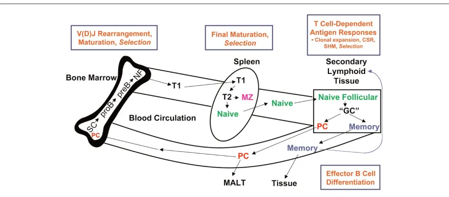
Under normal resting conditions, B cells follow a tightly regulated life cycle (Figure 1) with a large number of check points at indicated stages (dependent and antigen-independent selection) to prevent the development of autoimmunity [1]. In the bone marrow, B cells develop from stem cells through a series of precursor stages during which they rearrange their variable immunoglobulin (Ig) genes to generate a wide range of unique antigen-binding specificities. Immature CD10+ transitional B cells expressing surface
IgM/IgD emigrate from the bone marrow into the peripheral blood and then mature into naïve B cells. In the mouse, this occurs in the spleen, although the site of maturation in humans is not known [2]. After encountering antigen and T-cell help in follicles of secondary lymphoid organs, mature naïve B cells undergo germinal center (GC) reactions leading to their clonal expansion, somatic hypermutation of Ig gene rearrangements, and Ig heavy-chain class-switch recombination. Notably, these complex molecular processes are unique capacities of B cells and ensure specific higher avidity binding by the B-cell receptor (BCR) and also the production of antibodies with altered effector function. During the GC reaction, naïve antigen-specific B cells mature into either memory B cells or Ig-secreting plasma cells.
In mice, B1 B cells producing natural antibodies are impor-tant for the immediate defense against encapsulated bacteria. Whether they contribute to abnormalities of peripheral B cells in SLE [3] and primary Sjögren syndrome (pSS) [4] is not known. The reduced susceptibility of B1 B cells at mucosal sites after depletion by anti-CD20 therapy [5] suggests particular survival conditions of these cells in mice. The B1 B-cell equivalent subset and its role in human autoimmune
Review
B cells in autoimmunity
Thomas Dörner
1, Annett M Jacobi
1and Peter E Lipsky
21Charite Center 12 and 14, Charite University Hospital and DRFZ Berlin, Chariteplatz 01, 10098 Berlin, Germany
2National Institute of Arthritis and Musculoskeletal and Skin Diseases, National Institutes of Health, Bethesda, MD 20892-1560, USA
Corresponding author: Thomas Dörner, [email protected]
Published: 14 October 2009 Arthritis Research & Therapy2009, 11:247 (doi:10.1186/ar2780) This article is online at http://arthritis-research.com/content/11/5/247
© 2009 BioMed Central Ltd
diseases, however, remain to be delineated. Although there is an increase in CD5+B cells in both SLE and pSS, these cells
may represent an expended population of pre-naïve conven-tional B2 cells and not the human equivalent of B1 cells [6]. In addition, CD5 can be regarded as a B-cell activation marker in humans and there are no convincing data providing evidence that it can be used as a lineage marker as accepted in mice.
On the other hand, B2 B cells comprise the adaptive portion of humoral immune responses. B2 cells participate prefer-entially in T cell-dependent GC reactions, during which they can hypermutate their IgV gene rearrangements, switch Ig classes, and differentiate into memory cells and long-lived plasma cells. However, B2 cells can also be activated during T cell-independent responses [7]. B2 precursor cells are generated in the bone marrow and are subjected to central tolerance mechanisms. The immature survivors with functional BCRs leave the bone marrow and migrate to the periphery and are thought to be exposed to further selection (peripheral tolerance). Although it has been suggested that B2 B cells differentiate into either a mature follicular B-cell or a marginal zone (MZ) B-cell program [8], there are insufficient data that confirm this in humans. Alternatively, MZ B cells and B2 B cells can derive from precursor transitional 2 (T2) B cells that still need additional confirmation. Mouse models suggest that both B1 and MZ B-cell responses occur independently of T-cell help and B1 and MZ B cells are thought to be excluded from undergoing GC reactions. In conclusion, there appear to be substantial differences between mice and humans in terms of the specifics of the
differentiation of B-lineage cells. Moreover, the distinct contri-bution of B1, B2, and MZ B cell-equivalent subsets in human systemic autoimmunity as well as the role of T cell-indepen-dent (TI) and T cell-depencell-indepen-dent (TD) B-cell activation remain to be fully delineated. A more precise understanding of these processes in human autoimmunity would allow us a more targeted approach to treat specific autoimmune diseases.
Important for the interaction with T cells and the generation of GC reactions are a series of ligand-receptor interactions, including those mediated by CD154/CD40 and inducible co-stimulator ligand/inducible co-co-stimulator (ICOS-L/ICOS). Defects in these interactions have been shown to lead to hyper-IgM syndrome, resulting in impaired plasma cell and memory B-cell generation, including B lymphopenia and adult-onset common variable hypogamamglobulinemia, res-pectively [9,10]. Moreover, the presence of certain cytokines, such as interleukin (IL)-6, lymphotoxin-β, IL-4, and IL-21, is required in order to facilitate specific stages of B-cell differentiation by providing essential co-stimulatory signals.
[image:2.612.77.535.95.296.2]Because of the complexity of the abnormalities of immune regulation in systemic autoimmunity, a few key B-cell abnor-malities will be highlighted since they provide insight into the nature of perturbations of B-cell function that could contri-bute to autoimmunity, either in a causative manner or as a pathway that amplifies disease. In this regard, it is not clear whether the identified abnormalities of B cells in SLE are intrinsic or secondary to the disturbed internal milieu characteristic of SLE.
Figure 1
1. Defects in proper selection against
autoreactivity during B-cell development
A. Autoantibody production
Currently, the detailed nature of defects in immunologic check points during B-cell development in SLE is unclear. However, autoantibodies against double-stranded DNA (dsDNA) and nucleosomes, serologic hallmarks of lupus, and other nuclear antigens reflect the breakdown of immune tolerance. Notably, autoantibodies have been observed in some patients 6 to 10 years before the onset of the disease [11-13], indicating that the breakdown of tolerance may precede and not be secondary to disease activity in SLE. The processes of recombination and somatic hypermutation for affinity maturation in the bone marrow and subsequently in several lymphoid organs, respectively, are followed by strong selective pressures (‘check points’) under normal conditions to protect the body from the emergence of B cells with self-reactivity. In this regard, a number of check points in B-cell development have been proposed between immature and mature naïve B cells [14,15].
Examples of abnormalities in selection in patients with SLE have been reported. The idiotype defined by the 9G4 mono-clonal antibody that is encoded by VH4-34 heavy-chain gene rearrangements and frequently used by autoantibodies has been shown to circumvent negative selection in GCs in tonsils from SLE patients with subsequent expansion into the memory B-cell and plasma cell pool [16]. Moreover, the level of 9G4-expressing B cells as well as 9G4-containing anti-dsDNA antibodies is related to disease activity in SLE [17]. Compelling evidence of the failure of peripheral tolerance has also been found in an analysis of somatically mutated VH gene rearrangements encoding anti-DNA antibodies [18,19], where back-mutation clearly resulted in loss of binding activity. These data are consistent with the conclusion that most but possibly not all anti-DNA antibodies can arise by somatic mutation from precursors that lack autoantibody specificity. Therefore, the induction of some autoantibodies requires activation-induced cytidine deaminase for somatic hypermutation and Ig switching [20], and their development occurs de novo in the periphery fostered by a defect in peripheral tolerance.
B. Plasmablasts/plasma cells
Although we have acquired a broad knowledge in the use of autoantibodies in the diagnosis of autoimmunity, less is known about how autoantibodies are generated in humans. Clinical data suggest that in patients with active SLE there are short-lived plasmablasts that are CD27high/HLA-DRhigh
-producing anti-DNA antibodies and their frequency in the blood correlates with disease activity [17,21,22], whereas long-lived plasma cells (likely CD27highHLA-DRlow) produce
stable autoantibody titers, such as anti-Sm, -Ro, or -La, independently of disease activity. Experimental evidence from mice provided further evidence that plasmablasts and plasma cells reside in the spleen of lupus-prone mice [23] during
early stages of the disease and that only proliferating plasmablasts showed susceptibility to cyclophosphamide treatment. One critical question is whether these major subsets of Ig-producing cells are generated differently in autoimmunity [24].
Recently, in SLE patients, a more specific plasmablast subset that expresses HLA-DR very brightly and that clearly represents freshly generated plasmablasts was identified [22,25]. Notably, this cell fraction but not the remaining HLA-DRlow plasma cells correlated with lupus activity (Systemic
Lupus Erythematosus Disease Activity Index score) and anti-DNA titers, which indicates that they directly reflect the activity of SLE. Thus, there is an apparent defect of negative selection or regulation of newly generated plasmablasts in SLE, or alternatively, the process is normal but not appropriately terminated. In this context, proper regulation of antigen-specific plasmablast generation can be seen after secondary tetanus vaccination, when these cells appear in the circulation between days 6 to 8, but their appearance is downregulated as the immune response wanes [26]. The data on plasmablasts in murine and human lupus provide evidence that these cells are susceptible to anti-proliferative immunosuppressive agents, whereas more differentiated non-dividing plasma cells show resistance to these drugs [23]. Their distinct contribution to the disease and susceptibility to therapeutics remain to be assessed.
C. Memory B-cell compartments
There are a number of abnormalities of peripheral B-cell subsets in human SLE, including an expanded population of transitional B cells and post-switched CD27+B cells as well
as the appearance of a distinct population of CD27–/IgD–B
autoimmunity is emphasized by data showing that some memory B cells acquire polyreactivity and autoreactivity induced by somatic hypermutation [18,19,28].
D. Naïve B cells
Of further interest remains the question of the extent to which autoimmunity is already contained in the naïve B-cell repertoire of SLE before an encounter with antigen in the periphery. This is a difficult question to address in humans. SLE patients clearly have defects in check points which result in the appearance of an increased frequency of B cells capable of polyreactive autoantigen reactivity. Interestingly, a comprehensive study [15] showed that even inactive SLE patients fail to remove self-reactive BCRs expressed by naïve B cells. Consistent with this, earlier studies demonstrated that self-reactivity or loss of proper selection during early B-cell development from immature (CD10+ CD27– IgM+
B cells) to mature (CD10–CD27–IgM+B cells) naïve B cells
is a key feature in SLE [14]. Although these cells appeared to be influenced neither by antigen nor by T-cell help, the available data do not allow a conclusion about the detailed impact of a primary autoimmune predisposition or secondary influences such as cytokines or co-stimulatory signals. While it has been debated that polyreactive B cells can represent a source of autoantibody-secreting cells, formal evidence is lacking. On one hand, the IgM–/– mouse develops
‘auto-immunity’ [29]. By contrast, polyreactive IgG antibodies in SLE may impact on autoimmune manifestations [19]. Although it is tempting to conclude that SLE has defects in
‘check points against autoimmunity’ that explain the development of pathologic IgG autoantibodies, the possibility cannot be excluded that these ‘check points’ are active in SLE to prevent serious autoimmunity but are simply over-whelmed by chronic polyclonal B-cell activation. Alternatively, extensive cytokine release and enhanced co-stimulation may bypass these check points. It is also important to consider that B cells with a memory phenotype appear among CD27–
B cells previously considered to be exclusively naïve [30,31]. A subset of CD27–/IgD– B cells that express CD95 were
characterized as activated memory B cells with mutated and Ig-class-switched BCRs. Interestingly, their appearance correlated with lupus activity, which is in striking contrast to the CD27+ memory B-cell population that does not vary
much with disease activity. This CD27–memory subset was
found in patients with SLE but not in patients with infection. The role of this CD27– memory B-cell subset in the
patho-genesis of SLE remains to be determined.
E. Pre-naïve B cells
Recently, a unique pre-naïve peripheral B-cell population representing an intermediate stage between transitional and naïve B cells was identified at enhanced numbers in human SLE [6]. These cells are CD5+and express levels of CD38,
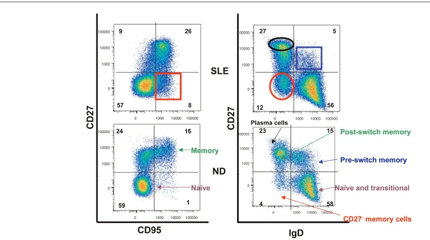
[image:4.612.58.486.97.336.2]CD10, CD9, and the ABCB1 transporter which are inter-mediate between transitional and naïve B cells. Therefore, these cells were considered pre-naïve B cells that could be induced to differentiate into naïve B cells in vitro. These pre-naïve B cells showed defective responses to BCR stimulation Figure 2
Major differences of peripheral B-cell compartments between systemic lupus erythematosus (SLE) patients and normal controls as shown in a representative dot plot. Please note the increased frequency of Ig-class-switched CD27+memory B cells and CD27–/IgD–B cells. ND, normal
but intact responses to CD40 ligation, whereas spontaneous apoptosis and cell death were enhanced compared with naïve B cells. Of note, B cell-activating factor/B-lymphocyte stimulator (BAFF/BLyS) was not an essential survival factor of these pre-naïve cells compared with naïve B cells. Finally, these cells had the capacity to differentiate into plasma cells after stimulation and the ability to function as antigen-presenting cells. The contribution of this population to lupus pathogenesis remains unknown. It is clear, however, that disturbances of early stages of B-lymphocyte homeostasis are also present in SLE and indicate that not only memory compartments are affected.
2. Aspects of disturbed immunregulation in
systemic lupus erythematosus
A. Regulation of B-cell activation by Fc receptors
The immune system has evolved to defend the organism against a variety of pathogens and applies threshold mecha-nisms for regulation. Independently of the co-stimulatory mechanisms, the coupling of activating and inhibitory receptors is able to use thresholds for immune cell activation. IgG immune complexes have long been recognized to have potent immunoregulatory functions ranging from a strong enhancement to complete suppression of antibody res-ponses [32] using selective engagement of specific FcγRs on discrete cell types, which result in either arrest or progression of an immune response. Four different classes of Fc receptors have been defined: FcγRI (CD64), FcγRII (CD32), FcγRIII (CD16), and FcγRIV [33]. Functionally, there are two different classes of Fc receptors: the activating and the
inhibitory receptors, which transmit their signals via immuno-receptor tyrosine-based activation (ITAMs) or immunoimmuno-receptor tyrosine-based inhibitory motifs (ITIMs). Co-expression of activating and inhibitory molecules on the same cell allows a balanced immune response, and the biochemical structure of IgG has substantial influence of the effects on Fc receptors, resulting in either a pro- or anti-inflammatory response [33].
B. The inhibitory Fcγγreceptor IIB
[image:5.612.85.538.105.314.2]The inhibitory Fcγreceptor IIB is part of the family of immune inhibitory receptors and its loss leads to autoimmunity and autoimmune disease [34,35]. FcγRIIB is a single-chain recep-tor that carries an ITIM in its cytoplasmic domain, a hallmark of this inhibitory protein family, and signals via inositol phosphatase SHIP (Src homology 2-containing inositol phosphatase) [36]. With the exception of T cells and natural killer cells, FcγRIIB is expressed on all cells of the immune system, including B cells, where it regulates activating signals delivered by immune complexes. As a consequence of its role in regulating BCR signals, which ultimately will decide whether a B cell will undergo proliferation and differentiation into an antibody-secreting plasma cell, FcγRIIB has been suggested to play an important role in maintaining peripheral tolerance [34,35]. The capacity of FcγRIIB to trigger B-cell apoptosis has been proposed to be another mechanism for controlling B-cell responses and maintaining self-tolerance. This hypothesis was supported by the generation of Fcγ RIIB-deficient mice that spontaneously develop a lupus-like disease characterized by the production of autoantibodies and premature death because of severe glomerulonephritis Figure 3
CD27+B cells with a memory phenotype are less susceptible to cyclophosphamide treatment in patients with systemic lupus erythematosus (SLE)
(n = 20). Severely active SLE patients undergoing monthly intravenous cyclophosphamide bolus therapy were followed for a period of 3 to 6 months. Whereas CD27–B cells and CD27++plasmablasts/plasma cells showed a decrease, the absolute numbers of CD27+memory B cells did
[37]. Recently, it was suggested that FcγRIIB co-ligation inhibits BLyS signaling and BLyS-R upregulation following BCR engagement [38].
C. Polymorphisms in the human FcγγRIIB promoter and autoimmunity
Polymorphisms in the human FcγRIIB promoter linked to lupus have been identified. One polymorphism leads to decreased transcription and surface expression of FcγRIIB on activated B cells of human lupus patients [39]. Another polymorphism in the transmembrane domain of FcγRIIB is linked to human lupus in several ethnic populations [33]. It has been suggested that this allelic variant of the inhibitory receptor loses its function because of the inability to associate with lipid rafts [40,41].
Autoreactive B cells can potentially be generated at several stages during B-cell development. There is accumulating evidence that FcγRIIB mediates its function during late antigen-dependent stages of B-cell maturation, thus representing a distal check point against autoimmunity [33]. Of note, FcγRIIB deficiency did not impact on early events in the bone marrow, such as receptor editing, nor did it prevent the development of IgM+ autoreactive B cells. After
class-switching to IgG, however, FcγRIIB was essential to prevent the expansion of autoreactive B cells and their maturation into plasma cells [33]. Considering the higher pathogenic potential of IgG compared with IgM antibody isotypes, this relatively late stage of FcγRIIB-mediated negative regulation has a major role in preventing the initiation of severe autoreactive processes.
Central check points, including receptor editing, deletion, and anergy of self-reactive BCR species, ensure that the majority of B cells with an autoreactive BCR are deleted in the bone marrow [42], which occurs independently of FcγRIIB. In contrast, autoreactive B cells can be generated de novoby somatic hypermutation, which is supported by the finding that many autoantibodies are encoded by somatically mutated VH gene rearrangements and switched Ig. Here, FcγRIIB might serve as the final barrier to prevent these B cells with potentially autoreactive BCR specificities from maturing into plasma cells.
3. Abnormalities of immune activation in
systemic lupus erythematosus
Co-stimulation results from a complex mix of factors involved in effective immune activation, which includes antigen presentation, the provision of soluble and insoluble co-stimulatory factors as well as the anatomic organization of secondary lymphoid organs, including the GCs. One important function of memory B cells is antigen presentation, which is facilitated by the expression of high-avidity BCR and also major histocompatibility complex class II molecules. Under sufficient co-stimulation, memory B cells can effec-tively present antigen primarily to memory T cells. In mice, MZ
B cells were even shown to provide co-stimulatory activating signals to naïve T cells [43]. In addition, CD80/CD86 expression on B cells has been shown to be indispensable for the activation of autoreactive T cells in a murine arthritis model [44]. The function of B cells as antigen-presenting cells has also been suggested by instructive data from animal models [45], in which a lupus-like disease developed when autoimmune-prone mice were reconstituted with B cells that lacked the ability to secrete Ig but not when they were deprived of B cells completely. These studies provided data on the possible role of antigen presentation by B cells and its pathogenic relevance.
Antigen presentation by B cells, in particular by memory B cells, may be important in the amplification and maintenance of autoimmunity after it has been initiated. Memory B-cell subsets in SLE [46] provide a sensitive pool of cells that immediately react to various stimuli, such as Toll-like receptor (TLR) ligands, IL-21, BAFF, IL-10, BCR activation, or co-stimulatory signals, resulting in the production of Ig-secreting cells. Although SLE memory B cells are mainly Ig-class-switched and show an activated phenotype, it remains to be determined whether these cells are also effective antigen-presenting cells. However, while the impact of individual stimuli is still a matter of debate, the decrease of IgM memory B cells [47] could represent the result of continuous memory B-cell activation by elevated BAFF, IL-21, or IL-10 levels [48-51].
[53,55] in myeloid cells suggests that BAFF may act as a kind of secondary cytokine translating mainly IFN and TNF effects.
An important immunoregulatory cytokine produced by B cells is IL-10, which is able to activate dendritic cells to be more effective antigen-presenting cells and, with the help of T cells, to enhance the differentiation of memory B cells into plasma cells in the presence of IL-2 and which may likely be an important factor of the immunoregulatory function of B cells [56]. Recently, it was suggested that triggering of TLRs can induce IL-10 production, which appears to play a role in protection from chronic inflammatory diseases. Therefore, immunoregulatory effects of B cells may result from their IL-10 production in autoimmune diseases. Such a regulatory function would have important implications for B-cell depletion therapies, which so far have not been substantiated by clinical trials. Whether low IL-10 levels after B-cell depletion ultimately lead to higher IFNγand TNFαexpression, translating into high BAFF levels, remains open.
With regard to the expression of co-stimulatory ligands involved in plasma cell and memory B-cell differentiation, it has been shown that T and B cells from SLE blood spon-taneously express CD154, which is an indicator that GCs in this disease abnormally release activated lymphocytes into the periphery and implies the presence of overactive GCs [57] or insufficient control mechanisms allowing an egress of premature memory B cells. Similarly, CD4+and CD8+T cells
from lupus patients show enhanced ICOS expression, whereas memory B cells downregulate ICOS-L, likely as a result of continuous interaction with T cells [58]. Since these interactions of the CD28 family are important for memory B-cell and plasma B-cell generation but not for the formation of GCs, they are consistent with the conclusion that there is overactivated adaptive immunity in SLE and that this repre-sents an important therapeutic target. All of these distur-bances of B-cell subsets in adults and children with lupus with a predominance of memory B cells may also contribute to the increases of plasmablasts during active lupus [21], which could be sufficiently blocked by anti-CD154 therapy [57]. Unfortunately, this therapy [59] had severe side effects that stopped the trials.
The role of the type I cytokine, IL-21, in the pathogenesis of SLE has been suggested by a number of recent findings. First, findings in both humans and mice have indicated an essential role for this cytokine in co-stimulating B cells to differentiate into plasma cells [29]. IL-21R–/– mice have a
diminished capacity to produce IgG1 in response to immuni-zation, whereas IL-21 transgenic mice develop hypergamma-globulinemia [60]. Overexpression of IL-21 is found in the BXSByaa and the sanroche murine models of SLE [61]. Finally, blocking of IL-21 activity successfully treats the manifestations of lupus in the MRL mouse, whereas crossing the BXSByaa mouse with the IL-21R–/– mouse prevents all
manifestations of SLE [62]. IL-21 is produced by CXCR5+
follicular helper T cells, which require ICOS stimulation for their generation [61]. These findings link ICOS and IL-21 in a definable pathway required for B-cell stimulation in secondary lymphoid organs and suggest that blocking IL-21 may be effective in human SLE. Formation of ectopic GCs has been identified in the kidneys of patients with SLE, in the salivary glands of patients with Sjögren syndrome, in the thymus in patients with myasthenia gravis, and in the central nervous system of patients with multiple sclerosis [4,58,63]. Although these aggregates of CD20+ B cells surrounded by T cells
and follicular dendritic cells have been found in only a fraction of patients, it is not known whether their formation is related to disease activity or T cell-dependent or -independent activation or whether they have the full capacity to select antigen-reactive cells and delete autoreactive B cells appropriately as in typical GCs.
Even though ectopic GCs have been linked to local over-activation of autoimmune B cells and plasma cells, conclusive evidence that they are required for the development of auto-immune diseases or are secondary to the deranged internal environment characteristic of these conditions has not been provided. Thus, no firm conclusion on the differences in the nature of classic GC versus ectopic GC in autoimmunity can be drawn since even classic GCs in tonsils from SLE patients were found to be defective in selection against 9G4 B cells as an indicator of anti-dsDNA activity [16].
An area of interest within the last decade has been the role of T cell-independent activation of B cells. While this can occur in vitro, ligating one of the receptors for BAFF/BLyS and APRIL, the transmembrane activator and calcium modulator and cyclophilin ligand interactor (TACI) [64-68], in only a subset of memory B cells [69], conclusive evidence concern-ing the role of this pathway in SLE is lackconcern-ing.
Another mechanism by which B cells can be activated in the absence of T cells is via TLR activation. TLRs are also known as ‘pathogen-associated molecular pattern receptors’ or ‘pattern recognition receptors’ and are expressed by nearly every cell in the body. TLR-7, TLR-8, and TLR-9 are the most important of these with respect to B-cell activation. Bacterial DNA is the natural ligand of TLR-9, and single-stranded RNA is the ligand of TLR-7 and TLR-8. All three receptor-ligand interactions apparently lead to activation of B cells by an
In conclusion, a complex interplay between a constantly over-activated immune system and apparent multiple abnormalities of B-cell development can be assumed in SLE. This permanent overactivation (of whatever cause) might over-whelm all possible check capacities of the immune system. Consistent with that, early check points before antigen or T-cell influences have been identified to be defective [14,15], classic GCs do not select properly [16], and ectopic GCs with a potential lack of selection are found in SLE that may permit the emergence of autoreactive cells.
4. Lessons from immune intervention trials
After therapeutic trials of anti-CD4 therapy in RA [73] and SLE [74] failed to show substantial clinical benefit, questions about the central role of CD4+ T cells were raised. Recentsuccess achieved by blocking T-cell co-stimulation with CTLA4Ig (abatacept) by antigen-presenting cells, including B cells, and the effects of blocking CD40/CD154 interactions on autoimmunity [57,59] have again implied a role for the regulatory interaction of immune cells in systemic auto-immune diseases. However, in SLE, the use of anti-CD154 (BG9588) led to some safety concerns with thromboembolic complications [59] in lupus nephritis, although clear immuno-logic effects were seen. Moreover, blockade of ICOS-L was shown in mice to reduce lupus nephritis [75].
The success of B cell-depleting therapy in ameliorating rheumatoid inflammation and joint destruction has docu-mented a role for B cells in RA but also in other autoimmune diseases, such as idiopathic thrombocytopenic purpura and SLE [76]. Interestingly, a reduction of expressed co-stimu-latory molecules such as CD80, CD86, and CD40L on T cells after B-cell depletion was observed in SLE [77], a reduced infiltration of CD68 macrophages was noted in the RA synovium [78], and an increase of regulatory T cells was seen in autoimmune thrombocytopenia [79]. It is clear from these clinical trial results that B cells together with other immune cells play important roles in autoimmunity. However, their role in the induction versus maintenance phase of the disease and the specific contributions of particular B-cell subsets have not been dissected in detail. Although previous data in lupus have shown that B-cell hyperactivity and the resulting autoantibody production are central elements of the immunopathogenesis of SLE, preliminary data of the use of rituximab as an anti-CD20 antibody in non-renal (EXPLORER trial) [80] and renal (LUNAR trial) SLE have reportedly failed the primary endpoints. Although it remains possible that anti-B-cell therapy is not sufficient to suppress lupus activity, a number of other confounding variables of SLE trial design may have substantially contributed (too short trial duration, allowance of substantial glucocorticoid usage, heterogeneity of the patient population, etc). Since a number of additional early trials in SLE, such as the use of abatacept and abetimus, have not provided convincing therapeutic effects, the most likely explanation is that lupus patients are hetero-geneous with regard to organ manifestations and
patho-physiology and require customized therapeutic strategies. Patients need to be stratified and characterized in detail prior to the choosing of a certain therapeutic approach. Parameters like the ‘interferon signature’, BLyS levels, and serologic and clinical findings should be considered and validated regarding their predictive value as biomarkers. In this regard, frequently detectable autoantibodies against DNA are produced by short-lived plasma cells versus anti-extractable nuclear antigens produced by apparently long-lived plasma cells. Given that both produce pathogenic autoantibodies, therapies should target both. Future assessments will be necessary to determine whether B cell-directed therapy can be effective clinically in SLE but also to test the hypothesis that specific B-cell abnormalities are essential for the pathogenesis of this disease. A further explanation of recent trial failure could be that the British Isles Lupus Assessment Group (BILAG) scoring system is less sensitive to changes in the BILAG B, which may preclude a sufficient discrimination of therapeutic success. Overall, the results of recent lupus trials challenge the rheumatology community to improve trial approaches in SLE, a patient population in need of improved therapies.
A different B cell-directed approach is targeting CD22 on B cells; CD22 is uniquely expressed on mature B cells but not on plasma cells or pre-B cells. The humanized anti-CD22 monoclonal antibody epratuzumab causes partial B-cell reduction in the blood, inhibits B-cell proliferation, and probably interferes with intracellular signaling. Preliminary results from an open-label phase IIa study indicate that epratuzumab is efficacious in SLE [81], with BILAG index scores improving by more than 50% in 77% of treated patients at week 6. A central question yet to be answered is how partial inhibition of B-cell activation may affect the clinical risk-benefit ratio as compared with total depletion as mediated by anti-CD20 antibodies. Partial inhibition might mean that B cells can still be activated by certain pathogens in this setting while epratuzumab (anti-CD22) inhibits activation of autoreactive B cells. A preferential reduction of naïve B cells in the peripheral blood under epratuzumab
This article is part of a special collection of reviews, The Scientific Basis of Rheumatology: A Decade of Progress, published to mark Arthritis Research &
Therapy’s 10th anniversary.
Other articles in this series can be found at: http://arthritis-research.com/sbr
treatment has been seen in clinical study. It has not been resolved whether this was caused by preferential depletion or apoptosis of naïve B cells or by enhanced migration of these cells from the blood into the tissue. In vitro data, however, suggested that this agent acts also by blocking proliferation of B cells [46], an effect that has been observed in patients with SLE but not in normal controls [46].
The current data are consistent with the conclusion that B-cell abnormalities in SLE can be targeted by cellular approaches, such as anti-B-cell therapy, but also inter-ventions on key cytokines, such as IFNα, or blocking co-stimulation. A critical question will be to identify a common denominator of B-cell activation as a target that allows sufficient and safe immune intervention.
Competing interests
TD declares associations with the following companies: Roche, Genentech, Immunomedics, UCB. AJ and PL declare that they have no competing interests.
References
1. Goodnow CC, Sprent J, Fazekas de St Groth B, Vinuesa CG: Cellular and genetic mechanisms of self tolerance and autoimmunity.Nature2005, 435:590-597.
2. Sims GP, Ettinger R, Shirota Y, Yarboro CH, Illei GG, Lipsky PE: Identification and characterization of circulating human transi-tional B cells.Blood2005, 105:4390-4398.
3. Milner EC, Anolik J, Cappione A, Sanz I: Human innate B cells: a link between host defense and autoimmunity?Springer Semin Immunopathol2005, 26:433-452.
4. Hansen A, Odendahl M, Reiter K, Jacobi AM, Feist E, Scholze J, Burmester GR, Lipsky PE, Dörner T: Diminished peripheral blood memory B cells and accumulation of memory B cells in the salivary glands of patients with Sjogren’s syndrome.
Arthritis Rheum2002, 46:2160-2171.
5. Gong Q, Ou Q, Ye S, Lee WP, Cornelius J, Diehl L, Lin WY, Hu Z, Lu Y, Chen Y, Wu Y, Meng YG, Gribling P, Lin Z, Nguyen K, Tran T, Zhang Y, Rosen H, Martin F, Chan AC: Importance of cellular microenvironment and circulatory dynamics in B cell immunotherapy.J Immunol2005, 174:817-826.
6. Lee J, Kuchen S, Fischer R, Chang S, Lipsky PE: Identification and characterization of a human CD5(+) pre-naive B cell pop-ulation.J Immunol2009, 182:4116-4126.
7. Won WJ, Laessing U, Bachmann M, Kearney J: Expression of CD36 by mouse marginal zone B cell. FASEB J 2005, 19: A358.
8. Tarlinton D: B-cell memory: are subsets necessary?Nat Rev Immunol2006, 6:785-790.
9. Peter HH, Warnatz K: Molecules involved in T-B co-stimulation and B cell homeostasis: possible targets for an immunologi-cal intervention in autoimmunity.Expert Opin Biol Ther2005, 5:S61-S71.
10. Warnatz K, Bossaller L, Salzer U, Skrabl-Baumgartner A, Schwinger W, van der Burg M, van Dongen JJ, Orlowska-Volk M, Knoth R, Durandy A, Draeger R, Schlesier M, Peter HH, Grim-bacher B: Human ICOS deficiency abrogates the germinal center reaction and provides a monogenic model for common variable immunodeficiency.Blood2006, 107:3045-3052. 11. Isenberg D, Rahman A: Systemic lupus erythematosus - 2005
annus mirabilis?Nat Clin Pract Rheumatol2006, 2:145-152. 12. van Gaalen FA, Linn-Rasker SP, van Venrooij WJ, de Jong BA,
Breedveld FC, Verweij CL, Toes RE, Huizinga TW: In undifferen-tiated arthritis autoantibodies to cyclic citrullinated peptides (CCP) predict progression to rheumatoid arthritis: a prospec-tive cohort study [abstract].Arthritis Rheum2003, 48:S108. 13. van Gaalen FA, Linn-Rasker SP, van Venrooij WJ, de Jong BA,
Breedveld FC, Verweij CL, Toes RE, Huizinga TW: Autoantibod-ies to cyclic citrullinated peptides predict progression to
rheumatoid arthritis in patients with undifferentiated arthritis -A prospective cohort study.Arthritis Rheum2004, 50:709-715. 14. Yurasov S, Wardemann H, Hammersen J, Tsuiji M, Meffre E, Pascual V, Nussenzweig MC: Defective B cell tolerance checkpoints in systemic lupus erythematosus.J Exp Med2005, 201:703-711. 15. Yurasov S, Tiller T, Tsuiji M, Velinzon K, Pascual V, Wardemann H,
Nussenzweig MC: Persistent expression of autoantibodies in SLE patients in remission.J Exp Med2006, 203:2255-2261. 16. Cappione A 3rd, Anolik JH, Pugh-Bernard A, Barnard J, Dutcher
P, Silverman G, Sanz I: Germinal center exclusion of autoreac-tive B cells is defecautoreac-tive in human systemic lupus erythemato-sus.J Clin Invest2005, 115:3205-3216.
17. Odendahl M, Jacobi A, Hansen A, Feist E, Hiepe F, Burmester GR, Lipsky PE, Radbruch A, Dörner T: Disturbed peripheral B lymphocyte homeostasis in systemic lupus erythematosus.J Immunol2000, 165:5970-5979.
18. Jahn S, Neimann B, Winkler T, Kalden JR, Vonbaehr R: Expan-sion of A B-lymphocyte clone producing Igm autoantibodies encoded by a somatically mutated V(H)J gene in the spleen of an autoimmune patient.Rheumatol Int1994, 13:187-196. 19. Zhang J, Jacobi AM, Wang T, Diamond B: Pathogenic
autoanti-bodies in systemic lupus erythematosus are derived from both self-reactive and non-self-reactive B cells. Mol Med
2008, 14:675-681.
20. Muramatsu M, Kinoshita K, Fagarasan S, Yamada S, Shinkai Y, Honjo T: Class switch recombination and hypermutation require activation-induced cytidine deaminase (AID), a poten-tial RNA editing enzyme.Cell2000, 102:553-563.
21. Jacobi AM, Odendahl M, Reiter K, Bruns A, Burmester GR, Rad-bruch A, Valet G, Lipsky PE, Dörner T: Correlation between cir-culating CD27(high) plasma cells and disease activity in patients with systemic lupus erythematosus.Arthritis Rheum
2003, 48:1332-1342.
22. Jacobi AM, Mei H, Hoyer BF, Mumtaz IM, Thiele K, Radbruch AH, Burmester GR, Hiepe F, Dörner T: HLA-DRhigh/CD27high
plas-mablasts indicate active disease in patients with SLE. Ann Rheum Dis2009, Feb 5. [Epub ahead of print].
23. Hoyer BF, Moser K, Hauser AE, Peddinghaus A, Voigt C, Eilat D, Radbruch A, Hiepe F, Manz RA: Short-lived plasmablasts and long-lived plasma cells contribute to chronic humoral autoim-munity in NZB/W mice.J Exp Med2004, 199:1577-1584. 24. Radbruch A, Muehlinghaus G, Luger EO, Inamine A, Smith KG,
Dörner T, Hiepe F: Competence and competition: the chal-lenge of becoming a long-lived plasma cell.Nat Rev Immunol
2006, 6:741-750.
25. Mei HE, Yoshida T, Sime W, Hiepe F, Thiele K, Manz RA, Rad-bruch A, Dörner T: Blood-borne human plasma cells in steady state are derived from mucosal immune responses. Blood
2009, 113:2461-2469.
26. Odendahl M, Mei H, Hoyer BF, Jacobi AM, Hansen A, Muehling-haus G, Berek C, Hiepe F, Manz R, Radbruch A, Dörner T: Gen-eration of migratory antigen-specific plasma blasts and mobilization of resident plasma cells in a secondary immune response.Blood2005, 105:1614-1621.
27. Dörner T, Lipsky PE: Molecular basis of immunoglobulin vari-able region gene usage in systemic autoimmunity.Clin Exp Med2005, 4:159-169.
28. Tiller T, Tsuiji M, Yurasov S, Velinzon K, Nussenzweig MC, Warde-mann H: Autoreactivity in human IgG(+) memory B cells.
Immunity2007, 26:205-213.
29. Ehrenstein MR, Cook HT, Neuberger MS: Deficiency in serum immunoglobulin (Ig)M predisposes to development of IgG autoantibodies.J Exp Med2000, 191:1253-1257.
30. Jacobi AM, Reiter K, Mackay M, Aranow C, Hiepe F, Radbruch A, Hansen A, Burmester GR, Diamond B, Lipsky PE, Dörner T: Acti-vated memory B cell subsets correlate with disease activity in systemic lupus erythematosus - Delineation by expression of CD27, IgD, and CD95.Arthritis Rheum2008, 58:1762-1773. 31. Wei C, Anolik J, Cappione A, Zheng B, Pugh-Bernard A, Brooks J,
Lee EH, Milner EC, Sanz I: A new population of cells lacking expression of CD27 represents a notable component of the B cell memory compartment in systemic lupus erythematosus.J Immunol2007, 178:6624-6633.
33. Nimmerjahn F, Ravetch JV: Fc-receptors as regulators of immu-nity.Adv Immunol2007, 96:179-204.
34. Bolland S, Ravetch JV: Spontaneous autoimmune disease in Fc gamma RIIB-deficient mice results from strain-specific epistasis.Immunity2000, 13:277-285.
35. Bolland S, Ravetch JV: Inhibitory pathways triggered by ITIM-containing receptors.Adv Immunol1999, 72:149-177. 36. Dörner T, Lipsky PE: Signalling pathways in B cells:
implica-tions for autoimmunity.Curr Top Microbiol Immunol 2006, 305: 213-240.
37. Takai T, Ono M, Hikida M, Ohmori H, Ravetch JV: Augmented humoral and anaphylactic responses in Fc gamma RII-defi-cient mice.Nature1996, 379:346-349.
38. Crowley JE, Stadanlick JE, Cambier JC, Cancro MP: Fc gamma RIIB signals inhibit BLyS signaling and BCR-mediated BLyS receptor up-regulation.Blood2009, 113:1464-1473.
39. Blank MC, Stefanescu RN, Masuda E, Marti F, King PD, Redecha PB, Wurzburger RJ, Peterson MG, Tanaka S, Pricop L: Decreased transcription of the human FCGR2B gene medi-ated by the-343 G/C promoter polymorphism and association with systemic lupus erythematosus.Hum Genet 2005, 117: 220-227.
40. Floto RA, Clatworthy MR, Heilbronn KR, Rosner DR, MacAry PA, Rankin A, Lehner PJ, Ouwehand WH, Allen JM, Watkins NA, Smith KG: Loss of function of a lupus-associated Fc gamma RIIb polymorphism through exclusion from lipid rafts. Nat Med2005, 11:1056-1058.
41. Floto RA, Clatworthy MR, Heilbronn KR, Rosner DR, MacAry PA, Rankin A, Lehner PJ, Ouwehand WH, Allen JM, Watkins NA, Smith KG: A polymorphism of the inhibitory receptor, Fc gamma RIIb, prevents its access to lipid rafts and alters macrophage responses to immune complexes and opsonized bacteria.Clin Immunol2005, 115:S214.
42. Meffre E, Davis E, Schiff C, Cunningham-Rundles C, Ivashkiv LB, Staudt LM, Young JW, Nussenzweig MC: Circulating human B cells that express surrogate light chains and edited receptors.
Nat Immunol2000, 1:207-213.
43. Attanavanich K, Kearney JF: Marginal zone, but not follicular B cells, are potent activators of naive CD4 T cells.J Immunol
2004, 172:803-811.
44. O’Neill SK, Cao YX, Hamel KM, Doodes PD, Hutas G, Finnegan A: Expression of CD80/86 on B cells is essential for autoreac-tive T cell activation and the development of arthritis. J Immunol2007, 179:5109-5116.
45. Chan OTM, Hannum LG, Haberman AM, Madaio MP, Shlomchik MJ: A novel mouse with B cells but lacking serum antibody reveals an antibody-independent role for B cells in murine lupus.J Exp Med1999, 189:1639-1647.
46. Jacobi AM, Goldenberg DM, Hiepe F, Radbruch A, Burmester GR, Dörner T: Differential effects of epratuzumab on periph-eral blood B cells of patients with systemic lupus erythemato-sus vererythemato-sus normal controls.Ann Rheum Dis2008, 67:450-457. 47. Wehr C, Eibel H, Masilamani M, Illges H, Schlesier M, Peter HH, Warnatz K: A new CD21(low) B cell population in the periph-eral blood of patients with SLE.Clin Immunol2004, 113: 161-171.
48. Briere F, Servetdelprat C, Bridon J, Saintremy JM, Banchereau J: Human interleukin-10 induces naive surface-immunoglobulin D+ (Sigd(+)) B-cells to secrete Igg1 and Igg3. J Exp Med
1994, 179:757-762.
49. Litinskiy MB, Nardelli B, Hilbert DM, He B, Schaffer A, Casali P, Cerutti A: DCs induce CD40-independent immunoglobulin class switching through BLyS and APRIL.Nat Immunol2002, 3:822-829.
50. Pène J, Gauchat JF, Lécart S, Drouet E, Guglielmi P, Boulay V, Delwail A, Foster D, Lecron JC, Yssel H: Cutting edge: IL-21 is a switch factor for the production of IgG(1) and IgG(3) by human B cells.J Immunol2004, 172:5154-5157.
51. Ettinger R, Sims GP, Fairhurst AM, Robbins R, da Silva YS, Spolski R, Leonard WJ, Lipsky PE: IL-21 induces differentiation of human naive and memory B cells into antibody-secreting plasma cells.J Immunol2005, 175:7867-7879.
52. Ronnblom L, Alm GV: A pivotal role for the natural interferon alpha-producing cells (plasmacytoid dendritic cells) in the pathogenesis of lupus.J Exp Med2001, 194:F59-F63. 53. Lindh E, Lind SM, Lindmark E, Hässler S, Perheentupa J, Peltonen
L, Winqvist O, Karlsson MC: AIRE regulates T-cell-independent
B-cell responses through BAFF. Proc Natl Acad Sci U S A
2008, 105:18466-18471.
54. Groom J, Kalled SL, Cutler AH, Olson C, Woodcock SA, Schnei-der P, Tschopp J, Cachero TG, Batten M, Wheway J, Mauri D, Cavill D, Gordon TP, Mackay CR, Mackay F: Association of BAFF/BLyS overexpression and altered B cell differentiation with Sjogren’s syndrome.J Clin Invest2002, 109:59-68. 55. Ohata J, Zvaifler NJ, Nishio M, Boyle DL, Kalled SL, Carson DA,
Kipps TJ: Fibroblast-like synoviocytes of mesenchymal origin express functional B cell-activating factor of the TNF family in response to proinflammatory cytokines.J Immunol2005, 174: 864-870.
56. Fillatreau S, Gray D, Anderton SM: Not always the bad guys: B cells as regulators of autoimmune pathology. Nat Rev Immunol2008, 8:391-397.
57. Grammer AC, Slota R, Fischer R, Gur H, Girschick H, Yarboro C, Illei GG, Lipsky PE: Abnormal germinal center reactions in sys-temic lupus erythematosus demonstrated by blockade of CD154-CD40 interactions.J Clin Invest2003, 112:1506-1520. 58. Hutloff A, Büchner K, Reiter K, Baelde HJ, Odendahl M, Jacobi A,
Dörner T, Kroczek RA: Involvement of inducible costimulator in the exaggerated memory B cell and plasma cell generation in systemic lupus erythematosus. Arthritis Rheum 2004, 50: 3211-3220.
59. Boumpas DT, Furie R, Manzi S, Illei GG, Wallace DJ, Balow JE, Vaishnaw A; BG9588 Lupus Nephritis Trial Group: A short course of BG9588 (anti-CD40 ligand antibody) improves serologic activity and decreases hematuria in patients with proliferative lupus glomerulonephritis.Arthritis Rheum2003, 48:719-727. 60. Ettinger R, Kuchen S, Lipsky PE: Interleukin 21 as a target of
intervention in autoimmune disease.Ann Rheum Dis2008, 67: 83-86.
61. Vinuesa CG, Cook MC, Angelucci C, Athanasopoulos V, Rui L, Hill KM, Yu D, Domaschenz H, Whittle B, Lambe T, Roberts IS, Copley RR, Bell JI, Cornall RJ, Goodnow CC: A RING-type ubiq-uitin ligase family member required to repress follicular helper T cells and autoimmunity.Nature2005, 435:452-458. 62. Ettinger R, Kuchen S, Lipsky PE: The role of IL-21 in regulating
B-cell function in health and disease.Immunol Rev2008, 223: 60-86.
63. Stott DI, Hiepe F, Hummel M, Steinhauser G, Berek C: Antigen-driven clonal proliferation of a cells within the target tissue of an autoimmune disease - The salivary glands of patients with Sjogren’s syndrome.J Clin Invest1998, 102:938-946. 64. Salzer U, Chapel HM, Webster AD, Pan-Hammarström Q,
Schmitt-Graeff A, Schlesier M, Peter HH, Rockstroh JK, Schnei-der P, Schäffer AA, Hammarström L, Grimbacher B: Mutations in TNFRSF13B encoding TACI are associated with common vari-able immunodeficiency in humans.Nat Genet 2005, 37: 820-828.
65. Salzer U, Grimbacher B: TACItly changing tunes: farewell to a yin and yang of BAFF receptor and TACI in humoral immu-nity? New genetic defects in common variable immunodefi-ciency.Curr Opin Allergy Clin Immunol2005, 5:496-503. 66. Salzer U, Chapel HM, Webster AD, Pan-Hammarström Q,
Schmitt-Graeff A, Schlesier M, Peter HH, Rockstroh JK, Schnei-der P, Schäffer AA, Hammarström L, Grimbacher B: Mutations in TNFRSF13B encoding TACI are associated with common vari-able immunodeficiency in humans.Nat Genet 2005, 37: 820-828.
67. Castigli E, Wilson SA, Garibyan L, Rachid R, Bonilla F, Schneider L, Geha RS: TACI is mutant in common variable immunodefi-ciency and IgA defiimmunodefi-ciency.Nat Genet2005, 37:829-834. 69. Castigli E, Wilson SA, Scott S, Dedeoglu F, Xu S, Lam KP, Bram
RJ, Jabara H, Geha RS: TACI and BAFF-R mediate isotype switching in B cells.J Exp Med2005, 201:35-39.
69. Evans CH, Ghivizzani SC, Oligino TJ, Robbins PD: Gene therapy for autoimmune disorders.J Clin Immunol2000, 20:334-346. 70. Christensen SR, Kashgarian M, Alexopoulou L, Flavell RA, Akira S,
Shlomchik MJ: Toll-like receptor 9 controls anti-DNA autoanti-body production in murine lupus. J Exp Med2005, 202: 321-331.
72. Bernasconi NL, Traggiai E, Lanzavecchia A: Maintenance of serological memory by polyclonal activation of human memory B cells.Science2002, 298:2199-2202.
73. Burmester GR, Horneff G, Emmrich F, Kalden JR: Treatment of rheumatoid-arthritis with an anti-Cd4 monoclonal-antibody.
Arthritis Rheum1991, 34:129-140.
74. Hiepe F, Volk HD, Apostoloff E, Vonbaehr R, Emmrich F: Treat-ment of severe systemic lupus-erythematosus with anti-Cd4 monoclonal-antibody.Lancet1991, 338:1529-1530.
75. Iwai H, Abe M, Hirose S, Tsushima F, Tezuka K, Akiba H, Yagita H, Okumura K, Kohsaka H, Miyasaka N, Azuma M: Involvement of inducible costimulator-B7 homologous protein costimula-tory pathway in murine lupus nephritis.J Immunol2003, 171: 2848-2854.
76. Dörner T, Burmester GR: New approaches of B-cell-directed therapy: beyond rituximab.Curr Opin Rheumatol2008, 20: 263-268.
77. Sfikakis PP, Boletis JN, Lionaki S, Vigklis V, Fragiadaki KG, Iniotaki A, Moutsopoulos HM: Remission of proliferative lupus nephri-tis following B cell depletion therapy is preceded by down-regulation of the T cell costimulatory molecule CD40 ligand: an open-label trial.Arthritis Rheum2005, 52:501-513. 78. Thurlings RM, Vos K, Wijbrandts CA, Gerlag DM, Tak PP:
Dynamics of the synovial B cell response to rituximab therapy in rheumatoid arthritis: evaluation of serial synovial biopsies during the first four months of treatment. Ann Rheum Dis
2007, 66:123.
79. Stasi R, Del Poeta G, Stipa E, Evangelista ML, Trawinska MM, Cooper N, Amadori S: Response to B-cell-depleting therapy with rituximab reverts the abnormalities of T-cell subsets in patients with idiopathic thrombocytopenic purpura. Blood
2007, 110:2924-2930.
80. Merrill JT, Neuwelt CM, Wallace DJ, Shanahan JC, Latinis KM, Oates JC, Gordon C, Hsieh H, Brunetta P: Efficacy and safety of rituximab in patients with moderately to severely active sys-temic lupus erythematosus (SLE): results from the random-ized, double-blind phase II/III study EXPLORER [abstract].
Arthritis Rheum2008, 58:4029-4030.