N A N O E X P R E S S
Open Access
Conversion of rod-shaped gold nanoparticles to
spherical forms and their effect on biodistribution
in tumor-bearing mice
Yasuyuki Akiyama
1, Takeshi Mori
1, Yoshiki Katayama
1,2,3and Takuro Niidome
1,2,3*Abstract
Gold nanorods that have an absorption band in the near-infrared region and a photothermal effect have been used as nanodevices for near-infrared imaging and thermal therapy. Choice of the optimal shape of gold nanorods which relates optical properties andin vivobiodistribution is important for their applications. In the present study, to investigate the relationship between the shape of gold nanorods and their biodistribution after intravenous injection, we first prepared two types of gold nanorods that had distinct aspect ratios but had the same volume, zeta potential, and PEG density on the gold surface. Biodistributions of the two types of gold nanorods after intravenous injection into tumor-bearing mice were then compared. Although a slight difference in accumulation in the spleen was observed, no significant difference was observed in the liver, lung, kidney, and tumors. These results suggest that biodistribution of the gold nanorods in the aspect ratio range of 1.7 to 5.0, diameter of 10 to 50 nm, and volume of approximately 4 × 10³nm3was dependent mainly on surface characteristics, PEG density, and zeta potential.
Keywords:Aspect ratio, Gold nanoparticles, Gold nanorods, Laser, Biodistribution
Background
Gold nanorods are tiny, rod-shaped gold particles. They have two distinctive different absorption bands derived from the transverse and longitudinal surface plasmon resonance of free electrons in the visible and near-infrared (NIR) regions, respectively [1,2]. Gold nanorods absorb NIR light, which is suitable for in vivo applica-tions such as imaging and for photoradiation therapy be-cause of maximal penetration of light into tissues [3]. Gold nanorods have a photothermal effect, i.e., the adsorbed light energy is converted to heat [4]. Therefore, gold nanorods are expected to be contrast agents for NIR imaging and exothermic nanodevices for photother-mal therapy.
Gold nanorods are prepared in the presence of the cat-ionic detergent 1-hexadecyltrimethylammonium chloride (cetyltrimethylammonium chloride; CTAB), which acts
as a stabilizer of gold nanorods. To apply gold nanorods as medical nanodevices, biocompatible gold nanorods have been prepared by coating with phosphatidylcholine [5] or by modifying gold nanorods with polyethylene gly-col (PEG) [6,7]. PEG-modified gold nanorods show high dispersion stability, high circulation stability in the blood after intravenous injection into mice [7], and accumula-tion in tumors mediated by the enhanced permeability and retention (EPR) effect [8,9]. The PEG-modified gold nanorods have been applied to NIR imaging and photo-dynamic/photothermal therapy of tumors [8,10-13].
The size and aspect ratio of gold nanorods are import-ant factors of their uptake into cells [14-16] and biodis-tribution [17-20]. Arnida et al. reported that rod-shaped gold nanoparticles accumulated more efficiently in tumors and less efficiently in the liver compared with sphere-shaped gold nanoparticles [20]. However, the compared gold nanorods and gold nanospheres had different volumes and zeta potentials that affected cellular uptake (e.g., phagocytosis) [21]. Therefore, if we examine the effect of the geometry of gold nanoparticles on biodistribution, other parameters should be equivalent.
* Correspondence:[email protected]
1
Department of Applied Chemistry, Faculty of Engineering, Kyushu University, 744 Motooka, Nishi-ku, Fukuoka 819-0395, Japan
2
Center for Future Chemistry, Kyushu University, 744 Motooka, Nishi-ku, Fukuoka 819-0395, Japan
Full list of author information is available at the end of the article
In the present study, we prepared two types of gold nanorods that had distinct aspect ratios but had an identi-cal volume, zeta potential, and surface structure. Biodistri-butions of these nanorods after intravenous injection into tumor-bearing mice were then compared.
Methods Animals
Animal experiments were undertaken according to the Guidelines for Animal Care and Use Committee, Kyushu University (Fukuoka, Japan). Male ddY mice (Kyudo Co., Ltd., Saga, Japan) aged 5 to 6 weeks were used. Mice were maintained in a temperature-controlled environ-ment at 24°C with a 12-h light-dark cycle. They were provided with drinking water and foodad libitum.
Preparation of gold nanorods of lower aspect ratio from higher ones
Gold nanorods prepared by our research team were obtained from Mitsubishi Materials Corp. (Tokyo, Japan) and Dai Nippon Toryo Co. Ltd. (Tokyo, Japan) [22]. The mean length and width of the as-obtained gold nanorods were 49.6 ± 9.7 nm and 10.6 ± 3.2 nm, respectively (aspect ratio, 5.0 ± 1.2). A suspension of gold nanorods containing CTAB was stored at a constant temperature (37°C). Gold nanorods were modified with PEG chains. Briefly, the gold nanorods in the suspension were centrifuged at 14,000 ×g for 10 min at room temperature, decanted, and re-suspended in water to remove excess CTAB. Thiol-terminated mPEG solution (molecular weight approxi-mately 5,000 Da; NOF Corporation, Tokyo, Japan) was added to the gold nanorod suspension at a PEG/gold molar ratio of 1.0. The mixture of PEG solution and gold nanorod suspension at a final gold concentration of 1 mM was stirred for 24 h at room temperature. NIR pulsed laser light (Nd:YAG 1064 nm, 750 mW, 20 Hz, approximately 6 mm in beam diameter) irradiated the mixture for 5 min. The mean length and width of gold nanorods irradiated with the NIR pulsed laser light were 27.0 ± 8.2 nm and 17.2 ± 3.7 nm, respectively (aspect ratio, 1.7 ± 0.9). A con-trol mixture was not irradiated with the NIR pulsed laser light. These mixtures were stirred for another 24 h at room temperature and centrifuged twice at 14,000 ×g for 10 min at room temperature, decanted, and re-suspended in water to remove remaining CTAB and excess PEG reagent.
Characterization of PEG-modified gold nanorods
The absorption spectra of gold nanorods in the visible-to-NIR light regions were measured with a V-670 spectropho-tometer (Jasco, Tokyo, Japan). Gold nanorods and the PEG layer on the gold nanorods were imaged with a transmis-sion electron microscope (JEM-2010, JEOL, Tokyo, Japan) after staining the PEG layer with 1% phosphotungstic acid
[23]. Particle size distribution, surface area, and volume were calculated from transmission electron microscopy (TEM) images of 100 particles. Amounts of PEG modified on the gold nanorods were evaluated by elemental analyses according to our previous publication [24]. Zeta potentials of gold nanorod suspensions in 0.2 M sodium phosphate buffer (pH 7.4) were evaluated using a Malvern Zetasizer Nano ZS (Malvern, Worcestershire, UK).
Biodistribution of gold nanorods
Tumor-bearing mice were used in biodistribution experiments and were prepared as follows: Colon-26 cells: mouse colon carcinoma cells were cultured in RPMI 1640 medium containing 10% fetal bovine serum, 100 U/mL penicillin, 100 μg/mL streptomycin, and 0.25μg/mL amphotericin B. Cells were incubated at 37°C under a 5% CO2-humidified atmosphere. Incubated colon-26 cells were injected subcutaneously into the abdo-men at 2 × 10⁶cells in 100μL of Hank's balanced salt so-lution per mouse (ddY, male, 5 weeks, 29 to 35 g). They were allowed to grow for 5 days, wherein the tumors reached approximately 5 to 7 mm in diameter. PEG-modified gold nanorods were injected into tumor-bearing mice via the tail vein. Doses of PEG-modified gold nanor-ods with an aspect ratio of 5.0 and PEG-modified gold nanorods with an aspect ratio of 1.7 were at 1.01 and 1.05 mM of gold concentration in 10μL/g body weight of 5% glucose solution, respectively. Mice were sacrificed 72 h after injection. Several hundred microliters of blood were collected from the heart and immediately mixed with 1 mg ethylenediamine tetra-acetic acid. The liver, lung, spleen, and kidney, as well as tumors, were collected. These samples were added to approximately 4 mL aqua regia and then heated overnight at 90°C to 100°C. Samples were diluted in 0.5 M HCl to the appropriate volume. Gold concentrations were quantified by inductively coupled plasma mass spectrometry (ICP-MS) using an Agilent 7500c system (Agilent. Tokyo, Japan). ICP-MS measurement was done with thallium as the internal standard.
Results and discussion
Preparation of PEG-modified gold nanorods with distinct aspect ratios
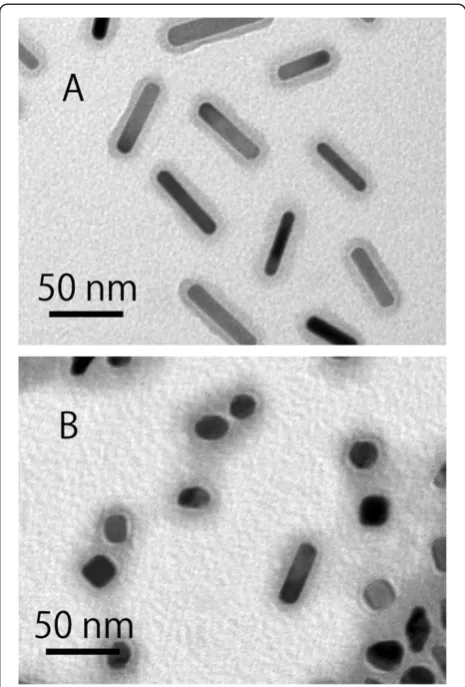
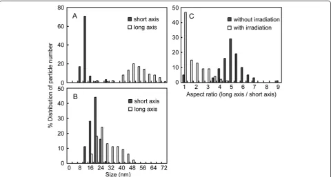
nanorods in the NIR region decreased and shifted to-wards a shorter wavelength (Figure 1). TEM images revealed that the irradiated gold nanorods showed mainly spherical or elliptical shapes and partly rod shapes (Figure 2). For gold nanorods with and without irradiation, PEG modified on the gold nanorods was confirmed from a gray layer of PEG stained with phos-photungstic acid. Particle sizes of gold nanorods in the TEM images were measured; then, their distributions were plotted (Figures 3A, B). Laser irradiation made the size of the short axis of the gold nanorod increase and the size of the long axis decrease. As a result, the size distribution of the short and long axes overlapped. As-pect ratios were calculated and then plotted (Figure 3C). Half of the irradiated gold nanorods were transformed to a spherical shape (aspect ratio, 1.0). The other irra-diated gold nanorods were transformed to an elliptical shape.
Several characteristics of the gold nanorods are summar-ized in Table 1. The particle length of the short axis and long axis increased from 10.6 to 17.2 nm and decreased from 49.6 to 27.0 nm by laser irradiation, respectively. Laser irradiation made the aspect ratio decrease from 5.0 to 1.7. The surface area and volume of the gold nanorods were calculated on the assumption that a nanoparticle with an aspect ratio of 1.0 was a sphere and a nanorod with an aspect ratio >1.0 was a cylinder with a hemisphere domain at both ends. The surface area decreased from 1.61 × 10³nm2to 1.42 × 10³nm2by laser irradiation. The volume did not change significantly after irradiation. Previ-ously, it was reported that irradiation of gold nanorods by pulsed laser light caused fragmentation into smaller particles [26] or fusion into larger particles [27,28]. However, in the irradiation condition described here,
neither fragmentation nor fusion of the gold nanorods was observed in TEM images. This was probably be-cause the intensity of the pulsed laser light was not sufficient to induce fragmentation of the gold nanor-ods, and the PEG layer on the gold nanorods disturbed the fusion between gold nanorods.
[image:3.595.305.538.88.432.2]The amount of modified PEG on gold nanorods was measured by elemental analyses (Table 1) [24]. The number of modified PEG molecules on one particle decreased from 9.50 × 10² to 8.25 × 10² by laser irradi-ation. The actual release of PEG from the gold surface to supernatant has been already confirmed at a similar condition in our previous report [24]. However, the area occupied by one PEG molecule as PEG density on the gold nanorods and the zeta potentials of the gold nanor-ods did not change after laser irradiation. These data suggested that the laser irradiation of gold nanorods resulted in transformation from a rod shape to a spher-ical shape without changing the volume, PEG density, or zeta potential.
[image:3.595.57.291.508.694.2]Figure 1Absorption spectra of PEG-modified gold nanorods treated without (A) and with (B) near-infrared pulsed laser light.
Biodistribution in tumor-bearing mice
We compared the biodistribution of PEG-modified gold nanorods with distinct aspect ratios but with the same volume, PEG density, and zeta potentials. Prepared gold nanorods with aspect ratios of 5.0 and 1.7 were injected intravenously into tumor-bearing mice to which colon-26 cells were subcutaneously injected. Mice were sacri-ficed 72 h after the injection, and the collected organs were dissolved using aqua regia. At this time point, it has been already shown that little amount of PEG-modified gold nanorods was observed in the blood and that most gold nanorods were distributed to organs [7,9]. Gold concentrations in the samples were measured by ICP-MS. Both types of injected gold nanorods accu-mulated mainly in the liver and spleen (Figure 4A). Most
gold nanorods with aspect ratios of 5.0 and 1.7 were probably cleared by the reticuloendothelial system (RES) in the liver and spleen [28].
[image:4.595.58.539.89.346.2]Figure 4B shows the concentration of accumulated gold nanorods in the organs as percentage of injected dose per gram of tissue or milliliter of blood. The con-centration is an important parameter for photothermal therapy and bioimaging mediated by gold nanorods. Ac-cumulation of gold nanorods with an aspect ratio of 1.7 in the spleen was slightly lower than that of gold nanor-ods with an aspect ratio of 5.0, although statistical sig-nificance was low (0.1 >p> 0.05; Figure 4B). In the case of the liver, lung, and kidney, no difference between the gold nanorods with aspect ratios of 1.7 and 5.0 was observed. No difference was observed in the tumors,
[image:4.595.58.539.600.725.2]Figure 3Size distribution and aspect ratios of PEG-modified gold nanorods.Size distribution of PEG-modified gold nanorods treated without (A) and with (B) near-infrared pulsed laser light. Closed and open bars show the size distributions of the short and long axes of gold nanorods, respectively. (C) Aspect ratios of PEG-modified gold nanorods without (closed bars) and with (open bars) pulsed laser light.
Table 1 Characteristics of PEG-modified gold nanorods treated without and with near-infrared pulsed laser light
Without irradiation With irradiation
Particle size of short axis (nm) 10.6 ± 3.2 17.2 ± 3.7
Particle size of long axis (nm) 49.6 ± 9.7 27.0 ± 8.2
Aspect ratio 5.0 ± 1.2 1.7 ± 0.9
Surface area (×10−3nm2) 1.61 ± 0.43 1.42 ± 0.40
Volume (×10−3nm3) 4.02 ± 1.81 4.32 ± 1.70a
Number of modified PEG on one particle (×10−2) 9.50 ± 4.28 8.25 ± 3.25
Area occupied by one PEG molecule (nm2) 1.82 ± 0.29 1.82 ± 0.33a
Zeta potential (mV) 0.48 ± 1.66 1.83 ± 3.34a
suggesting that a change in the shape of the gold nanor-ods was not sufficient to influence the EPR effect in the tumors. The slight difference observed in the spleen may have been due to physical filtration in this organ as observed for larger particles (>200 nm) [29,30]. It may not have been due to changes in the contribution of the RES in the spleen because a significant difference in the accumulation in the liver (where many Kupffer cells (macrophage-like cells) constitute the RES at sinusoids) was not observed. These results suggested that the shape of the gold nanorods barely affected the biodistribution of gold nanorods after intravenous injection into tumor-bearing mice except for that in the spleen (though the change in biodistribution was slight). Previously, we com-pared biodistributions of several types of gold nanorods,
which have different zeta-potentials and PEG densities on the gold surface but have the same aspect ratio [8,9]. In those studies, the surface structure of the gold nanorods strongly affected their biodistributions after intravenous injection. Therefore, it can be concluded that the biodistri-bution would be more affected primarily by the surface characteristics of the gold nanorods than the aspect ratio.
Conclusions
In the present study, the shape of the original gold nanorods was changed by irradiation with NIR light to a spherical form. Then, we obtained two types of PEG-modified gold nanorods with distinct aspect ratios but with the same volume, PEG density, and zeta potential. The change in the aspect ratio of the gold nanorods did not affect the biodistribution appreciably. These results suggest that the biodistribution of gold nanorods in the aspect ratio range of 1.7 to 5.0 and size of 10 to 50 nm at a volume of approximately 4 × 10³nm3is dependent mainly on the surface characteristics, PEG density, and zeta potential. If the application of gold nanorods to therapeutic use is considered, the gold nanorods circu-lating in the blood or other tissues such as tumors are irradiated with NIR light. Irradiation with the pulsed laser light will induce the shape change of the gold nanorods and release of molecules bound on the gold nanorods without changing their biodistribution. On the other hand, irradiation with continuous wave laser light can heat the gold nanorods continuously without changing the shape of the gold nanorods [24]. Thus, combinations of gold nanorods and different modes of light irradiation pro-vide us various drug delivery/release and heating systems, and it will be an advantage of gold nanorods over other types of nanoparticles. The results obtained in this study will be used as basic information on thein vivoapplication not only of gold nanorods with various aspect ratios, but also of other types of anisotropic nanoparticles.
Abbreviations
CTAB: cetyltrimethylammonium chloride 1-hexadecyltrimethylammonium chloride; EPR: enhanced permeability and retention; ICP-MS: inductively coupled plasma mass spectrometry; NIR: near infrared; PEG: polyethylene glycol.
Competing interests
The authors declare that they have no competing interests.
Authors’contributions
YA designed and carried out all the experiments and drafted the manuscript. MT and KY supported the experiments. TN provided the idea, supervised the study, and wrote the manuscript. All authors read and approved the final manuscript.
Acknowledgments
[image:5.595.56.291.89.453.2]This research was supported by a Grant-in-Aid for Scientific Research (B, grant number no. 22300158) from the Japan Society for the Promotion of Science (JSPS) and by a grant from the Precursory Research for Embryonic Science and Technology (PRESTO) of the Japan Science and Technology Agency (JST).
Author details
1
Department of Applied Chemistry, Faculty of Engineering, Kyushu University, 744 Motooka, Nishi-ku, Fukuoka 819-0395, Japan.2Center for Future
Chemistry, Kyushu University, 744 Motooka, Nishi-ku, Fukuoka 819-0395, Japan.3International Research Center for Molecular Systems, Kyushu
University, 744 Motooka, Nishi-ku, Fukuoka 819-0395, Japan.
Received: 28 August 2012 Accepted: 4 October 2012 Published: 11 October 2012
References
1. Yu YY, Chang SS, Lee CL, Wang CRC:Gold nanorods: electrochemical synthesis and optical properties.J Phys Chem B1997,101:6661–6664. 2. Link S, Mohamed MB, El-Sayed MA:Simulation of the optical absorption
spectra of gold nanorods as a function of their aspect ratio and the effect of the medium dielectric constant.J Phys Chem B1999,
103:3073–3077.
3. Weissleder R:A clearer vision for in vivo imaging.Nat Biotechnol2001,
19:316–317.
4. Chou CH, Chen CD, Wang CRC:Highly efficient, wavelength-tunable, gold nanoparticle based optothermal nanoconvertors.J Phys Chem B2005,
109:11135–11138.
5. Takahashi H, Niidome Y, Niidome T, Kaneko K, Kawasaki H, Yamada S:
Modification of gold nanorods using phosphatidylcholine to reduce cytotoxicity.Langmuir2006,22:2–5.
6. Liao L, Hafner JH:Gold nanorod bioconjugates.Chem Mater2005,
17:4636–4641.
7. Niidome T, Yamagata M, Okamoto Y, Akiyama Y, Takahashi H, Kawano T, Katayama Y, Niidome Y:PEG-modified gold nanorods with a stealth character forin vivoapplications.J Control Release2006,114:343–347. 8. Niidome T, Akiyama Y, Yamagata M, Kawano T, Mori T, Niidome Y, Katayama Y:
Poly(ethylene glycol)-modified gold nanorods as a photothermal nanodevice for hyperthermia.J Biomater Sci Polym Ed2009,20:1203–1215. 9. Akiyama Y, Mori T, Katayama Y, Niidome T:The effects of PEG grafting
level and injection dose on gold nanorod biodistribution in the tumor-bearing mice.J Control Release2009,139:81–84.
10. Niidome T, Akiyama Y, Shimoda K, Kawano T, Mori T, Katayama Y, Niidome Y:
In vivo monitoring of intravenously injected gold nanorods using near-infrared light.Small2008,4:1001–1007.
11. Dickerson EB, Dreaden EC, Huang X, El-Sayed IH, Chu H, Pushpanketh S, McDonald JF, El-Sayed MA:Gold nanorod assisted near-infrared plasmonic photothermal therapy (PPTT) of squamous cell carcinoma in mice.Cancer Lett2008,269:57–66.
12. von Maltzahn G, Park JH, Agrawal A, Bandaru NK, Das SK, Sailor MJ, Bhatia SN:Computationally guided photothermal tumor therapy using long-circulating gold nanorod antennas.Cancer Res2009,69:3892–3900. 13. Jang B, Park JY, Tung CH, Kim IH, Choi Y:Gold nanorod–photosensitizer
complex for near-infrared fluorescence imaging and photodynamic/ photothermal therapyin vivo.ACS Nano2011,5:1086–1094. 14. Chithrani BD, Ghazani AA, Chan WCW:Determining the size and shape
dependence of gold nanoparticle uptake into mammalian cells.Nano Lett2006,6:662–668.
15. Alkilany AM, Nagaria PK, Hexel CR, Shaw TJ, Murphy CJ, Wyatt MD:Cellular uptake and cytotoxicity of gold nanorods: molecular origin of cytotoxicity and surface effects.Small2009,5:701–708.
16. Qiu Y, Liu Y, Wang L, Xu L, Bai R, Ji Y, Wu X, Zhao Y, Li Y, Chen C:Surface chemistry and aspect ratio mediated cellular uptake of Au nanorods.
Biomaterials2010,31:7606–7619.
17. Sonavane G, Tomoda K, Makino K:Biodistribution of colloidal gold nanoparticles after intravenous administration: effect of particle size.
Colloid Surf B-Biointerfaces2008,66:274–280.
18. De Jong WH, Hagens WI, Krystek P, Burger MC, Sips AJ, Geertsma RE:
Particle size-dependent organ distribution of gold nanoparticles after intravenous administration.Biomaterials2008,29:1912–1919. 19. Zhang G, Yang Z, Lu W, Zhang R, Huang Q, Tian M, Li L, Liang D, Li C:
Influence of anchoring ligands and particle size on the colloidal stability andin vivobiodistribution of polyethylene glycol-coated gold nanoparticles in tumor-xenografted mice.Biomaterials2009,
30:1928–1936.
20. Arnida, Janát-Amsbury MM, Ray A, Peterson CM, Ghandehari H:Geometry and surface characteristics of gold nanoparticles influence their
biodistribution and uptake by macrophages.Eur J Pharm Biopharm2011,
77:417–423.
21. Thiele L, Merkle HP, Walter E:Phagocytosis and phagosomal fate of surface-modified microparticles in dendritic cells and macrophages.
Pharm Res2003,20:221–228.
22. Niidome Y, Nishioka K, Kawasaki H, Yamada S:Rapid synthesis of gold nanorods by the combination of chemical reduction and photoirradiation processes.Chem Commun2003, 2376–2377.
23. Khan TN, Mobbs RH, Price C, Quintana JR, Stubbersfield RB:Synthesis and colloidal behaviour of a polystyrene-b-poly(ethylene oxide) block copolymer.Eur Polym J1987,23:191–194.
24. Yamashita S, Niidome Y, Katayama Y, Niidome T:Photochemical reaction of poly(ethylene glycol) on gold nanorods induced by near infrared pulsed-laser irradiation.Chem Lett2009,38:226–227.
25. Chang SS, Shih CW, Chen CD, Lai WC, Wang CRC:The shape transition of gold nanorods.Langmuir1999,15:701–709.
26. Link S, Burda C, Mohamed MB, Nikoobakht B, El-Sayed MA:Laser photothermal melting and fragmentation of gold nanorods energy and laser pulse width dependence.J Phys Chem A1999,103:1165–1170. 27. Chandrasekharan N, Kamat PV, Hu J, Jones G II:Dye-capped gold
nanoclusters: photoinduced morphological changes in gold/rhodamine 6 G nanoassemblies.J Phys Chem B2000,104:11103–11109.
28. Takahashi H, Niidome Y, Yamada S:Controlled release of plasmid DNA from gold nanorods induced by pulsed near-infrared light.Chem Commun2005, 2247–2249.
29. Moghimi SM, Hunter AC, Murray JC:Long-circulating and target-specific nanoparticles: theory to practice.Pharmacol Rev2001,53:283–318. 30. Chen LT, Weiss L:The role of the sinus wall in the passage of
erythrocytes through the spleen.Blood1973,41:529–537.
doi:10.1186/1556-276X-7-565
Cite this article as:Akiyamaet al.:Conversion of rod-shaped gold
nanoparticles to spherical forms and their effect on biodistribution in tumor-bearing mice.Nanoscale Research Letters20127:565.
Submit your manuscript to a
journal and benefi t from:
7Convenient online submission
7Rigorous peer review
7Immediate publication on acceptance
7Open access: articles freely available online
7High visibility within the fi eld
7Retaining the copyright to your article