pISSN 2320-1770 | eISSN 2320-1789
Research Article
Insulin resistance and its relation to inflammatory status and
serum lipids among young women with PCOS
Asmathulla S
1*, Rupa Vani K
2, Kripa S
3, Rajarajeswari R
1INTRODUCTION
Polycystic ovarian syndrome (PCOS) is a complex and heterogeneous disorder of women within reproductive age group. It affects around 5 to 6 % of women1 and its prevalence is on the rise among adolescent girls.2 PCOS is characterized by polycystic ovaries, hyperandrogenism, and chronic anovulation complicated by varying degrees of insulin resistance, hyperinsulinemia and abdominal obesity. It is one of the most common causes of anovulatory infertility associated with long-term consequences such as type 2 diabetes mellitus, endometrial hyperplasia, and coronary artery disease.3
Insulin resistance (IR) and hyperinsulinemia may play an important role in pathophysiology of PCOS.4 All women with PCOS are therefore at risk to develop impaired glucose tolerance and type-2 diabetes mellitus (T2DM). Due to the strong association between insulin resistance and PCOS, the Androgen Excess Society had recommended for screening all PCOS patients with two hour oral glucose tolerance test every 2 years and annually if evidence of impaired glucose tolerance or additional risk factors for emergence of T2DM is identified.5
PCOS is accompanied by a low grade chronic inflammation as it was found to have significantly elevated levels of high sensitive C-reactive protein (hs-1
Department of Biochemistry, Sri Manakula Vinayagar Medical College and Hospital, Puducherry, India 2
Department of Obstetrics and Gynaecology, Sri Manakula Vinayagar Medical College and Hospital, Puducherry, India 3II year MBBS, Sri Manakula Vinayagar Medical College and Hospital, Puducherry, India
Received: 20 May 2013
Accepted: 15 June 2013
*Correspondence:
Dr. Asmathulla S,
E-mail: asmath.ulla@rediffmail.com
© 2013 S Asmathulla et al. This is an open-access article distributed under the terms of the Creative Commons Attribution Non-Commercial License, which permits unrestricted non-commercial use, distribution, and reproduction in any medium, provided the original work is properly cited.
ABSTRACT
Background: The incidence of polycystic ovarian syndrome (PCOS) is increasing among young women. PCOS women have decreased insulin sensitivity independent of body mass index with increase in lipid levels. Studies on measuring inflammatory status in PCOS showed varying results. The inter-relationship between inflammatory status, insulin resistance and lipid levels among PCOS women was studied.
Methods: Twenty PCOS women and 20 healthy controls of age 18-25 years were recruited. Fasting blood samples were collected for estimation of serum glucose, insulin levels, lipid levels and C-reactive protein (CRP) concentration. Insulin resistance was determined by homeostasis model assessment (HOMA-IR) index.
Results: PCOS women had significant increase in fasting insulin, HOMA-IR and triglycerides compared to healthy controls. HOMA-IR was positively associated with serum triglycerides, VLDL levels and CRP levels among PCOS subjects. Total Cholesterol was positively associated with CRP. Regression analysis showed HOMA-IR as a sole parameter strongly linked with PCOS women. This indicates that, IR is an independent pathogenic variable linked with PCOS which in turn showed positive association with CRP and triglycerides.
Conclusion: IR is the hallmark of PCOS among young adolescent women. IR is associated with elevated CRP and triglyceride levels. Taking measures to increase insulin sensitivity, may help in altering dyslipidemia and inflammatory status, thereby reducing CVD risk among young women with PCOS.
CRP) compared to healthy controls.6 In an attempt to identify the possible cardiovascular disease (CVD) risk factors in PCOS subjects, Boulman et al found that CRP was the only factor elevated. This finding had given special role that it may be used as a marker of early cardiovascular risk in these patients.7 Chronic inflammatory status was found to be more profound in obese PCOS patients than non-obese PCOS patients.8
Altered lipid levels is another CVD risk associated with PCOS patients. Total cholesterol, triglycerides and LDL cholesterol were found to be elevated in both obese and non-obese PCOS patients compared to controls.9 Anuradha et al have identified that dyslipidemia was more evident among PCOS women with insulin resistance compared to PCOS women without insulin resistance and this dyslipidemic status was independent of obesity.10 The clustering of occurrence of lipid risk factors make PCOS individuals to have increased risk of coronary diseases.11 The most consistent alterations in lipid metabolism associated with metabolic syndrome are elevated triglyceride and low HDL concentration.12
The interrelation between insulin resistance, hs-CRP and dyslipidemia was extensively studied in multiple disorders like diabetes mellitus, chronic renal failure and metabolic syndrome etc. In an attempt to study the inter-relation between insulin resistance and inflammatory markers in PCOS, only interleukin-6 was found to have positive correlation with Homeostasis Model Assessment – Insulin Resistance (HOMA-IR) index, even though hs-CRP was elevated in PCOS patients compared to controls in Egyptian population.8 But studies demonstrating the relation of IR with inflammatory status and serum lipid levels in PCOS are not done to our knowledge in Indian population. In view of this we attempted to explore the relationship between CRP and dyslipidemia with insulin resistance in women with PCOS and compare it with normal healthy controls.
METHODS
This study was conducted in Department of Biochemistry in association with Department of Obstetrics and Gynaecology, Sri Manakula Vinayagar Medical College & Hospital (SMVMCH), Puducherry. After obtaining approval by Institute Human Ethical Committee, 20 women with PCOS (According to Androgen Excess Society criteria 2006)1 and 20 normal healthy women of age group 20-30 years were enrolled. Three milliliters of fasting blood samples were collected from cases and controls, centrifuged at 2500g for 5 minutes to separate the serum. Serum glucose, hs-CRP levels and lipid profile were analysed immediately and the rest of the sample was stored at -20ºC for Insulin assay.
Biochemical Analysis
Serum total cholesterol was measured by cholesterol oxidase - peroxidase method and triacylglycerol (TAG)
level was measured by glycerol kinase - peroxidase method. HDL-cholesterol was measured by divalent cation precipitation method. VLDL cholesterol level was calculated by dividing the triacylglycerol concentration by 5. LDL-cholesterol was calculated using Friedwald’s formulae [TC-(VLDL+HDL)].13 Serum glucose was measured by glucose oxidase - peroxidase method. All these parameters were analysed using reagent kits adapted to automated chemistry analyzer, Cobas mira plus, Rosche Diagnostics. hs-CRP was quantified by latex (slide) agglutination process with the reagents from Beacon Diagnostics, Navsari, India. Serum Insulin was measured by solid phase competitive chemiluminescent enzyme immune assay using the reagents supplied by Siemens health care diagnostics, USA adapted to Immulite-1000 Automated Chemiluminescent Analyser. Homeostasis model assessment (HOMA) index was calculated [fasting glucose (mmol/L) x fasting insulin/22.5] to assess insulin resistance.14
Statistical Analysis
All parameters were expressed as mean ± standard deviation. Unpaired student‘t’ test was used to compare the significance between controls and PCOS cases. Pearson’s correlation analysis was performed to assess the interrelation between the various parameters. A ‘p’ them 20 were having PCOS and 20 were normal healthy controls.
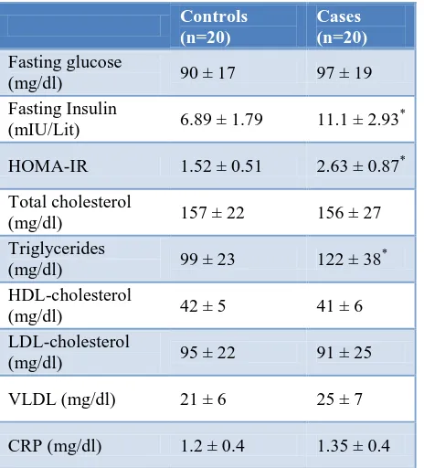
Table 1 displays the results of serum fasting glucose, fasting insulin, lipid profile and hs-CRP levels among PCOS subjects and healthy controls. Serum fasting insulin, insulin resistance (HOMA-IR) and triglycerides were significantly higher in PCOS subjects compared to the normal healthy controls. Fasting serum glucose and CRP levels were increased in PCOS subjects compared to controls but not statistically significant.
When correlation analysis was done between IR and other biochemical variables among PCOS, we found that insulin resistance as calculated by HOMA-IR was positively associated with serum triglycerides and VLDL levels (Table 2). HOMA-IR and Total Cholesterol were found to be positively correlated with hs-CRP as shown in Table 3. No correlation was found between other lipid risk factors and CRP.
pathogenic variable linked with PCOS which in turn showed positive association with CRP and triglycerides.
Table 1: Comparison of biochemicalparameters between PCOS subjects and controls.
Controls (n=20)
Cases (n=20) Fasting glucose
(mg/dl) 90 ± 17 97 ± 19
Fasting Insulin
(mIU/Lit) 6.89 ± 1.79 11.1 ± 2.93 *
HOMA-IR 1.52 ± 0.51 2.63 ± 0.87*
Total cholesterol
(mg/dl) 157 ± 22 156 ± 27
Triglycerides
(mg/dl) 99 ± 23 122 ± 38
*
HDL-cholesterol
(mg/dl) 42 ± 5 41 ± 6
LDL-cholesterol
(mg/dl) 95 ± 22 91 ± 25
VLDL (mg/dl) 21 ± 6 25 ± 7
CRP (mg/dl) 1.2 ± 0.4 1.35 ± 0.4
HOMA-IR: Homeostasis model assessment – Insulin resistance
*
p<0.05 compared to controls; p value calculated by student ‘t’ test
Table 2: Correlation analysis between biochemical variables with insulin resistance among PCOS
subjects.
HOMA - IR
r-value p value
Fasting glucose 0.574* 0.001
Insulin (mIU/Lit) 0.805* 0.001
Total cholesterol 0.382 0.096
Triglycerides 0.615* 0.004
HDL-cholesterol 0.065 0.787
LDL-cholesterol 0.209 0.376
VLDL 0.615* 0.004
CRP 0.455 0.049
p value is calculated by Pearsons method. p <0.05 is considered statistically significant; HOMA-IR: Homeostasis model assessment – Insulin resistance
Table 3: Correlation analysis between biochemical variables with inflammatory status among PCOS
subjects.
CRP
r-value p value
Fasting glucose 0.222 0.34
Insulin (mIU/Lit) 0.387 0.092
HOMA-IR 0.445* 0.049
Total cholesterol 0.466* 0.038
Triglycerides 0.267 0.25
HDL-cholesterol 0.279 0.23
LDL-cholesterol 0.357 0.12
VLDL 0.267 0.25
p value is calculated by Pearsons method. p <0.05 is considered statistically significant; HOMA-IR: Homeostasis model assessment – Insulin resistance
Table 4: Binary regression analysis of factors associated with subjects with polycystic ovarian
syndrome.
OR (95% CI) p value
HOMA-IR 0.004 – 0.286 0.002
Total Cholesterol 0.987 – 1.074 0.173
Triglycerides 0.986 – 1.022 0.705
C-Reactive Protein 0.111 – 8.857 0.993
OR: odds ratio; HOMA-IR: Homeostasis model assessment- Insulin resistance
DISCUSSION
insulin signaling pathway and may be related to constitutive serine phosphorylation of the insulin receptors.18
Studies have shown that there is an increased prevalence of cardiovascular disease in women with PCOS.19 CRP is a globally accepted indicator of cardiovascular disease (CVD) risk.20 CRP has been shown to be elevated in PCOS subjects compared to controls in various studies, but this effect was found to disappear after adjusting for BMI indicating that BMI is a major factor associated with elevation of CRP among PCOS subjects.21 This is supported by other studies where age and BMI were contributing factors for elevation of CRP levels.22,23 Even in our study, as the cases and controls were age and BMI matched, we did not find any significant elevation of hs-CRP levels in PCOS cases compared to controls. Ji Young Oh et al showed a lower CRP levels in Asian women when compared to Caucasian and Hispanic women.21 This may also be a reason for not getting significant elevation in CRP levels in our study.
In the present study, we found a significant positive correlation between hs-CRP levels and IR. On binary regression analysis, IR only was identified as a persistent associated parameter with PCOS. This indicates that IR linked with PCOS pathogenesis might be the cause for the association of CRP with PCOS. This may be due to the lack of inhibitory effect of insulin on synthesis of acute phase proteins like CRP by hepatocytes.24
Dyslipidemia is another accepted indicator of CVD risk. Approximately 70% of PCOS patients exhibit an abnormal serum lipid profile.25 In the present study, PCOS women showed significant elevation in serum triglyceride level compared to healthy subjects whereas no difference was seen in total cholesterol, LDL and HDL levels. Triglycerides were also positively associated with IR. IR leads to increased catecholamines induced lipolysis in adipocytes, increasing serum free fatty acid (FFA) levels. This increase in FFA level stimulates VLDL production in liver leading to hypertriglyceridemia.26 Another mechanism postulated by Wetterau and coworkers explains that insulin is needed for the repression of microsomal triglyceride protein, which is responsible for the secretion of apo B and VLDL and ultimately leading to hypertriglyceridemia.27
In conclusion, IR is the hallmark of PCOS among young adolescent women. IR is associated with elevated CRP levels and hypertriglyceridemia. Taking measures to increase insulin sensitivity, may help in altering dyslipidemia and inflammatory status, thereby reducing CVD risk among young women with PCOS.
ACKNOWLEDGEMENTS
We sincerely thank all women who gave consent and participated in this study. We are grateful to The Management and Research Committee of Sri Manakula
Vinayagar Medical College and Hospital, for funding this project. We express our sincere thanks to Mr. Srinivasan for his technical support in measuring the laboratory parameters of this project.
Funding:Institute
Conflicts of interest:There are no conflicts of interest to declare
Ethical approval: The study was approved by the Institutional ethics committee
REFERENCES
1. Padubidri VG, Daftary SN. Disorders of the Ovary and Benign Tumours. In: Howkins and Bourne eds. Shaws textbook of gynaecology. 15th ed. Haryana, India: Elsevier Publication; 2011:369-70.
2. Nidhi R, Padmalatha V, Nagarathna R, Amritanshu R. Prevalence of polycystic ovarian syndrome in Indian adolescents. J Pediatr Adolesc Gynecol 2011;24(4):223-7.
3. Allahbadia GN, Merchant R. Polycystic ovary syndrome in the Indian Subcontinent. Semin Reprod Med 2008;26(1):22-34.
4. Dunaif A. Insulin resistance and polycystic ovary syndrome: mechanism and implication for pathogenesis. Endocr Rev 1997;18960:774-800. 5. Salley KE, Wickham EP, Cheang KI, Essah
PA, Karjane NW, Nestler JE. Glucose intolerance in polycystic ovary syndrome–a position statement of the Androgen Excess Society. J Clin Endocrinol Metab 2007;92(12):4546-56.
6. Flavia Tosi, Romolo Dorizzi, Roberto Castello, Claudio Maffeis, Giovanna Spiazzi, iacomo Zoppini, Michele Muggeo and Paolo Moghetti. Body fat and Insulin Resistance independently predict increased serum C-Reactive protein in hyperandrogenic women with polycystic ovary syndrome. Eur J Endocrinol 2009;161(5):737-45. 7. Boulman N , Levy Y, Leiba R, Shachar S, Linn R,
Zinder O and Blumenfeld Z.Increased C-Reactive Protein Levels in the Polycystic Ovary Syndrome: a marker of cardiovascular disease. J Clin Endocrinol Metab 2004; 89(5):2160–2165.
8. Samy N, Hashim M, Sayed M, Said M. Clinical significance of inflammatory markers in polycystic ovary syndrome: their relationship to insulin resistance and body mass index. Dis Markers 2009; 26(4):163-70.
9. Mahabeer S, Naidoo C, Norman RJ, Jialal I, Reddi K, Joubert SM. Metabolic profiles and lipoprotein lipid concentrations in non-obese and obese patients with polycystic ovarian disease. Horm Metab Res 1990;22(10):537-40.
11. Ford ES. The metabolic syndrome and mortality from cardio vascular disease and all-causes: finding from the National Health and Nutrition Examination Survey II Mortality study. Atherosclerosis 2004;173:307-312.
12. Holvoet P, Kritchevsky SB, Tracy RP, Mertens A, Rubin SM, Butler J, et al. The metabolic syndrome, circulating oxidized LDL and risk of myocardial infarction in well- functioning elderly people in the health, ageing and body composition. Diabetes 2004;53(4):1068-1073.
13. Friedewald WT, Levy RI, Fredrickson DS. Estimation of the concentration of low-density lipoprotein cholesterol in plasma, without use of the preparative ultracentrifuge. Clin Chem 1972;18:499–502
14. Matthews DR, Hosker JP, Rudenski AS, Naylor BA, Treacher DF, Turner RC. Homeostasis model assessment: insulin resistance and beta-cell function from fasting plasma glucose and insulin concentration in man. Diabetologia 1985;28(7):412– 9.
15. Mukherjee S, Maitra A. Molecular and genetic factors contributing to insulin resistance in polycystic ovary syndrome. Indian J Med Res 2010;131:743-760.
16. Dunaif A, Xia J, Book CB, Schenker E and Tang Z. Excessive insulin receptor serine phosphorylation in cultured fibroblasts and in skeletal muscle. A potential mechanism for insulin resistance in the polycystic ovary syndrome. J Clin Invest 1995;96:801- 810.
17. Rosenbaum D, Haber RS, Dunaif A. Insulin resistance in polycystic ovary syndrome: ecreased expression of GLUT4 glucose transporters in adipocytes. Am J Physiol 1993;264:197- 202. 18. Talbot JA, Bicknell EJ, Rajkhowa M, Krook A,
O’Rahilly S, Clayton RN. Molecular scanning of the insulin receptor gene in women with polycystic
ovarian syndrome. J Clin Endocrinol Metab 1996;81(5):1979-83
19. Wild S, Pierpoint T, McKeigue P, Jacobs H. Cardiovascular disease in women with polycystic ovary syndrome at long-term follow-up: a retrospective cohort study. Clin Endocrinol 2000;52(5):595–600.
20. Verma S, Anderson TJ. Fundamentals of endothelial function for the clinical cardiologist. irculation 2002;105:546–549.
21. Oh JY, Lee JA, Lee H, Oh JY, Sung YA, and Chung H. Serum C-Reactive Protein Levels in Normal Weight Polycystic Ovary Syndrome. Korean J Intern Med 2009;24(4):350-355.
22. Saijo Y, Kiyota N, Kawasaki Y, Miyazaki Y, Kashimura J, Fukuda M, et al. Relationship between C reactive protein and visceral adipose tissue in healthy Japanese subjects. Diabetes Obes Metab 2004;6(4):249-258.
23. Pieroni L, Bastard JP, Piton A, Khalil L, Hainque B, Jardel C. Interpretation of circulating C-reactive protein levels in adults: body mass index and gender are a must. Diabetes Metab 2003;29:133-138. 24. Campos SP, Baumann H. Insulin is a prominent
modulator of the cytokine- stimulated expression of acute-phase plasma protein genes. Mol Cell Biol 1992;12(4):1789-97.
25. Third Report of the National Cholesterol Education Program (NCEP). Expert panel on detection, evaluation, and treatment of high blood cholesterol in adults (Adult Treatment Panel III) final report. Circulation 2002;106(25):3143-3421.
26. Diamanti-Kandarakis E, Papavassiliou AG, Kandarakis SA, Chrousos GP. Pathophysiology and types of dyslipidemia in PCOS. Trends Endocrinol Metab 2007;18(7):280-285.
27. Wetterau JR, Lin MC, Jamil H. Microsomal triglyceride transfer protein. Biochim Biophys Acta 1997;1345:136-150.
DOI: 10.5455/2320-1770.ijrcog20130913