pISSN 2320-1770 | eISSN 2320-1789
Research Article
Correlation of doppler studies at 34 weeks of gestation with perinatal
outcome in high risk pregnancies
Apoorva Reddy
1*, Renuka Malik
1, Shibani Mehra
1, Pushpa Singh
1, Lakshman Ramachandran
2INTRODUCTION
Antepartum detection of the fetus at risk of death or compromise in utero remains a major challenge in modern obstetrics. The waveform analysis of the feto- maternal circulation by Doppler ultrasound has therefore
become a quick and a simple way of screening and identifying fetal compromise.1
The diameters and flow volumes of all the vessels of the feto- maternal circulation increases significantly as the gestational age advances. However there is an initial fall
1
Department of Obstetrics & Gynecology, PGIMER & Dr. RML Hospital, Delhi, India
2Department of Internal Medicine, Jaypee Hospital, Noida, India
Received: 19 September 2015
Accepted: 30 October 2015
*Correspondence:
Dr. Apoorva Reddy,
E-mail: [email protected]
Copyright: © the author(s), publisher and licensee Medip Academy. This is an open-access article distributed under the terms of the Creative Commons Attribution Non-Commercial License, which permits unrestricted non-commercial use, distribution, and reproduction in any medium, provided the original work is properly cited.
ABSTRACT
Background: Antepartum detection of the fetus at risk of death or compromise in utero remains a major challenge in modern obstetrics. The waveform analysis of the feto- maternal circulation by Doppler ultrasound has therefore become a quick and a simple way of screening and identifying fetal compromise.The main objective of the study is to evaluate Doppler flow indices as an index for assessing fetal well being in high risk pregnancies and to determine the predictive value of various Doppler parameters with perinatal outcome.
Methods: The study was a prospective cohort study were forty pregnant women with a high risk factor (intrauterine
growth restriction, pre eclampsia and gestational diabetes mellitus) and forty pregnant women with no high risk(controls) were selected at 34 weeks of gestation. Both the groups underwent an obstetrical ultrasound with color Doppler examination of bilateral uterine arteries, umbilical artery and middle cerebral artery. Abnormality was serially monitored and pregnancy terminated in the presence of absent, reversal of end diastolic flow in umbilical artery or non-reassuring tests of fetal wellbeing.
Results: Uterine artery S/D abnormality was seen in 32.5% of high risk cases were as abnormal umbilical artery S/D was seen in 25% and abnormal Pulsatility Index (PI) in 15% cases. Middle cerebral artery flow was abnormal in only 17.5% cases. Abnormality in the uterine artery flow correlated well with the incidence of preterm delivery (69.2%), need for cesarean section (53.8%) and length of Neonatal intensive care unit(NICU) stay >48 hours (69.23%). Abnormal umbilical artery flow was associated with a significant increase in the incidence of preterm delivery(75%), small for gestational age babies(93.75%) and length of NICU stay >48 hours (93.75%). There was no significant correlation seen with isolated abnormal middle cerebral artery flow.
Conclusions: Both uterine and umbilical artery Doppler velocities correlate well with the perinatal outcome but abnormal uterine artery Doppler predicts adverse neonatal outcome better than the fetal vessels as it discriminates fetuses at risk because of abnormal placental vascularisation from those who are at risk due to other causes.
in the blood flow when adjusted to millilitres/kilogram body weight with a minimum at 30 weeks of gestation after which it rises till term.2
There is ample evidence that Doppler interrogation of feto- maternal circulation can reliably predict adverse perinatal outcome in a high risk pregnancy viz. intra uterine growth restriction and pre-eclampsia. The umbilical artery and the middle cerebral artery Doppler are also used in pregnancies at a risk of having an anemic fetus.3
Doppler assessment of the utero-placental circulation hence plays an important role in screening for impaired placentation and its resultant complications of pre-eclampsia, intrauterine growth restriction and perinatal death.
METHODS
The study was a prospective controlled non randomized cohort study conducted in a Tertiary care center in Delhi, India from 2009-11. The study was approved by the institutional ethics and research review board.
A total of 80 pregnant women were selected at 34 weeks of gestation and 40 of them had a high risk factor (preeclampsia, intrauterine growth restriction and gestational diabetes mellitus). Pre-eclampsia was defined as blood pressure of ≥140/90 mmHg or diastolic BP ≥110 mmHg on two occasions 4 hours apart after 20 weeks of gestation with proteinuria ≥300 mg/24 hours or ≥1 + by quantitative dip stick test in the absence of urinary infection. Intra uterine growth restriction (IUGR) was defined as weight at a particular gestational age being less than the 10th percentile4 for that gestational age. Gestational Diabetes Mellitus was defined according to the Carpenter and Coustan criteria5 for diagnosis of gestational diabetes mellitus.
The remaining 40 women who had no obstetrics disorder or any risk factor were selected as controls. Pregnant women with fetal congenital anomalies and multiple pregnancies were excluded from the study. A detailed history and examination was done on all patients recruited in the study. Both the study groups underwent routine antenatal screening.
A grey scale ultrasound with color Doppler using a trans abdominal probe of frequency 3.5 MHz was performed in both the groups. Doppler flow of 4 vessels namely the right and the left uterine artery, the umbilical artery and the middle cerebral artery were obtained and the recordings were taken. An abnormality of the Doppler flow was serially monitored.
Abnormal Doppler flow values were taken for that particular gestational age as established by published normograms.6,7,8,9 Presence of a diastolic notch (at least
two readings) or S/D >95th percentile in uterine artery was considered abnormal. Abnormal umbilical artery Doppler was taken as Pulsatility Index (PI) >95th percentile or S/D ratio >95th percentile or absent or reversed End Diastolic flow. Abnormal middle cerebral artery flow was taken as PI <5th percentile.
Termination of pregnancy was considered in the presence of any of the following: absent or reversal end diastolic flow in umbilical artery, non reassuring tests of fetal wellbeing and worsening of maternal condition. The obstetric and the perinatal outcome in both the groups were noted.
All the above data was compiled and statistically analysed using Stat View 4.5 (Berkeley, California) and IBM - SPSS Statistics v19.0.0 Multilingual. Data was compared using Chi- squared test and Fischer exact test with a two tailed P value <0.05 was considered significant. Multivariate step wise forward regression analysis was used to determine the correlation of various Doppler parameters with the individual perinatal outcome.
RESULTS
Of all the high risk cases 35% had growth restricted fetuses, 22.5% had a hypertensive disorder and 15% had gestational diabetes mellitus. The remaining 27.5% had a combination of the above risk factors.
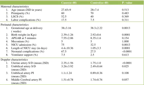
No significant difference in the maternal characteristics of the cases and the control group was noted (Table 1).
Neonatal complication rates were significantly higher in the cases as compared to the controls (Table 2).
Abnormal uterine artery Doppler flow was noted in 32.5% of patients in the high risk group and 5% of the patients in the control group. This difference was statistically significant (Table 1). There was a significant increase in the incidence of preterm delivery (69.2%), emergency cesarean section (53.8%), length of neonatal intensive care unit(NICU) stay (69.23%) and need for ventilator support (19.23%) when uterine artery Doppler abnormality was seen(Table 3).
Table 1: Comparison of Maternal, perinatal and Doppler characteristics between case and control groups.
Cases(n=40) Control(n=40) P- value
Maternal characteristics
1. Age (mean±2SD in years) 2. Primiparity (%)
3. LSCS (%)
4. Labor complications (%)
27.65±9 60 52.5 17.5
28±7.4 70 40 7.5
0.513 0.483 0.369 0.311
Perinatal characteristics
1. Gestational age at delivery ( weeks)
2. Birth weight (in Kgs) 3. APGAR at 5 minutes 4. Meconium (%) 5. NICU admission (%)
6. Length of NICU stay (in days) 7. Neonatal complications (%) 8. Ventilator support (%)
38±3.16
2.39±1.26 7.55±2.06 5
75 4.4±10.36 67.5 7.5
38.3±2.22
2.92±0.6 8.35±1.6 5 32.5 1.05±3.3 27.5 2.5
0.0013
0.0001 0.154 1.000 0.0013 0.0001 <0.0001 0.613
Doppler characteristics
1. Uterine artery S/D (mean±2SD) 2. Umbilical artery S/D
(mean±2SD) 3. Umbilical artery PI
(mean±2SD)
4. Middle Cerebral artery PI (mean±2SD)
2.35±1.56 3.26±3.92
1.1±1.24
1.51±0.78
1.75±1.0 2.49±0.64
0.89±0.36
1.74±0.76
<0.0001 0.025
0.108
[image:3.595.51.549.418.574.2]0.057
Table 2: Neonatal complications
Complication Cases (%) Controls (%)
Jaundice 47.5 (19) 10 (4)
Birth asphyxia 30 (12) 10 (4)
Polycythemia 12.5 (5) 0
Sepsis 5 (2) 0
Weight Loss 2.5 (1) 2.5 (1)
Feed Intolerance 2.5 (1) 2.5 (1)
Hypoglycemia 2.5 (1) 0
Respiratory Distress Syndrome 2.5 (1) 0
Hypoxic Encephalopathy 2.5 (1) 0
Necrotizing Enterocolitis 2.5 (1) 0
Anemia 0 2.5 (1)
Numbers in parenthesis indicate n. Overlapping may be present
Table 3: Perinatal outcome with abnormal Uterine artery Doppler
Parameter Sensitivity(%) Specificity(%) PPV*(%) NPV†(%)
Preterm delivery 50 81.81 69.2 66.67
Small for gestation 41.67 81.25 76.92 48.14
APGAR at 5 minutes < 7 64.28 74.24 34.62 90.74 No. of days in NICU >48 hrs 40.9 77.78 69.23 51.85
Ventilator support 83.33 71.62 19.23 98.15
*
[image:3.595.57.545.614.693.2]Table 4: Perinatal outcome with abnormal Umbilical artery Doppler
Parameter Sensitivity(%) Specificity(%) PPV*(%) NPV†(%)
Preterm delivery 33.33 90.90 75 62.5
Small for gestation 31.25 96.88 93.75 48.44
APGAR at 5 minutes < 7 35.71 83.33 31.25 85.94 No. of days in NICU >48 hrs 34.09 97.22 93.75 54.69
Ventilator support 66.67 83.78 25 96.88
*
Positive predictive value; †Negative predictive value
Table 5: Perinatal outcome with abnormal Middle cerebral artery Doppler
Parameter Sensitivity(%) Specificity(%) PPV*(%) NPV†(%)
Preterm delivery 11.11 77.27 28.57 51.51
Small for gestation 16.67 81.25 57.14 39.39
APGAR at 5 minutes < 7 14.29 81.81 14.29 81.81 No. of days in NICU >48 hrs 18.18 83.3 57.14 45.45
Ventilator support 0 81.08 0 90.90
*
Positive predictive value; †Negative predictive value
An abnormal MCA Doppler was observed in 17.5% of the high risk and 2.5% of the control group (Table 1). Although correlation of abnormal MCA Doppler was seen with perinatal morbidities but none of them was significant. However, as with the other Doppler parameters, the specificity and the negative predictive value of abnormal MCA Doppler was high (Table 5).
Only one case of absent end diastolic flow in the umbilical artery was seen. No case of reversal of end diastolic flow in umbilical artery was noted.
DISCUSSION
The introduction of real time ultrasound has enabled the observation of changes in fetal growth during pregnancy, thereby making it possible to detect antenatal fetuses at increased risk of being small for gestational age. However this antenatal detection cannot distinguish a normal small fetus from a compromised small fetus and its associated risks of stillbirth, perinatal mortality and neonatal handicap.
The present prospective cohort study was in continuum with the numerous studies done in assessing the role of various color Doppler parameters in the prediction of an adverse perinatal outcome.
Doppler interrogation of the uterine artery provides important information on the conversion process of spiral arteries into uteroplacental arteries. This conversion becomes abnormal in growth restricted pregnancies and pre- eclampsia. In the present study, cut off for uterine artery S/D ratio at 34 weeks was taken as 2.6 in accordance with Fleischer et al. 6
We found a uterine artery Doppler abnormality in 32.5% of the high risk patients at 34 weeks as against 5% in the control group. A higher figure was seen in the study by T
Ghi et al (51.5%) on late onset preeclampsia.10 In low risk cases studied by Irion et al11 an abnormal uterine artery S/D was seen in 10% cases.
Increase in the uterine artery volume flow and consequently a decrease in the S/D ratio with increasing gestation was observed by Konje et al in their study.12 However their study shows that although the quantified volume flow in both SGA and AGA fetuses increases throughout gestation, the increase in the SGA group was less marked.12 In accordance with this, the mean uterine artery S/D ratio at 34 weeks in the high risk and control groups of the present study was 2.35 and 1.75 respectively.
Studies have shown that Doppler velocimetry of fetal vessels, which have been used extensively to reduce the risk of perinatal morbidity in high risk babies loses its predictive ability when approaching term.13 Normal umbilical artery in many instances has been seen in high risk cases beyond 34 weeks of gestation inspite of adverse perinatal outcome. In accordance with this , we have found that the uterine artery Doppler predicts the adverse neonatal outcome in terms of premature delivery, small for gestational age babies, low APGAR at 5 minutes, need for NICU admission and ventilator support better than the fetal vessel Doppler velocimetry.
The umbilical artery Doppler is valuable in investigation of high risk pregnancy, although it is not a good screening tool in the low risk population. Perinatal morbidity and mortality is higher in high risk group with abnormal umbilical artery Doppler flow. In clinical practice several umbilical Doppler indices have been used for fetal surveillance. In the present study we used 2 indices: S/D ratio and Pulsatility index.
[image:4.595.52.549.213.293.2]S/D ratio 95th percentile in high risk pregnancy was given as 3 at 34 weeks by Cameron et al.8
Using these cut-offs, 25% of high risk cases and 5% of the controls had an abnormal S/D ratio at 34 weeks. Similarly, Dubey et al14 found an abnormal umbilical artery S/D in 29.2% of high risk cases. 15% of the high risk patients had an abnormal PI and in the control group this value was 2.5%. Similar figures were observed by Vergani et al15 who had 16% of patients in adverse outcome group having umbilical PI >95th percentile as opposed to 3% in the good outcome group.
A statically significant correlation was seen between abnormal umbilical artery Doppler flow, the incidence of SGA, the length of the NICU stay and the need for ventilator support. Previous studies by McCowan et al16 & Gudmundsson et al17 have also shown similar results.
Thus the resistance and flow in the umbilical artery is a good indicator of placental function and neonatal morbidity. However the presence of normal umbilical artery Doppler does not rule out the chance of perinatal morbidity as the changes in the Doppler indices do not occur in mild forms of placental insufficiency and they become abnormal only in advanced stages of placental dysfunction.
Velocimetry of the uterine and the umbilical arteries are considered to be more accurate than that of MCA.18 Fetal brain sparing indicates redistribution of blood flow to the baby’s vital organs. The involvement of cerebral vessels is a compensatory mechanism in response to decreased placental flow.
We have noted the mean MCA PI at 34 weeks as 1.51in the high risk and 1.74 in the control group. Harrington et al,19 who in their longitudinal analysis of Doppler circulation, showed that the MCA means were different in the high risk group than the normal group.
Though MCA indices are not useful parameters for identification of growth restricted fetuses at risk, abnormal MCA PI values were highly suggestive of poor perinatal outcomes in fetuses with abnormal uterine and umbilical velocimetry.
Doppler studies identify uteroplacental insufficiency and its effects on fetus. Hence, in the present study constant surveillance including tests for fetal wellbeing and monitoring of maternal condition were done along with serial Doppler scans. The decision for the timing and mode of termination of pregnancy was considered by the obstetrician taking into account all the above mentioned factors.
CONCLUSIONS
All the Doppler parameters have a high negative predictive value but a low positive predictive value and
sensitivity. Both uterine and umbilical artery Doppler velocities correlate well with the perinatal outcome but abnormal uterine artery Doppler predicts adverse neonatal outcome better than the fetal vessels as it discriminates fetuses at risk because of abnormal placental vascularisation from those who are at risk due to other causes. However, long term studies of the antenatal Doppler parameters, their neurological and metabolic impact on the fetus need to be undertaken for the last word on this issue.
Funding: No funding sources Conflict of interest: None declared
Ethical approval: The study was approved by the Institutional Ethics Committee
REFERENCES
1. Dubay P, Chaturvedi P. Doppler Waveform Analysis in High Risk Pregnancy. Asian J Obs Gynae Practice. 2003;7(5):23-6.
2. Konje JC, Abrams K, Bell SC, de Chazal RC, Taylor DJ. The application of color power angiography to the longitudinal quantification of blood flow volume in the fetal middle cerebral arteries, ascending aorta, descending aorta, and renal arteries during gestation. Am J Obstet Gynecol. 2000;182(2):393-400.
3. Giancarlo M. Doppler ultrasonography in obstetrics: from the diagnosis of fetal anemia to the treatment of intrauterine growth-restricted fetuses. Am J Obstet Gynecol. 2009;200(6):613.e1-613.e9.
4. Alexander GR, Himes JH, Kaufmann RB, Mor J, Kogan M. A United States national reference for fetal growth. Obstet Gynecol. 1996;87:163.
5. Carpenter MW, Coustan DR .Criteria for screening tests for gestational diabetes. Am J Obstet Gynecol. 1982;144:768-73.
6. Fleischer A, Schulman H, Farmakides G, Bracero L, Blattner P, Randolph G. Uterine artery waveforms and intrauterine growth retardation. Am J Obstet Gynecol. 1985;151:502-5.
7. Vergani P, Roncaglia N, Locatelli A, Andreotti C, Crippa I, Pezzullo JC et al. Antenatal predictors of neonatal outcome in fetal growth restriction with absent end-diastolic flow in the umbilical artery. Am J Obstet Gynecol. Sept 2005;193 (3):1213-8.
8. Cameron AD, Nicholson MD, Nimrod CA, Harder JR, Davies DM. Doppler waveforms in the fetal aorta and umbilical artery in patients with hypertension in pregnancy. Am J Obstet Gynecol.1988;158:339-45. 9. Harrington K, Carpenter RG, Nguyen M, Campbell
S. Changes observed in Doppler studies of fetal circulation in pregnancies complicated by preeclampsia or delivery of a small for gestational age baby: Cross sectional analysis. Ultrasound Obstet Gynecol. 1995;6:19.
preeclampsia. Am J Obstet Gynecol. 2009;201:36.e1-5.
11. Irion O, Masse J, Forest JC, Moutquin JM. Prediction of pre-eclampsia, low birthweight for gestation and prematurity by uterine artery blood flow velocity waveforms analysis in low risk nulliparous women. BJOG. 1998;105:422-9.
12. Konje JC, Howarth ES, Kaufmann P, Taylor DJ. Longitudinal quantification of uterine artery blood volume flow changes during gestation in pregnancies complicated by intrauterine growth restriction. BJOG. 2003;110:301–5.
13. Vergani P, Roncaglia N, Andreotti C, Arreghini A, Teruzzi M, Pezzullo JC, et al. Prognostic value of uterine artery Doppler velocimetry in growth-restricted fetuses delivered near term. Am J Obstet Gynecol. 2002;187(4):932-6.
14. Dubey P, Pandey K, Gupta N. Color Doppler evaluation of Uteroplacental Circulation in Management of High risk Pregnancies. Asian J Obs Gynae Practice. 2009;4:19-22.
15. Vergani P, Andreotti C, Roncaglia N, Zani G, Pozzi E, Pezzullo JC, et al. Doppler predictors of adverse neonatal outcome in the growth restricted fetus at 34 weeks' gestation or beyond. Am J Obstet Gynecol. 2003;189(4):1007-11.
16. McCowan LME, Harding JE, Stewart AW. Umbilical artery Doppler studies in small for gestational age babies reflect disease severity. BJOG. 2000;107:916-25.
17. Gudmundsson S, Marsal K. Blood velocity waveforms in the fetal aorta and umbilical artery as predictors of fetal outcome: a comparison. Am J Perinatol. 1991;8:1–6.
18. Simanaviciute D, Gudmundsonn S. Fetal middle cerebral to uterine artery Pulsatility index ratios in normal and pre eclamptic pregnancies. Ultrasound Obstet Gynecol. 2006;28:794-801.
19. Harrington K, Thompson MO, Carpenter RG, Nguyen M, Campbell S. Doppler fetal circulation in pregnancies complicated by pre-eclampsia or delivery of a small for gestational age baby: 2. Longitudinal analysis. BJOG. 1999;106:453-66. 20. Redman CWG. Examination of the placental
circulation by Doppler ultrasound. BMJ. 1989;298:621-2.