Original Research Article
Study of the role of hysteroscopy with dilation and curettage in post-
menopausal bleeding: a cross sectional study
Atul Padmawar*, Ankita Khande
INTRODUCTION
Abnormal uterine bleeding is defined as any type of bleeding in which the duration, frequency or amount is excessive for an individual patient.1 In normal to 12-week
size uterus, the cause of abnormal bleeding often remains obscure. Goals of clinical management are primarily dependent upon attaining a correct etiological diagnosis. The history, physical examination and pelvic examination attempt to determine the site of the bleeding and its source. Information gathered from this will suggest what direction the investigation would take and the treatment
modality. Ultrasonography clearly depicts the uterine contour, any lesion in the myometrium like fibroid and the status of the ovary, but fails to provide adequate information regarding the endometrium. The endometrial pathology like small submucous fibroid, endometrial hyperplasia is missed sometimes by ultrasound.2
Dilatation and curettage is a blind procedure done without knowing the exact location of the lesion or the pathology of the endometrium and the endometrium has to be sent to the pathologist to study histological patterns and for the report.3 Hysteroscopy has ushered a new era
in the evaluation of abnormal uterine bleeding. By direct ABSTRACT
Background: Abnormal uterine bleeding is defined as any type of bleeding in which the duration, frequency or amount is excessive for an individual patient. Hysteroscopy has ushered a new era in the evaluation of abnormal uterine bleeding. With this background, we conducted a study to find the role of hysteroscopy with dilation and curettage in post-menopausal bleeding.
Methods: It was an observational study conducted in the department of obstetrics and gynecology of a tertiary care hospital in Maharashtra. Post-menopausal patients with abnormal uterine bleeding, admitted in the period of March 2018 to September 2019 were included in the study. A pretested and predesigned questionnaire was used to collect the data. Hysteroscopy with dilation and curettage was done and samples were sent for histopathology.
Results: Bleeding was present in all the 30 cases. About 33.33% had pain abdomen, 56.67% had hot flushes, 10.00% had mass and 10% had other symptoms in the present study. The sensitivity, specificity, positive predictive value, negative predictive value and diagnostic accuracy of hysteroscopy in diagnosing malignancy was 75%, 96.15%, 75%, 96.15% and 93.33% respectively.
Conclusions: To conclude the most common cause of abnormal uterine bleeding in post-menopausal women in our study was polyp. The sensitivity and specificity of the hysteroscopy in diagnosing the malignant lesions is 75% and 96.15% respectively which corroborated with other study findings. Hysteroscopy with dilation and curettage is better alternative to hysterectomy and biopsy in most of the cases.
Keywords: Dilation and curettage, Hysteroscopy, Post-menopausal bleeding DOI: http://dx.doi.org/10.18203/2320-1770.ijrcog20201787
Department of Obstetrics and Gynecology, Shri Vasantrao Naik Government Medical College, Yavatmal, Maharashtra, India
Received: 13 February 2020
Accepted: 06 March 2020
*Correspondence:
Dr.Atul Padmawar,
E-mail: [email protected]
visualization of the uterine cavity it is able to pin point the aetiology in majority of the cases. It can accurately detect endometrial hyperplasia and aids in the early diagnosis of endometrial carcinoma and uterine polyps. The judicious use of hysteroscopy to manage this medical entity adds a new dimension in handling this often-perplexing problem.4
METHODS
It was a observational study conducted in the department of obstetrics and gynecology of a tertiary care hospital in Maharashtra. Post-menopausal patients with abnormal uterine bleeding, admitted in the period of March 2018 to September 2019 were included in the study. Women with abnormal uterine bleeding due to injuries of the female reproductive tract, not willing to participate in the study, already diagnosed cases of carcinoma were excluded and omen of hormone replacement therapy were excluded from the study. Study included 30 cases of post-menopausal uterine bleeding admitted under our department during the study period. Since, we have limitation of duration of the study so we included most of the cases in the study.
Data was collected using a pretested and predesigned questionnaire. The questionnaire consisted of sociodemographic factors like age, occupation, educational status, socioeconomic status etc. It also included the general physical examination findings, systemic examination findings of the study subjects. Associated morbidities like diabetes, hypertension, IHD and tuberculosis were also noted. The socioeconomic status was classified into classes based on the modified BG Prasad classification for the year 20195. The hemoglobin values were categorized based on the world health organization guidelines for classification of anaemia6. Further, we also noted the chief complaints and did a detailed gynecological examination of the subjects. Appropriate investigations were done before the start of the procedure.
Hysteroscopy was video-assisted and carried out with normal saline solution used as liquid distension medium; a 5-mm sheathed hysteroscope, with a working channel, was used for each examination. After hysteroscopic inspection, an endometrial sampling targeted under vision was performed by mechanical or electrosurgical instrumentation. When extensive features of hyper- plastic or neoplastic growth were observed, we combined a blind sampling procedure with dilatation- curettage. All cases diagnosed with carcinoma histopathologically were referred to an onco-surgeon for further evaluation.
Statistical analysis
The data was collected, compiled and analyzed using EPI info (version 7.2). The qualitative variables were expressed in terms of percentages. The quantitative variables were both categorized and expressed in terms of
percentages or in terms of mean and standard deviations. The diagnostic performance was expressed in terms of sensitivity, specificity, positive predictive value, negative predictive value and diagnostic accuracy. Histopathology was considered gold standard for the diagnosis. All tables were graphically represented.
Statistical analysis
All analysis was 2 tailed and the significance level was set at 0. 05.
RESULTS
[image:2.595.307.550.292.581.2]Study included 30 study subjects.
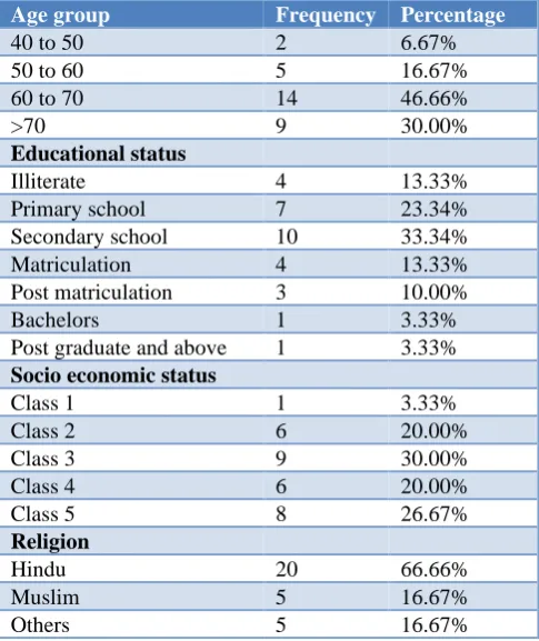
Table 1: Demographic characteristics of the study subjects.
Age group Frequency Percentage
40 to 50 2 6.67%
50 to 60 5 16.67%
60 to 70 14 46.66%
>70 9 30.00%
Educational status
Illiterate 4 13.33%
Primary school 7 23.34%
Secondary school 10 33.34%
Matriculation 4 13.33%
Post matriculation 3 10.00%
Bachelors 1 3.33%
Post graduate and above 1 3.33%
Socio economic status
Class 1 1 3.33%
Class 2 6 20.00%
Class 3 9 30.00%
Class 4 6 20.00%
Class 5 8 26.67%
Religion
Hindu 20 66.66%
Muslim 5 16.67%
Others 5 16.67%
Majority of them were in the age range of 60 to 70 years, were Hindus, were educated up to secondary school and were class 3 according to BG Prasad classification of socio-economic status (Table 1).
Figure 1 shows bleeding was present in all the 30 cases. About 33.33% had pain abdomen, 56.67% had hot flushes, 10.00% had mass and 10% had other symptoms in the present study.
cases diagnosed by hysteroscopy one case had complex hyperplasia and two cases were diagnosed to have carcinoma. Based on the hysteroscopic findings, 10% were normal, 40% were polyp, 23.33% had myoma, 16.67% had hyperplasia and 10% had carcinoma in the present study. Based on the histopathological diagnosis, 40% were polyp, 23.33% were myoma, 13.33% were simple hyperplasia, 6.67% had complex hyperplasia, 6.67% had carcinoma and 10% had atrophy of endometrium (Table 2).
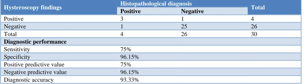
[image:3.595.311.545.71.218.2]The sensitivity, specificity, positive predictive value, negative predictive value and diagnostic accuracy of hysteroscopy in diagnosing malignancy was 75%, 96.15%, 75%, 96.15% and 93.33% respectively (Table 3).
[image:3.595.58.542.313.415.2]Figure 1: Distribution of the study subjects based on the chief complaints (n = 30).
Table 2: Histopathological correlation with hysteroscopy findings.
Hysteroscopy findings
Histopathological diagnosis
Polyp Myoma Simple
hyperplasia
Complex hyperplasia/
atypical Carcinoma
Atrophy/ not satisfactory
Normal 0 0 0 0 0 3
Polyp 12 0 0 0 0 0
Myoma 0 7 0 0 0 0
Hyperplasia 0 0 4 1 0 0
Carcinoma 0 0 0 1 2 0
Table 3: Diagnostic performance of hysteroscopy with histopathology.
Hysteroscopy findings Histopathological diagnosis Total
Positive Negative
Positive 3 1 4
Negative 1 25 26
Total 4 26 30
Diagnostic performance
Sensitivity 75%
Specificity 96.15%
Positive predictive value 75%
Negative predictive value 96.15%
Diagnostic accuracy 93.33%
DISCUSSION
Hysteroscopy is a novel method in which has been used to diagnose and treat the cause of abnormal uterine bleeding in post-menopausal women. With this basic background we conducted a study to understand the role of hysteroscopy in our patients with post-menopausal bleeding.
All the cases which were normal on hysteroscopy were having a tropic endometrium, all polyps and myomas were histopathologically the same. Among 5 cases of hyperplasia one case had complex or atypical hyperplasia
and four cases had simple hyperplasia. Among the three cases diagnosed by hysteroscopy one case had complex hyperplasia and two cases were diagnosed to have carcinoma. Elfayomy A et al, reported that endometrial polyps were the most common hysteroscopic pathologies observed in 25 cases (30.1%), while endometrial hyperplasia and carcinoma diagnosed visually by the hysteroscopes in 18 (21.7%) and 11 cases (13.3%), respectively.7 The hysteroscopic findings and the
histological diagnoses were compared in 83 cases. Of note, hyperplastic endometrium was confirmed by histology in 23 (27.8%), 13 cases had been suspected by the hysteroscope, the other 10 cases were not suspected during visual diagnosis of the uterine cavity. On the other
0 20 40 60 80 100
Pain abdomen
Bleeding per vagina
Hot flushes Mass Generalised
fatigue Others (Fever, nausea, vomiting)
33.33 100
56.67
10
73.33
10 P
[image:3.595.51.553.448.586.2]hand, the hysteroscopic findings were not suspicious for malignancy in seven cases proved by histological examination. All six myomas at hysteroscopy were confirmed by biopsy.
Kormazer E et al reported that thirty women had irregular endometrium by hysteroscopic impression; 10 of them had simple endometrial hyperplasia, 1 of them had EIN and 2 of them had endometrial carcinoma in pathological assessment.8 Hysteroscopy diagnosed all intrauterine
space occupying lesions (76 endometrial polyp, 17 submucosal fibroid) but D and C missed 25 of these women (atrophy). Alanis Fuentes J et al, correlated results between hysteroscopic findings and histopathological biopsy diagnosis obtained were: atrophy (157 cases, 63.3%), polyps (76 cases, 77.55%), endometrial cancer (14 cases, 93.05%), endometrial hyperplasia (10 cases, 90.63%) and miomatosis (three cases, 16.20%).9
The sensitivity, specificity, positive predictive value, negative predictive value and diagnostic accuracy of hysteroscopy in diagnosing malignancy was 75%, 96.15%, 75%, 96.15% and 93.33% respectively. Elfayomy A et al reported that in benign endometrial lesions, the sensitivity of the hysteroscopic view was 94.7%, specificity was 97.8%, positive (PPV) and negative (NPV) predictive values were 97.3 and 95.7%, respectively.7 On the other hand, hysteroscopy
demonstrated an overall sensitivity, specificity, PPV, and NPV of 56.5, 91.6, 72.2, and 84.6%, respectively, in endometrial hyperplasia, whereas the same parameters for endometrial cancer were 50, 94.2, 63.6, and 90.2% Dinic P et al, conducted a study to define the significance of hysteroscopy as a diagnostic procedure for the evaluation of patients with postmenopausal bleeding.10 The
sensitivity of hysteroscopy in the detection of intrauterine pathology was 100%, the specificity 81%, the positive predictive value 92% and the negative predictive value 100%.
Loiacono R et al, conducted a study compares hysteroscopic and histo-pathological results in postmenopausal women with abnormal uterine bleeding (AUB) and asymptomatic postmenopausal women with a thickened endometrium.11 In the AUB group, if the
hysteroscopy results were normal, a sensitivity, specificity, positive predictive value (PPV) and negative predictive value (NPV) of 100, 95, 71 and 100%, respectively, were achieved, while in the asymptomatic group these values were 100, 97, 90 and 100%, respectively. For both the group with polyps and that with myomas, the sensitivity, specificity, PPV and NPV were 100%. For endometrial hyperplasia, hysteroscopy showed sensitivity, specificity, PPV and NPV of 81, 96, 87 and 93%, respectively, in the AUB group, while in the asymptomatic group, the sensitivity was 60%, the specificity and PPV were 100%, and the NPV was 98%. The sensitivity of hysteroscopy for endometrial cancer was 63%, the specificity 97%, the PPV 77%, and the
NPV 95%. Tinelli R et al, inferred that hysteroscopy was technically successful in all women and revealed sensitivity of 98%, specificity of 91%, a positive predictive value of 88%, a negative predictive value of 98%, and diagnostic accuracy of 94%.12
Garuti G et al, reported that the sensitivity, specificity, and negative and positive predictive values of hysteroscopy to foresee a diagnosis of infiltrating carcinoma were 84.6%, 100%, 87.5%, and 100%, respectively.13 Kormazer E et al, reported that the
hysteroscopy revealed sensitivity, specificity, positive predictive value and negative predictive value as 76.4%, 76.9%, 73.1%, 79.8%, respectively.8 Sarvi F et al,
conducted a study to investigate the histologic and hysteroscopic findings of post-menopausal women with uterine bleeding.14
In the AUB group sensitivity, specificity, and positive and negative predictive values of hysteroscopy for normal findings were 98%, 100%, 100% and 90%, respectively. In the asymptomatic group, the same parameters were 98%, 100%, 100% and 85%, respectively. The sensitivity, specificity, and positive and negative predictive values of hysteroscopy for polyps and myomas were 100%. Also, the sensitivity, specificity, and positive and negative predictive values were 100% in hyperplasia cases found during hysteroscopy in both groups. Kanani K et al, conducted a study the hysteroscopic evaluation of abnormal uterine bleeding in reproductive and post-menopausal women.15 Comparison
of hysteroscopic and histopathologic results show that the sensitivity of hysteroscopy in detection of intrauterine pathology was 95.4%, specificity 96.4%, positive predictive value was 95.4% and the negative predictive value was 96.4%.
Valson H et al, conducted a study hysteroscopic evaluation of abnormal uterine bleeding in reproductive and post-menopausal women.16 Both hysteroscopy and
(D and C) were accurate when an abnormality was diagnosed, giving a specificity of 96.4% and 96.4% respectively and positive predictive value of (95.2% versus 94.4%). The ability to diagnose a lesion was more with hysteroscopy i.e. sensitivity in comparison to curettage, (90.9% versus 77.4%), while a negative diagnosis was less wrongly made with hysteroscopy in comparison to curettage (false negative rate: 9.1% versus 22.7%). In intracavitary lesions like polyp, hysteroscopy gave 100% accuracy.
CONCLUSION
better alternative to hysterectomy and biopsy in most of the cases.
Funding: No funding sources Conflict of interest: None declared
Ethical approval: The study was approved by the Institutional Ethics Committee
REFERENCES
1. Whitaker L, Critchley HOD. Abnormal uterine bleeding. Best Pract Res Clin Obstet Gynaecol. 2016;34:54-65.
2. Goldstein SR. Abnormal uterine bleeding: the role of ultrasound. Radiol Clin North Am. 2006;44(6):901-10.
3. Su H, Huang L, Huang KG, Yen CF, Han CM, Lee CL. Accuracy of hysteroscopic biopsy, compared to dilation and curettage, as a predictor of final pathology in patients with endometrial cancer. Taiwan J Obstet Gynecol. 2015;54(6):757-60. 4. Sinha P, Yadav N, Gupta U. Use of hysteroscopy in
abnormal uterine bleeding: an edge over histopathological examination. J Obstet Gynaecol India. 2018;68(1):45-50.
5. Pandey VK, Aggarwal P, Kakkar R. Modified BG Prasad socio-economic classification, Update-2019. Indian J Community Heal. 2019;31(1):150-2. 6. World Health Organization guidelines for
classification of anaemia, 2014. Available at: https://www.who.int/vmnis/indicators/haemoglobin.p df. Accessed on 10th January 2020.
7. Elfayomy AK, Habib FA, Alkabalawy M. Role of hysteroscopy in the detection of endometrial pathologies in women presenting with postmenopausal bleeding and thickened endometrium. Arch Gynecol Obs. 2012;285:839-43. 8. Korkmazer E, Solak N, Üstünyurt E. Hysteroscopic
assessment of postmenopausal endometrial thickening. Prz Menopauzalny. 2014;13(6):330-3. 9. Alanis Fuentes J, Martinez Gutlerrez M, Mata M.
Hysteroscopy findings in patients with postmenopausal genital bleeding. Ginecol Obs Mex. 2007;75(5):253-8.
10. Dinic P, Ljubic A, Kopitovic V, Antic V, Stamenovic S, Pjevic A. The role of hysteroscopy in diagnosis and treatment of postmenopausal bleeding. Vojn Pregl. 2013;70(8):1-2.
11. Loiacono R, Trojano G, Gaudio N, Kardhashi A, Deliso M, Gaetano F, et al. Novel insights from clinical practice hysteroscopy as a valid tool for endometrial pathology in patients with postmenopausal bleeding or asymptomatic patients with a thickened endometrium: hysteroscopic and histological results. Gynecol Obs Invest. 2015;79:210-6.
12. Tinelli R, Tinelli FG, Cicinelli E, Malvasi A, Tinelli A. The role of hysteroscopy with eye-directed biopsy in postmenopausal women with uterine bleeding and endometrial atrophy. Menopause. 2008;15(4):737-42.
13. Garuti G, Mirra M, Luerti M. Hysteroscopic view in atypical endometrial hyperplasias: a correlation with pathologic findings on hysterectomy specimens. J Minim Invasive Gynecol. 2006;13:325-30.
14. Sarvi F, Alleyassin A, Aghahosseini M, Ghasemi M, Gity S. Hysteroscopy: a necessary method for detecting uterine pathologies in post-menopausal women with abnormal uterine bleeding or increased endometrial thickness Histeroskopi. Turk J Obs Gynecol. 2016;13:183-8.
15. Kanani K, Panchal P, Shah S, Mehta A, Bhagde A. Evaluation of abnormal uterine bleeding by hysteroscopy and curettage. Int J Sci Res. 2017;6(8):417-9.
16. Valson H, Kulkarni C, Mukerjee S, Gowda SN, Valson H, Contracept JR, et al. The role of diagnostic hysteroscopy in abnormal uterine bleeding and its histopathological correlation following blind dilatation and curettage. Int J Reprod Contracept Obs Gynecol. 2016;5(3):609-14.