Scholarship@Western
Scholarship@Western
Electronic Thesis and Dissertation Repository
10-29-2019 4:15 PM
The Neural Circuitry of Sensory Processing in Post-traumatic
The Neural Circuitry of Sensory Processing in Post-traumatic
Stress Disorder
Stress Disorder
Sherain HarricharanThe University of Western Ontario
Supervisor Lanius, Ruth A.
The University of Western Ontario Graduate Program in Neuroscience
A thesis submitted in partial fulfillment of the requirements for the degree in Doctor of Philosophy
© Sherain Harricharan 2019
Follow this and additional works at: https://ir.lib.uwo.ca/etd
Part of the Psychiatric and Mental Health Commons
Recommended Citation Recommended Citation
Harricharan, Sherain, "The Neural Circuitry of Sensory Processing in Post-traumatic Stress Disorder" (2019). Electronic Thesis and Dissertation Repository. 6695.
https://ir.lib.uwo.ca/etd/6695
This Dissertation/Thesis is brought to you for free and open access by Scholarship@Western. It has been accepted for inclusion in Electronic Thesis and Dissertation Repository by an authorized administrator of
ii
Background: Traumatic experiences can have severe emotional and psychological consequences, which may affect the capacity to process both internal and external sensory
information. Such aberrations may have cascading effects in individuals with post-traumatic
stress disorder (PTSD), where alterations in sensory processing may hinder the capacity for
higher-order executive functions, including emotion regulation. Delineating the neural
circuitry of subcortical and cortical structures thought to be central to sensory processing is
therefore critical to the study of PTSD and may help to develop an understanding of the
neurobiological mechanisms underlying this often debilitating disorder.
Methods: Various neuroimaging approaches were employed to investigate sensory processing in PTSD, its dissociative subtype, and healthy controls. First, resting-state
connectivity patterns of subcortical brainstem structures linked to interoceptive and
exteroceptive sensory processing, including the periaqueductal gray and the vestibular nuclei,
were examined (chapters 2 and 3). In addition, given that the insula is critical for relaying
exteroceptive and interoceptive sensory information to other neurocognitive networks in the
brain, resting-state whole brain seed-based connectivity patterns of different insula
subregions were investigated (chapter 4). Furthermore, machine learning analyses were used
to assess the utility of insula subregion resting-state connectivity patterns as a diagnostic
predictor for classifying PTSD, its dissociative subtype, and healthy controls. Finally, a
task-based paradigm using oculomotor stimuli with simultaneous traumatic autobiographical
memory recall was employed to examine cortical brain structures involved in the
iii
Results and Discussion: As compared to controls,widespread periaqueductal gray
connectivity was observed with cortical structures associated with emotional reactivity and
defensive responding in PTSD and its dissociative subtype at rest. In addition, as compared
to controls, decreased vestibular nuclei connectivity with cortical structures essential to
exteroceptive sensory processing and multisensory integration was observed in individuals
with the PTSD dissociative subtype. Moreover, PTSD showed limited cortical insula
subregion resting-state connectivity with frontal lobe structures involved in the central
executive network, which may be associated with impairment of higher-order executive
functions, including emotion regulation, in PTSD. Finally, exposure to simultaneous
exteroceptive and interoceptive sensory stimuli through oculomotor eye movements
performed simultaneous to traumatic memory recall engaged the dorsal attentional network
and default-mode frontoparietal networks that have been demonstrated to work in tandem to
facilitate connectivity with structures in the central executive network, including the
dorsolateral and dorsomedial prefrontal cortex, necessary for multisensory integration and
emotion regulation. This effect was greater in individuals with PTSD and may provide a
neurobiological account for how oculomotion may influence the frontoparietal cortical
representation of traumatic memories. Overall, the findings of this dissertation reveal that
individuals with PTSD experience aberrations in the neural circuitry necessary for processing
both interoceptive and exteroceptive sensory information. We hypothesize that these
observed alterations in interoceptive and exteroceptive neural processing may underlie, in
part, the emotion dysregulation and maladaptive responses to chronic stress, including
iv
Keywords
post-traumatic stress disorder, dissociation, sensory processing, neuroimaging, brainstem, periaqueductal gray, vestibular system, insula, prefrontal cortex, frontoparietal network, multisensory integration, executive function, emotion regulation
Summary for Lay Audience
Post-traumatic stress disorder (PTSD) is a psychiatric disorder that is triggered by an
individual experiencing or witnessing a traumatic event, which may precipitate persistent
flashbacks and severe anxiety, causing individuals to be fearful and hypervigilant of their
surroundings. Approximately 14-30% of traumatized individuals may present with a
dissociative subtype of PTSD, which is often associated with repeated trauma or childhood
trauma. These patients may present with additional symptoms, including depersonalization
and derealization, where they may feel as if the world or self is “dream-like” and not real
and/or describe “out-of-body” experiences. This dissertation explores potential neural
alterations that may signify how traumatized individuals with PTSD and its dissociative
subtype experience sensations differently, whether they are from the outside world (i.e.
touch, auditory, visual sensations) or from the internal body (i.e. emotions, visceral
sensations). It is hypothesized that alterations in neural pathways important for the
processing of these sensations may have cascading effects on the performance of
higher-order cognitive functions, including emotion regulation. Various functional magnetic
resonance imaging techniques were employed to examine brain structures critical to sensory
v
The cumulative findings from this dissertation have been summarized into a theoretical
framework that hypothesizes a neurobiological account through which sensory processing
vi
Co-Authorship Statement
Sherain Harricharan, Maria Densmore, Dr. Andrew Nicholson, Dr. Mischa Tursich, Dr. Jean Théberge, Dr. Margaret McKinnon, Dr. Paul Frewen, Dr. Richard Neufeld, Dr. Daniela Rabellino, Dr. Allan Schore Dr. Janine Thome, Dr. Bessel Van der Kolk, Dr. Ruth Lanius
As author of this thesis and the primary author of the four experimental chapters, Sherain
Harricharan was responsible for designing the experiments, data analysis, and writing the
completed thesis and manuscripts for each experiment. Maria Densmore helped supervise the
statistical analysis used to investigate neuroimaging data (Chapters 2-5) and assisted in data
analysis for Chapter 5. Dr. Andrew Nicholson supervised the machine learning neuroimaging
analysis used in Chapter 4 and was consulted about the neuroscientific inferences made from
experimental findings in Chapters 3 and 4. Dr. Mischa Tursich helped design the task-based
paradigm used in Chapter 5. Dr. Jean Théberge oversaw the implementation of brain imaging
acquisition methods used to collect neuroimaging data in Chapters 2-5. Dr. Margaret
McKinnon was consulted about the neuroscientific inferences made from the experimental
findings for Chapter 5 and helped revise the experimental manuscripts for Chapter 2-5. Dr.
Paul Frewen was consulted about the neuroscientific inferences made from the experimental
findings for Chapter 2,4 and 5. Dr. Richard Neufeld was consulted about the statistical
analysis used to study neuroimaging data in Chapters 3 and 4. Dr. Daniela Rabellino and Dr.
Allan Schore were consulted about the neuroscientific inferences made from the
experimental findings for Chapter 2. Dr. Janine Thome was consulted about the
neuroscientific inferences made from the experimental findings for Chapter 4. Dr. Bessel
vii
findings for Chapter 5. Dr. Ruth Lanius supervised all stages of this thesis and assisted in
viii
Acknowledgments
Foremost, I would like to thank my supervisor, Dr. Ruth Lanius. Thank you for taking a
chance on me and providing me with an amazing opportunity that ignited my passion for
studying post-traumatic stress disorder. I feel incredibly proud of the work done in our lab
and it is because of the confidence and trust you instill in your staff and students.
Next, I would like to thank Suzy Southwell and Stephanie Nevill for all the hard work they
do in recruiting and assessing participants for our numerous studies. In addition, I would like
to thank Maria Densmore for her guidance and mentorship to ensure neuroimaging statistical
models are implemented correctly. I would also like to thank Nancy Mazza for all the
administrative work she does to make sure everything in our lab runs smoothly.
I would also like to acknowledge Dr. Margaret McKinnon and Dr. Paul Frewen for their
guidance and mentorship throughout all stages of this dissertation, I am very grateful to have
learned from you both.
I feel so grateful to have learned from the post-doctoral fellows and students in our lab,
including Dr. Daniela Rabellino, Dr. Janine Thome, Dr. Andrew Nicholson, Chantelle Lloyd
and Braeden Terpou. You are all brilliant, but more importantly, you are all incredible people
who always manage to keep everything in perspective.
In addition, I would like to thank all my family and friends for their support throughout the
past few years. In particular, to my sisters, Sharada and Monisha Harricharan, thank you for
ix
Finally, this body of work is dedicated to the memory of my beloved parents, Dr. Rajendra
and Indira Harricharan, whose light will always guide and motivate me throughout
x
Table of Contents
Abstract ... ii
Summary for Lay Audience ... iv
Co-Authorship Statement... vi
Acknowledgments... viii
Table of Contents ... x
List of Tables ... xv
List of Figures ... xvi
List of Appendices ... xviii Chapter 1 ... 1
1 « Introduction and Overview» ... 1
1.1 « Brainstem Sensory Processing » ... 4
1.2 « Cortical Sensory Processing » ... 7
1.3 « Objectives » ... 10
1.4 « References » ... 11
Chapter 2 ... 26
2 « fMRI functional connectivity of the periaqueductal gray in PTSD and its dissociative subtype» ... 26
2.1 « Introduction» ... 26
2.2 «Methods»... 29
2.2.1 Clinical and Demographic Information ... 29
2.2.2 Data Acquisition ... 32
2.2.3 Resting-State fMRI Data Preprocessing ... 33
2.2.4 Seed-Based Regions of Interest ... 34
xi
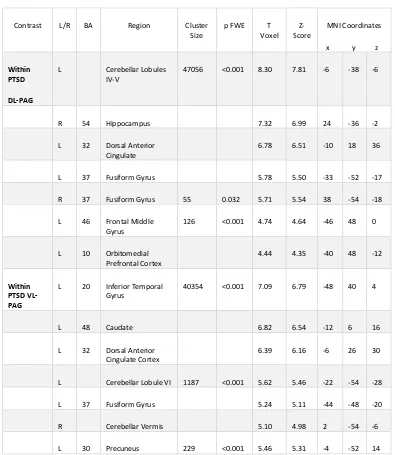
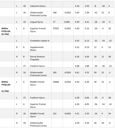
2.3 «Results» ... 38
2.3.1 Clinical and Demographic Measures ... 38
2.3.2 Full Factorial Design... 38
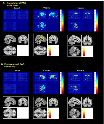
2.3.3 Functional Connectivity of DL-PAG and VL-PAG within Participant Groups ... 39
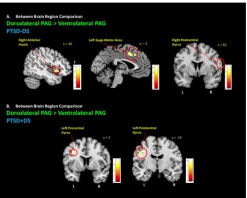
2.3.4 Functional Connectivity Differences between Participant Groups ... 46
2.3.5 Clinical Score Correlations with Functional Connectivity Patterns in PTSD Patients ... 49
2.4 Discussion ... 49
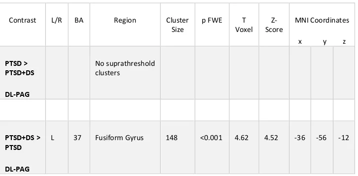
2.4.1 PAG Connectivity with Brain Regions Involved in Autonomic Control . 50 2.4.2 PAG Connectivity with the Fusiform Gyrus ... 52
2.4.3 PAG Connectivity with the Cerebellum ... 53
2.4.4 PAG Connectivity with Motor Regions ... 54
2.4.5 PAG Connectivity with Regions Involved in Depersonalization ... 55
2.4.6 Limitations and Future Directions ... 56
2.4.7 Conclusions ... 57
2.5 References ... 58
Chapter 3 ... 72
3 « Sensory overload and imbalance: Resting-state vestibular connectivity in PTSD and its dissociative subtype» ... 72
3.1 « Introduction » ... 73
3.2 Methods... 77
3.2.1 Clinical and Demographic Information ... 77
3.2.2 Data Acquisition ... 80
3.2.3 Resting-State fMRI Data Preprocessing ... 80
3.2.4 fMRI Statistical Analyses ... 82
xii
3.3.1 Overview ... 85
3.3.2 Clinical and Demographic Measures ... 85
3.3.3 Full Factorial Design... 86
3.4 Discussion ... 95
3.4.1 Vestibular Nuclei and Parieto-Insular Vestibular Cortex Connectivity ... 96
3.4.2 Vestibular Nuclei and Dorsolateral Prefrontal Cortex Functional Connectivity ... 100
3.4.3 Limitations and Future Directions ... 102
3.4.4 Conclusions ... 103
3.5 References ... 104
Chapter 4 ... 118
4 « PTSD and its dissociative subtype through the lens of the insula: Anterior and posterior insula resting-state functional connectivity and its predictive validity using machine learning» ... 118
4.1 « Introduction » ... 119
4.2 Methods... 123
4.2.1 Clinical and Demographic Information ... 123
4.2.2 Data Acquisition ... 127
4.2.3 Resting-State fMRI Data Preprocessing ... 128
4.2.4 fMRI Statistical Analyses ... 129
4.2.5 Multiclass Gaussian Process Classification Machine Learning... 131
4.3 Results ... 132
4.3.1 Overview ... 132
4.3.2 Clinical and Demographic Measures ... 134
4.3.3 Full Factorial Design... 134
4.3.4 Between-Group Functional Connectivity ... 135
xiii
4.3.6 Machine Learning Results ... 160
4.4 Discussion ... 162
4.4.1 Insula Subregion Connectivity in PTSD ... 163
4.4.2 Insula Subregion Connectivity in PTSD+DS ... 165
4.4.3 Limitations and Future Directions ... 167
4.4.4 Conclusions ... 167
4.5 References ... 168
Chapter 5 ... 185
5 « A pilot study examining overlapping frontoparietal networks in response to oculomotion and traumatic autobiographical memory retrieval: Implications for eye movement desensitization and reprocessing » ... 185
5.1 « Introduction » ... 186
5.1.1 Dorsal Attentional Network ... 187
5.1.2 Frontoparietal Executive Control Network ... 190
5.1.3 The Role of Oculomotion in Integration of Autobiographical Memories ... 191
5.1.4 Objectives ... 192
5.2 Methods... 193
5.2.1 Clinical and Demographic Information ... 193
5.2.2 Data Acquisition ... 196
5.2.3 Eye Movement Scan Procedure ... 197
5.2.4 fMRI Preprocessing ... 200
5.2.5 fMRI Statistical Analysis ... 200
5.3 Results ... 202
5.3.1 fMRI Statistical Analyses ... 202
5.3.2 Psychophysiological Interactions... 203
xiv
5.4.1 Top-Down Emotion Regulation ... 212
5.4.2 Dissociative Symptoms May Impede Emotion Regulation ... 216
5.4.3 Limitations and Future Directions ... 217
5.4.4 Conclusions ... 218
5.5 References ... 218
Chapter 6 ... 228
6 « Discussion of Findings and Conclusions» ... 228
6.1 Interoceptive Sensations ... 230
6.2 Exteroceptive Sensations ... 231
6.3 Interoceptive Inference ... 233
6.4 Multisensory Integration and the Embodied Self ... 234
6.5 Conclusions ... 236
6.6 References ... 237
Appendices ... 242
xv
List of Tables
Table 2.1 Clinical and Demographic Information ... 31
Table 2.2 Within Group DL-PAG and VL-PAG Connectivity Patterns in PTSD Patients .... 40
Table 2.3 DL-PAG versus VL-PAG Functional Connectivity Patterns ... 41
Table 2.4 Between-Group PAG Functional Connectivity Patterns ... 46
Table 3.1 Clinical and Demographic Information ... 79
Table 3.2 Between-Group LVN and RVN Functional Connectivity Patterns ... 90
Table 3.3 LVN versus RVN Functional Connectivity Within Participant Groups ... 93
Table 4.1 Clinical and Demographic Information ... Error! Bookmark not defined. Table 4.2 Healthy Controls versus PTSD and PTSD+DS Insula Subregion Functional Connectivity ... 136
Table 4.3 PTSD versus PTSD+DS and Healthy Controls Insula Subregion Functional Connectivity ... 143
Table 4.4 PTSD+DS versus PTSD and Healthy Controls Insula Subregion Functional Connectivity ... 150
Table 4.5 Clinical Score Correlations with Insula Subregion Connectivity Patterns in PTSD Patients ... 157
Table 5.1 Clinical and Demographic Information ... 194
xvi
List of Figures
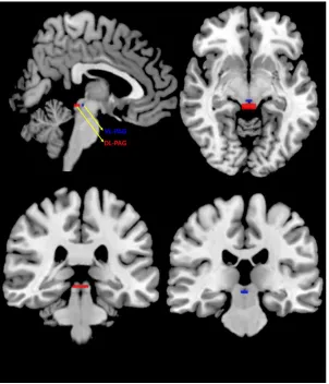
Figure 2.1 Dorsolateral and Ventrolateral PAG Regions of Interest.. ... 35
Figure 2.2 Within Group Dorsolateral and Ventrolateral PAG Functional Connectivity Patterns. ... 44
Figure 2.3 Dorsolateral PAG Connectivity with Premotor Region. ... 45
Figure 2.4 PTSD+DS Ventrolateral PAG Connectivity with Brain Regions Implicated in Depersonalization. ... 48
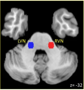
Figure 3.1 Vestibular Nuclei Seed Regions-of-Interest ... 83
Figure 3.2 Within-Group Vestibular Nuclei Functional Connectivity Patterns ... 88
Figure 3.3 Multisensory Integration. ... 101
Figure 4.1 Summary Figure of Right Ventral Anterior and Posterior Insula Functional Connectivity Patterns ... 133
Figure 4.2 Healthy Control Insula Subregion Connectivity Patterns ... 141
Figure 4.3 PTSD Insula Subregion Connectivity Patterns.ss ... 149
Figure 4.4 PTSD+DS Insula Subregion Connectivity Patterns. ... 156
Figure 4.5 Clinical Score Correlations with Insula Subregion Functional Connectivity Patterns in PTSD Patients. ... 159
Figure 4.6 Multiclass Gaussian Process Classification Machine Learning Analysis. ... 161
Figure 5.1 Oculomotor Network. ... 189
Figure 5.2 Experimental Paradigm. ... 199
xvii
field [SEF; (x: 2, y: 2, z: 62)] seed regions during the traumatic memory retrieval condition. ... 209
Figure 5.4 Explorative negative functional connectivity correlations with clinical
dissociative measures in the right supplementary eye field psychophysiological interaction during the traumatic memory retrieval condition.. ... 211
xviii
List of Appendices
Appendix A: Supplementary Data for Chapter 2 ... 239
Appendix B: Supplementary Data for Chapter 3 ... 24242
Appendix A: Supplementary Material for Chapter 2 ... 242
Appendix A: Supplementary Material for Chapter 2 ... 242
Chapter 1
1
« Introduction and Overview»
Traumatic experiences are associated with not only drastic emotional and
psychological consequences, but may also provoke aberrations in neural pathways
essential to the cognitive control of stress. Critically, traumatic stress is thought to disrupt
physiological homeostasis, with associated alterations in arousal, including hyperarousal
and hypoarousal states (Brown et al., 1985; D’Andrea et al., 2013; Frewen & Lanius,
2006; Pitman et al., 2012; Southwick et al., 1999; Vieweg et al., 2006; Yehuda et al.,
2015). Post-traumatic stress disorder (PTSD), arising in response to traumatic stressors, is
a disorder characterized by extreme arousal states, emotion dysregulation, and commonly
persistent negative alterations in cognition and mood (American Psychiatric Association,
2013). In addition, individuals with PTSD may experience intrusive memories of past
traumatic experiences and may show persistent hypervigilance concerning their
surroundings, even in the absence of threat (American Psychiatric Association, 2013;
Ehlers & Clark, 2000; Taylor, Kuch, Koch, Crockett, & Passey, 1998; Van der Kolk &
McFarlane, 1998, Yehuda et al., 2015). Notably, approximately 14-30% of traumatized
individuals present with the dissociative subtype of PTSD characterized by
depersonalization and derealization symptoms associated with emotional detachment and
hypoarousal (Armour, Karstoft, & Richardson, 2014; Blevins, Weathers, & Witte, 2014;
Bremner & Southwick, 1992; Briere, Scott, & Weathers, 2005; Cloitre, Petkova, Wang,
& Lu, 2012; Feeny, Zoellner, Fitzgibbons, & Foa, 2000; Frewen & Lanius, 2006;
Hansen, Ross, & Armour, 2017; Lanius et al., 2010; Pain, Bluhm, & Lanius, 2010; Sierra
In addition to these core cognitive and affective symptoms, individuals with
PTSD have shown alterations in sensory processing patterns, often showing extreme
hypersensitivity to reminders related to traumatic memories (Engel-Yeger, Palgy-Levin,
& Lev-Wiesel, 2013; Grillon & Morgan III, 1999; Näätänen & Alho, 1995; Shalev et al.,
2000). Here, it is possible that a compromised ability to utilize both internal and external
sensory information necessary for multisensory integration at the level of the cortex may
negatively influence the capacity to carry out higher-order executive functions.
Accordingly, delineating the neural circuitry of subcortical and cortical structures central
to sensory processing among individuals with PTSD is necessary to elucidate the
neurobiological mechanisms underlying this psychiatric disorder. It is this topic that the
current thesis addresses.
Sensory processing provides a contextual framework through which an individual
may develop an internal depiction of the external world. Moreover, understanding the
transmisson of incoming internal and external sensory information to higher-order areas
of brain is central to the study of executive functions, including goal-oriented action,
response inhibition, and emotion regulation (Alvarez & Emory, 2006; Chan, Shum,
Toulopoulou, & Chen, 2008; Fernandez-Duque, Baird, & Posner, 2000; Mazoyer et al.,
2001). Damasio & Carvalho (2013) theorized that affective feelings and sensations are
mental experiences of bodily states driven by alterations in physiological homeostasis,
which can potentiate large-scale neural systems that involve all areas of the brain,
including the brainstem, the limbic system and the cortex. Together, the subcortical and
executive functions such as decision making and emotion regulation (Bechara, Damasio
& Damasio, 2000). Here, it is critical to note that the initiation of higher-order executive
functions is thought, under some theories, to be dependent upon the raw affect and
sensations evoked at the level of the brainstem (Buck, 1999; Damasio, 1998; Davidson &
Irwin, 1999; Davidson, Jackson, & Kalin, 2000; Koelsch, 2015; Northoff et al., 2006).
For example, Paul MacLean (1990) described initially the Triune Brain Concept,
which categorizes the brain into three distinct areas: the reptilian brain, the mammalian
brain, and the human brain that together involve the brainstem, the limbic brain, and the
cortex, respectively. According to MacLean, the reptilian brain is integral to generating
raw affect and coordinating innate, reflexive responses in response to threat and
evolutionarily-relevant stimuli. By contrast, whereas the mammalian brain is thought to
evaluate subjective feelings, such as pleasure or distress, the human brain is thought
responsible for carrying out higher-order executive functions that fit into mental
constructs shaped by past experience (Schacter, Addis, & Buckner, 2007). Moreover, Jaak Panksepp (2004) expanded upon MacLean’s Triune Brain Concept, emphasizing in
particular the importance of the brainstem in affective neuroscience by suggesting that the midbrain’s role in generating raw affect can be divided into several primary process
emotional systems in the brain that carry out basal brain functioning. These systems are
thought to evoke both positive and negative affect, including care, play, lust, seek, rage,
fear, and panic (Panksepp, 1992, 1998, 2005). Together, these emotional systems are
thought to originate in medial subcortical structures, including the periaqueductal grey in
the brainstem, and are thought crucial for sensory and higher-order self-referential
the posterior cingulate cortex (Northoff et al., 2006). Here, both Northoff & Panksepp
(2008) suggest that emotionally salient stimuli may engage primitive affective responses
that originate at subcortical brainstem structures, hypothesizing further that these
structures may lay the foundation for neural transmission to both the limbic system and
the cortex.
1.1
« Brainstem Sensory Processing »
Numerous accounts suggest that sensory information derived from interoceptive and
exteroceptive processes enters the brain at the level of the brainstem initially (Craig,
2002; Craig, 2003; Critchley, 2009; Khalsa et al., 2018; Medford & Critchley, 2010;
Pezzulo, Rigoli, & Friston, 2015; Simmons et al., 2013; Stein, 1998). At the brainstem
level, incoming sensory information may, in turn, engage primary process emotional
systems described by Panksepp (2004) to elicit raw affective responses, such as panic or
rage (Cameron, 2001; Muir, Madill, & Brown, 2017; Owens, Allen, Ondobaka, &
Friston, 2018; Wiens, 2005; Zaki, Davis, & Ochsner, 2012). Northoff and colleagues
(2006) identified specific midbrain structures in the brainstem, including the
periaqueductal gray, the ventral tegmentum areas and the superior colliculus as key for
engaging primary process emotional systems that translate incoming sensory input to the
viscerosensory and the medial prefrontal cortices for self-referential processing. The
viscerosensory cortex includes the anterior cingulate cortex, the anterior insula, and the
ventromedial prefrontal cortex, with these structures thought collectively to monitor
continuously autonomic, metabolic, and immunological resources in the body necessary
to maintain physiological homeostasis. These cortical areas are further hypothesized to
grey, in order to initiate allostatic effects to maintain internal homeostasis (Barrett &
Simmons, 2015; Critchley et al. 2004; Wiens, 2005). Consistent top-down viscerosensory
cortical projections to the brainstem are thought to help minimize hyperreactivity to
continuous sensory input, as humans are thought to have developed interoceptive coding,
which predicts interoceptive input based on past experiences, such that allostatic effects
are only initiated when there are prediction errors that can disrupt physiological
homeostasis (Barrett & Simmons, 2015; Craig, 2002; Critchley, Mathias, & Dolan, 2001;
Critchley, Wiens, Rotshtein, Öhman, & Dolan, 2004; Critchley, 2005; Füstös, Gramann,
Herbert, & Pollatos, 2012; Herbert & Pollatos, 2012; Pollatos, Gramann, & Schandry,
2007).
When an individual perceives a threat that requires an immediate response, raw
affect generated at the level of the brainstem can trigger adaptive survival instincts that
evoke innate defensive behaviours (Holstege, 2014; Jansen, Van Nguyen, Karpitskiy,
Mettenleiter, & Loewy, 1995; Liddell et al., 2005; Siegel & Victoroff, 2009). This
response is often accompanied by a disruption of homeostasis within the brain and body
and is associated frequently with altered functioning of the sympathetic and
parasympathetic branches of the autonomic nervous system (Goldstein, 1987 Jansen et
al., 1995; Selye, 1973; Porges, 2009). Critically, sudden autonomic nervous system
changes may induce a stress response, which can elicit extreme hyperarousal and
hypoarousal states (Jansen et al., 1995; Paulus & Stein, 2006; Porges, 2009). On balance,
the evidence reviewed here suggests that alterations in arousal observed among
dysregulating the neural circuitry underlying transmission of affective information from
the brainstem to the cortex.
Notably, persistent alterations in arousal may predispose traumatized individuals
to be hypervigilant of their surroundings for fear of encountering trauma-related reminders. This state of defensive posturing may compromise further one’s ability to
interpret external sensory information continuously received at the supraliminal and
subliminal level (Bryant et al., 2008; Felmingham et al., 2009), thus disrupting awareness of one’s position in gravitational space (Ionta et al., 2011; Medford & Critchley, 2010).
Here, the vestibular system is thought to be imperative not only for maintaining one’s physical equilibrium but also aids in establishing the spatial orientation of one’s position
in gravitational space. External vestibular sensory information, necessary to spatial
orienting, is thought to be relayed from the inner ear to the brainstem vestibular nuclei
before eventually reaching the parieto-insular vestibular cortex (Day & Fitzpatrick, 2005;
De Waele, Baudonnière, Lepecq, Tran Ba Huy, & Vidal, 2001; Guldin & Grüsser, 1998;
Miller et al., 2008). This parieto-insular vestibular cortex spans primarily the posterior
insula and the temporoparietal junction, which are thought critical for receiving both
interoceptive and exteroceptive input, respectively (De Waele et al., 2001; Lenggenhager
& Lopez, 2015). Whereas the posterior insula is thought to be important for receiving
internal viscerosensory information, the temporoparietal junction is thought to be involved in understanding one’s self-location and self-orientation in space (Craig, 2003;
Heydrich & Blanke, 2013; Lanius et al., 2005; Simmons et al., 2013; Suzuki et al., 2013).
Importantly, if traumatized individuals experience sustained hypervigilance to their
how the cortex receives information from the internal viscera but may also have cascading effects on brain structures involved in locating one’s self in space.
Taken together, the literature reviewed here points to the brainstem as being
critical for receiving incoming raw interoceptive and exteroceptive sensory information.
Moreover, given that the brainstem is a critical relay point in the brain for neural
transmission to the cortex, examining brainstem connectivity patterns in traumatized
individuals may elucidate the underlying neural pathways associated with the alterations
in cognitions and mood observed in PTSD.
1.2
« Cortical Sensory Processing »
When interoceptive and exteroceptive information is relayed from the brainstem to the
cortex, it may have cascading effects on the three cortically-driven neurocognitive
intrinsic networks within the brain: (1) the salience network; (2) the default-mode
network; and (3) the central executive network (Menon, 2011; Seeley et al., 2007). The
salience network involves the dorsal anterior cingulate cortex and the frontoinsular cortex
and is thought to assist in filtering relevant interoceptive, autonomic, and emotional
information (Menon, 2011; Seeley et al., 2007). By contrast, the default-mode network
encompasses medial cortical structures, including the medial prefrontal cortex, the
hippocampus, the precuneus and the posterior cingulate, and is thought to be critical for
mediating self-referential processes relating to introspection and autobiographical
memory (Menon, 2011; Seeley et al., 2007). Finally, the central executive network is
thought to form a frontoparietal system, including the dorsolateral and the dorsomedial
to facilitate higher-order executive functions and goal-directed behaviours, including
emotion regulation (Menon, 2011; Seeley et al., 2007).
Importantly, the insula is critical to mediating switching between the default
mode- and executive networks (Mennon & Uddin, 2010). Here, it is thought that relevant
viscerosensory information is filtered to the insular cortex for interoceptive processing,
thus helping to identify emotional feeling states underlying incoming sensory information
(Chang et al., 2013; Couto et al., 2013). In turn, viscerosensory information relayed to the
insula is hypothesized to activate both salience processing and central-executive networks
to facilitate higher-order executive functions that assist in coordinating goal-directed
action to relevant external stimuli (Duerden et al., 2013; Kober et al., 2008; Menon &
Uddin, 2010). Here, the lateral frontoparietal central executive network converges
multiple modalities of sensory information (i.e., visual, spatial, emotional) into a coherent
multisensory perception about the environment (Ghazanfar & Schroeder, 2006; Maculoso
& Driver, 2005; Senkowski et al., 2008). Finally, when the default-mode network is
eventually reactivated after responding to salient stimuli, it assists in integrating this
sensory information into a contextual meaning that can be incorporated subsequently into the embodied representation of one’s self (Couto et al., 2013; Menon & Uddin, 2010).
As described above, one of the hallmark symptoms of PTSD involves alterations
in cognition and mood, where individuals with PTSD frequently experience persistent
negative trauma-related emotions and associated changes in perception of the self and the
world (Cox, Resnick, & Kilpatrick, 2014; Foa, Ehlers, Clark, Tolin, & Orsillo, 1999;
Frewen, Thornley, Rabellino, & Lanius, 2017). Here, cognitive functions such as emotion
integration of internal and external sensory information, as described above, is necessary
to form a coherent perception of one’s self and surroundings (Boden, Bonn-Miller,
Kashdan, Alvarez, & Gross, J.J., 2012; Cloitre, Miranda, Stovall-McClough, & Han,
2005; Ehring & Quack, 2010; Ford, 2017). Indeed, several neurophysiological studies in
PTSD reveal that PTSD is often associated with extreme sensory processing patterns,
including sensory hypersensitivity to stimuli associated with traumatic memories (such as
specific sounds, images, touch stimulation) (Engel-Yeger, Palgy-Levin, & Lev-Wiesel,
2013; Grillon & Morgan III, 1999; Näätänen & Alho, 1995; Shalev et al., 2000). It is
possible that such hypersensitivity may disrupt interoceptive signaling in individuals with
PTSD, thus altering the neural trajectory required for translation of viscerosensory
information from the brainstem to areas in the cortex linked to emotion regulation,
including the insula and the frontoparietal executive control network. In line with this
hypothesis, neuroimaging studies in individuals with PTSD point clearly to a decreased
capacity for emotion regulation, where emotional stress may alter cognitive networks that
process information about perception, salience processing and creating goal-oriented
responses. This research points to aberrations at the prefrontal cortex that may play a role
in disrupting emotion processing among individuals with PTSD, which may alter
semantic encoding of traumatic memories and the cognitive control of behavioural
responses to emotionally salient stimuli (Brown & Morey, 2012; Frewen et al., 2008;
Hayes, VanElzakker, Shin, 2012; Helpman et al., 2016; Rolle, Chick, Trivedi, Monuszko,
& Etkin, 2019).
Accordingly, delineating further the neural underpinnings of sensory processing at
appears necessary to enhance our understanding the neurobiological mechanisms
underlying this debilitating disorder.
1.3
« Objectives »
In keeping with this central objective, this thesis aimed to investigate the neural circuitry
underlying brain structures thought to be involved in sensory processing in PTSD and its
dissociative subtype. Firstly, whole brain resting state functional connectivity patterns of
brainstem structures central to interoceptive and exteroceptive processing, including the
periaqueductal gray (Chapter 2) and vestibular nuclei (Chapter 3), were examined.
Secondly, given that the insula has been identified a critical node for relaying incoming
exteroceptive and interoceptive sensory information to other neurocognitive networks in
the brain, insula resting-state connectivity patterns with the whole brain were investigated
(Chapter 4). Here, machine learning analyses were used to assess the utility of insula
resting-state connectivity patterns as a diagnostic predictor for discriminating between
individuals with PTSD, its dissociative subtype, and healthy individuals (Chapter 4).
Finally, a task-based paradigm was employed to investigate the neural mechanisms
associated with the presentation of both exteroceptive and interoceptive stimuli in
individuals with PTSD (Chapter 5). Here, we evaluated the neural circuitry underlying
horizontal eye movements (exteroceptive sensory stimulus) during simultaneous
traumatic autobiographical memory recall (interoceptive sensory stimulus) in an effort to
delineate the neurobiological mechanisms through which exteroceptive and interoceptive
sensory processing may converge to engage higher-order executive functions, such as
1.4
« References »
Alvarez, J. A., & Emory, E. (2006). Executive function and the frontal lobes: a meta
analytic review. Neuropsychology review, 16(1), 17-42.
American Psychiatric Association. (2013). Diagnostic and statistical manual of mental
disorders (5th ed.). Arlington, VA: American Psychiatric Publishing.
Armour, C., Karstoft, K. I., & Richardson, J. D. (2014). The co-occurrence of PTSD and
dissociation: Differentiating severe PTSD from dissociative-PTSD. Social
Psychiatry and Psychiatric Epidemiology, 49(8), 1297-1306.
Barrett, L. F., & Simmons, W. K. (2015). Interoceptive predictions in the brain. Nature
Reviews Neuroscience, 16(7), 419-429.
Bechara, A., Damasio, H., & Damasio, A. R. (2000). Emotion, decision making and the
orbitofrontal cortex. Cerebral cortex, 10(3), 295-307.
Blevins, C. A., Weathers, F. W., & Witte, T. K. (2014). Dissociation and posttraumatic
stress disorder: A latent profile analysis. Journal of Traumatic Stress, 27(4), 388
396.
Boden, M. T., Bonn-Miller, M. O., Kashdan, T. B., Alvarez, J., & Gross, J. J. (2012). The
interactive effects of emotional clarity and cognitive reappraisal in posttraumatic
stress disorder. Journal of Anxiety Disorders, 26(1), 233-238.
Bremner, J. D., & Southwick, S. (1992). Dissociation and posttraumatic stress disorder in
Briere, J., Scott, C., & Weathers, F. (2005). Peritraumatic and persistent dissociation in
the presumed etiology of PTSD. American Journal of Psychiatry, 162(12), 2295
2301.
Brown, M. R., Fisher, L. A., Webb, V., Vale, W. W., & Rivier, J. E. (1985).
Corticotropin-releasing factor: a physiologic regulator of adrenal epinephrine
secretion. Brain research, 328(2), 355-357.
Brown, V. M. & Morey, R. (2012). Neural systems for cognitive and emotional
processing in posttraumatic stress disorder. Frontiers in psychology, 3, 449.
Buck, R. (1999). The biological affects: a typology. Psychological review, 106(2), 301
336.
Cameron, O. G. (2001). Interoception: the inside story—a model for psychosomatic
processes. Psychosomatic medicine, 63(5), 697-710.
Chan, R. C., Shum, D., Toulopoulou, T., & Chen, E. Y. (2008). Assessment of executive
functions: Review of instruments and identification of critical issues. Archives of
clinical neuropsychology, 23(2), 201-216.
Chang, L. J., Yarkoni, T., Khaw, M. W., & Sanfey, A. G. (2012). Decoding the role of
the insula in human cognition: functional parcellation and large-scale reverse
Cloitre, M., Miranda, R., Stovall-McClough, K. C., & Han, H. (2005). Beyond PTSD:
Emotion regulation and interpersonal problems as predictors of functional
impairment in survivors of childhood abuse. Behavior Therapy, 36(2), 119-124.
Cloitre, M., Petkova, E., Wang, J., & Lu, F. (2012). An examination of the influence of a
sequential treatment on the course and impact of dissociation among women with
PTSD related to childhood abuse. Depression and Anxiety, 29(8), 709-717.
Couto, B., Salles, A., Sedeño, L., Peradejordi, M., Barttfeld, P., Canales-Johnson, A., ...
& Favaloro, R. (2013). The man who feels two hearts: the different pathways of
interoception. Social cognitive and affective neuroscience, 9(9), 1253-1260.
Cox K. S., Resnick H. S., & Kilpatrick D. G. (2014). Prevalence and correlates of
posttraumautic distorted beliefs: Evaluating DSM-5 PTSD expanded cognitive
symptoms in a national sample. Journal of Traumatic Stress, 27, 299–306.
Craig, A. D. (2002). How do you feel? Interoception: the sense of the physiological
condition of the body. Nature reviews neuroscience, 3(8), 655-656.
Craig, A. D. (2003). Interoception: the sense of the physiological condition of the
body. Current opinion in neurobiology, 13(4), 500-505.
Critchley, H. D. (2005). Neural mechanisms of autonomic, affective, and cognitive
Critchley, H. D. (2009). Psychophysiology of neural, cognitive and affective integration:
fMRI and autonomic indicants. International Journal of Psychophysiology, 73(2),
88-94.
Critchley, H. D., Mathias, C. J., & Dolan, R. J. (2001). Neural activity in the human brain
relating to uncertainty and arousal during anticipation. Neuron, 29(2), 537-545.
Critchley, H. D., Wiens, S., Rotshtein, P., Öhman, A., & Dolan, R. J. (2004). Neural
systems supporting interoceptive awareness. Nature neuroscience, 7(2), 189-195.
d'Andrea, W., Pole, N., DePierro, J., Freed, S., & Wallace, D. B. (2013). Heterogeneity
of defensive responses after exposure to trauma: Blunted autonomic reactivity in
response to startling sounds. International Journal of Psychophysiology, 90(1),
80-89.
Damasio, A. R. (1998). Emotion in the perspective of an integrated nervous
system. Brain research reviews, 26(2-3), 83-86.
Damasio, A., & Carvalho, G. B. (2013). The nature of feelings: evolutionary and
neurobiological origins. Nature Reviews Neuroscience, 14(2), 143.
Daniels, J. K., Frewen, P., Theberge, J., & Lanius, R. A. (2016). Structural brain
aberrations associated with the dissociative subtype of post‐traumatic stress
disorder. Acta Psychiatrica Scandinavica, 133(3), 232-240.
Davidson, R. J., & Irwin, W. (1999). The functional neuroanatomy of emotion and
Davidson, R. J., Jackson, D. C., & Kalin, N. H. (2000). Emotion, plasticity, context, and
regulation: perspectives from affective neuroscience. Psychological
bulletin, 126(6), 890-909.
Day, B. L., & Fitzpatrick, R. C. (2005). The vestibular system. Current biology, 15(15),
R583-R586.
De Waele, C., Baudonnière, P., Lepecq, J., Huy, P. T. B., & Vidal, P. (2001). Vestibular
projections in the human cortex. Experimental brain research, 141(4), 541-551.
Ehlers, A., & Clark, D. M. (2000). A cognitive model of posttraumatic stress
disorder. Behaviour research and therapy, 38(4), 319-345.
Ehring, T., & Quack, D. (2010). Emotion regulation difficulties in trauma survivors: The
role of trauma type and PTSD symptom severity. Behavior therapy, 41(4), 587
598.
Engel-Yeger, B., Palgy-Levin, D., & Lev-Wiesel, R. (2013). The sensory profile of
people with post-traumatic stress symptoms. Occupational Therapy in Mental
Health, 29(3), 266-278.
Feeny, N. C., Zoellner, L. A., Fitzgibbons, L. A., & Foa, E. B. (2000). Exploring the roles
of emotional numbing, depression, and dissociation in PTSD. Journal of
traumatic stress, 13(3), 489-498.
Fernandez-Duque, D., Baird, J. A., & Posner, M. I. (2000). Executive attention and
Foa E. B., Ehlers A., Clark D. M., Tolin D. F., & Orsillo S. M. (1999). The Posttraumatic
Cognitions Inventory (PTCI): Development and validation. Psychological
Assessment, 11, 303–314.
Ford, J. D. (2017). Treatment implications of altered affect regulation and information
processing following child maltreatment. Psychiatric Annals, 35(5), 410-419.
Frewen, P. A., & Lanius, R. A. (2006). Toward a psychobiology of posttraumatic self
dysregulation: Reexperiencing, hyperarousal, dissociation, and emotional
numbing. Annals of the New York Academy of Sciences, 1071(1), 110-124.
Frewen, P. A., Lanius, R. A., Dozois, D. J., Neufeld, R. W., Pain, C., Hopper, J. W., ... &
Stevens, T. K. (2008). Clinical and neural correlates of alexithymia in
posttraumatic stress disorder. Journal of Abnormal Psychology, 117(1), 171-181.
Frewen, P., Thornley, E., Rabellino, D., & Lanius, R. (2017). Neuroimaging the
traumatized self: fMRI reveals altered response in cortical midline structures and
occipital cortex during visual and verbal self- and other-referential processing in
women with PTSD. European journal of psychotraumatology, 8(1), 1314164.
Füstös, J., Gramann, K., Herbert, B. M., & Pollatos, O. (2012). On the embodiment of
emotion regulation: interoceptive awareness facilitates reappraisal. Social
cognitive and affective neuroscience, 8(8), 911-917.
Goldstein, D. S. (1987). Stress-induced activation of the sympathetic nervous
Grillon, C., & Morgan III, C. A. (1999). Fear-potentiated startle conditioning to explicit
and contextual cues in Gulf War veterans with posttraumatic stress
disorder. Journal of abnormal psychology, 108(1), 134-142.
Guldin, W. O., & Grüsser, O. J. (1998). Is there a vestibular cortex?. Trends in
neurosciences, 21(6), 254-259.
Hansen, M., Ross, J., & Armour, C. (2017). Evidence of the dissociative PTSD subtype:
A systematic literature review of latent class and profile analytic studies of
PTSD. Journal of Affective Disorders, 213, 59-69.
Hayes, J. P., VanElzakker, M. B., & Shin, L. M. (2012). Emotion and cognition
interactions in PTSD: a review of neurocognitive and neuroimaging
studies. Frontiers in integrative neuroscience, 6, 89.
Helpman, L., Marin, M. F., Papini, S., Zhu, X., Sullivan, G. M., Schneier, F., ... &
Lindquist, M. A. (2016). Neural changes in extinction recall following prolonged
exposure treatment for PTSD: a longitudinal fMRI study. Neuroimage:
clinical, 12, 715-723.
Herbert, B. M., & Pollatos, O. (2012). The body in the mind: on the relationship between
interoception and embodiment. Topics in cognitive science, 4(4), 692-704
Heydrich, L., & Blanke, O. (2013). Distinct illusory own-body perceptions caused by
damage to posterior insula and extrastriate cortex. Brain, 136(3), 790-803.
Holstege, G. (2014). The periaqueductal gray controls brainstem emotional motor
Ionta, S., Heydrich, L., Lenggenhager, B., Mouthon, M., Fornari, E., Chapuis, D., ... &
Blanke, O. (2011). Multisensory mechanisms in temporo-parietal cortex support
self-location and first-person perspective. Neuron, 70(2), 363-374.
Jansen, A. S., Van Nguyen, X., Karpitskiy, V., Mettenleiter, T. C., & Loewy, A. D.
(1995). Central command neurons of the sympathetic nervous system: basis of the
fight-or-flight response. Science, 270(5236), 644-646.
Khalsa, S. S., Adolphs, R., Cameron, O. G., Critchley, H. D., Davenport, P. W.,
Feinstein, J. S., ... & Meuret, A. E. (2018). Interoception and mental health: a
roadmap. Biological Psychiatry: Cognitive Neuroscience and
Neuroimaging, 3(6), 501-513.
Koelsch, S., Jacobs, A. M., Menninghaus, W., Liebal, K., Klann-Delius, G., von Scheve,
C., & Gebauer, G. (2015). The quartet theory of human emotions: an integrative
and neurofunctional model. Physics of life reviews, 13, 1-27.
Lanius, R. A., Vermetten, E., Loewenstein, R. J., Brand, B., Schmahl, C., Bremner, J. D.,
& Spiegel, D. (2010). Emotion modulation in PTSD: Clinical and neurobiological
evidence for a dissociative subtype. American Journal of Psychiatry, 167(6), 640
647.
Lanius, R. A., Williamson, P. C., Boksman, K., Densmore, M., Gupta, M., Neufeld, R.
W., ... & Menon, R. S. (2002). Brain activation during script-driven imagery
induced dissociative responses in PTSD: a functional magnetic resonance imaging
Lanius, R. A., Williamson, P. C., Bluhm, R. L., Densmore, M., Boksman, K., Neufeld, R.
W., ... & Menon, R. S. (2005). Functional connectivity of dissociative responses in
posttraumatic stress disorder: a functional magnetic resonance imaging
investigation. Biological psychiatry, 57(8), 873-884.
Lenggenhager, Bigna; Lopez, Christophe (2015). Vestibular Contributions to the Sense
of Body, Self, and Others. In: Metzinger, Thomas; Windt, Jennifer M. Open
MIND. Frankfurt am Main: MIND Group, pp.1-38.
Liddell, B. J., Brown, K. J., Kemp, A. H., Barton, M. J., Das, P., Peduto, A., ... &
Williams, L. M. (2005). A direct brainstem–amygdala–cortical ‘alarm’system for
subliminal signals of fear. Neuroimage, 24(1), 235-243.
MacLean, P. D. (1990). The triune brain in evolution: Role in paleocerebral functions.
New York, NY: Springer Science & Business Media.
Mazoyer, B., Zago, L., Mellet, E., Bricogne, S., Etard, O., Houdé, O., ... & Tzourio
Mazoyer, N. (2001). Cortical networks for working memory and executive
functions sustain the conscious resting state in man. Brain researchbulletin, 54(3),
287-298.
Medford, N., & Critchley, H. D. (2010). Conjoint activity of anterior insular and anterior
cingulate cortex: awareness and response. Brain Structure and Function, 214(56),
535-549.
Menon, V. (2011). Large-scale brain networks and psychopathology: a unifying triple
Menon, V., & Uddin, L. Q. (2010). Saliency, switching, attention and control: a network
model of insula function. Brain Structure and Function, 214(5-6), 655-667.
Miller, W. L., Maffei, V., Bosco, G., Iosa, M., Zago, M., Macaluso, E., & Lacquaniti, F.
(2008). Vestibular nuclei and cerebellum put visual gravitational motion in
context. Journal of neurophysiology, 99(4), 1969-1982.
Muir, K., Madill, A., & Brown, C. (2017). Individual differences in emotional processing
and autobiographical memory: interoceptive awareness and alexithymia in the
fading affect bias. Cognition and Emotion, 31(7), 1392-1404.
Näätänen, R., & Alho, K. (1995). Mismatch negativity-a unique measure of sensory
processing in audition. International Journal of Neuroscience, 80(1-4), 317-337.
Nieuwenhuys, R. (2012). The insular cortex: a review. Progress in brain research, 195,
123-163.
Northoff, G., Heinzel, A., De Greck, M., Bermpohl, F., Dobrowolny, H., & Panksepp, J.
(2006). Self-referential processing in our brain—a meta-analysis of imaging
studies on the self. Neuroimage, 31(1), 440-457.
Northoff, G., & Panksepp, J. (2008). The trans-species concept of self and the
subcortical–cortical midline system. Trends in cognitive sciences, 12(7), 259-264.
Owens, A. P., Allen, M., Ondobaka, S., & Friston, K. J. (2018). Interoceptive inference:
from computational neuroscience to clinic. Neuroscience & Biobehavioral
Pain, C., Bluhm, R. L., & Lanius, R. A. (2010). 23 Dissociation in Patients With Chronic
PTSD: Hyperactivation and Hypoactivation Patterns, Clinical and Neuroimaging
Perspectives. Dissociation and the dissociative disorders: DSM-V and beyond.
New York, NY: Springer.
Panksepp, J. (1992). A critical role for" affective neuroscience" in resolving what is basic
about basic emotions.
Panksepp, J. (1998). The periconscious substrates of consciousness: Affective states and
the evolutionary origins of the SELF. Journal of consciousness studies, 5(5-6),
566-582.
Panksepp, J. (2004). Affective neuroscience: The foundations of human and animal
emotions. New York, NY: Oxford university press.
Panksepp, J. (2005). Affective consciousness: Core emotional feelings in animals and
humans. Consciousness and cognition, 14(1), 30-80.
Paulus, M. P., & Stein, M. B. (2006). An insular view of anxiety. Biological
psychiatry, 60(4), 383-387.
Pezzulo, G., Rigoli, F., & Friston, K. (2015). Active Inference, homeostatic regulation
and adaptive behavioural control. Progress in neurobiology, 134, 17-35.
Pitman, R. K., Rasmusson, A. M., Koenen, K. C., Shin, L. M., Orr, S. P., Gilbertson, M.
W., ... & Liberzon, I. (2012). Biological studies of post-traumatic stress
Pollatos, O., Gramann, K., & Schandry, R. (2007). Neural systems connecting
interoceptive awareness and feelings. Human brain mapping, 28(1), 9-18.
Porges, S. W. (2009). The polyvagal theory: new insights into adaptive reactions of the
autonomic nervous system. Cleveland Clinic journal of medicine, 76(Suppl 2),
S86-S90.
Rolle, C., Chick, C. F., Trivedi, H. M., Monuszko, K., & Etkin, A. (2019). Transcranial
magnetic stimulation demonstrates a role for the ventrolateral prefrontal cortex in
emotion perception. Psychiatry Research, 112515.
Schacter, D. L., Addis, D. R., & Buckner, R. L. (2007). Remembering the past to imagine
the future: the prospective brain. Nature reviews neuroscience, 8(9), 657-661.
Seeley, W. W., Menon, V., Schatzberg, A. F., Keller, J., Glover, G. H., Kenna, H., ... &
Greicius, M. D. (2007). Dissociable intrinsic connectivity networks for salience
processing and executive control. Journal of Neuroscience, 27(9), 2349-2356.
Selye, H. (1973). The evolution of the stress concept. American Scientist, 61, 692-699.
Shalev, A. Y., Peri, T., Brandes, D., Freedman, S., Orr, S. P., & Pitman, R. K. (2000).
Auditory startle response in trauma survivors with posttraumatic stress disorder: a
prospective study. American Journal of Psychiatry, 157(2), 255-261.
Siegel, A., & Victoroff, J. (2009). Understanding human aggression: New insights from
Sierra, M., & Berrios, G. E. (1998). Depersonalization: neurobiological
perspectives. Biological psychiatry, 44(9), 898-908.
Simmons, W. K., Avery, J. A., Barcalow, J. C., Bodurka, J., Drevets, W. C., &
Bellgowan, P. (2013). Keeping the body in mind: insula functional organization
and functional connectivity integrate interoceptive, exteroceptive, and emotional
awareness. Human brain mapping, 34(11), 2944-2958.
Southwick, S. M., Bremner, J. D., Rasmusson, A., Morgan III, C. A., Arnsten, A., &
Charney, D. S. (1999). Role of norepinephrine in the pathophysiology and
treatment of posttraumatic stress disorder. Biological psychiatry, 46(9), 1192
1204.
Stein, B. E. (1998). Neural mechanisms for synthesizing sensory information and
producing adaptive behaviors. Experimental Brain Research, 123(1-2), 124-135.
Stein, D. J., Koenen, K. C., Friedman, M. J., Hill, E., McLaughlin, K. A., Petukhova, M.,
... & Bunting, B. (2013). Dissociation in posttraumatic stress disorder: evidence
from the world mental health surveys. Biological psychiatry, 73(4), 302-312.
Steuwe, C., Lanius, R. A., & Frewen, P. A. (2012). Evidence for a dissociative subtype of
PTSD by latent profile and confirmatory factor analyses in a civilian sample.
Depression and anxiety, 29(8), 689-700.
Suzuki, K., Garfinkel, S. N., Critchley, H. D., & Seth, A. K. (2013). Multisensory
integration across exteroceptive and interoceptive domains modulates
Taylor, S., Kuch, K., Koch, W. J., Crockett, D. J., & Passey, G. (1998). The structure of
posttraumatic stress symptoms. Journal of abnormal psychology, 107(1), 154.
Van der Kolk, B. A., & McFarlane, A. C. (1998). The Black Hole of Trauma (p. 487) in
Traumatic stress: The effects of overwhelming experience on mind, body, and
society. Guilford Press.
Vieweg, W. V. R., Julius, D. A., Fernandez, A., Beatty-Brooks, M., Hettema, J. M., &
Pandurangi, A. K. (2006). Posttraumatic stress disorder: clinical features,
pathophysiology, and treatment. The American journal of medicine, 119(5), 383
390.
Wiens, S. (2005). Interoception in emotional experience. Current opinion in
neurology, 18(4), 442-447.
Wolf, E. J., Lunney, C. A., Miller, M. W., Resick, P. A., Friedman, M. J., & Schnurr, P.
P. (2012). The dissociative subtype of PTSD: A replication and
extension. Depression and anxiety, 29(8), 679-688.
Yehuda, R., Hoge, C. W., McFarlane, A. C., Vermetten, E., Lanius, R. A., Nievergelt, C.
M., ... & Hyman, S. E. (2015). Post-traumatic stress disorder. Nature Reviews
Disease Primers, 1, 15057.
Zaki, J., Davis, J. I., & Ochsner, K. N. (2012). Overlapping activity in anterior insula
Zilverstand, A., Parvaz, M. A., & Goldstein, R. Z. (2017). Neuroimaging cognitive
reappraisal in clinical populations to define neural targets for enhancing emotion
Chapter 2
2
« fMRI functional connectivity of the periaqueductal
gray in PTSD and its dissociative subtype»
Chapter 2 has been published in its entirety as:
Harricharan, S., Rabellino, D., Frewen, P. A., Densmore, M., Théberge, J., McKinnon, M. C., Schore, A.N., & Lanius, R. A. (2016). fMRI functional connectivity of the
periaqueductal gray in PTSD and its dissociative subtype. Brain and
behavior, 6(12), e00579.https: doi.org/10.1002/brb3.579
2.1
« Introduction»
Posttraumatic stress disorder (PTSD) involves re-experiencing, avoidance, and
hyperarousal symptoms, where individuals tend to be hypervigilant of their surroundings
to ensure their own safety and to avoid exposure to threatening stimuli (Dalgleish,
Moradi, Taghavi, Nesha-Doost, & Yule, 2001; Taylor, Kuch, Koch, Crockett, & Passey,
1998; Ehlers & Clark, 2000; American Psychological Association, 2013). When a threat
is detected, PTSD patients may display hyperarousal symptoms associated with active
defensive fight and flight circuitry of the sympathetic nervous system as evidenced by
increased heart rate, skin conductance, and blood pressure (Pole, 2007). By contrast,
patients with the less common dissociative subtype of PTSD (14%) (Stein et al., 2013),
characterized by symptoms of depersonalization, often exhibit passive or submissive
defensive responses accompanied by autonomic blunting (Corrigan, Fisher, & Nutt,
2011; Lanius et al., 2005; Lanius, Bluhm, Lanius, & Pain, 2006, Lanius et al., 2010).
Schauer & Elbert (2010) recently proposed a defense cascade model aimed at
dissociative states in humans exposed to trauma are associated with a transition from
fight or flight defensive responses to more primitive animal defensive responses. These
defensive responses, evoked in passive or submissive responses to threats, include
unresponsive immobility, emotional blunting and analgesia (Baldwin, 2013; Nijenhuis,
Vanderlinden, & Spinhoven, 1998; Porges, 1995). Interestingly, Bandler and colleagues
(2000) propose that the periaqueductal gray (PAG), a small structure in the midbrain that
consists of multiple subdivisions that oppose each other in function, is a central structure
for mediating autonomic responses and is thus responsible for coordinating defensive
reactions when confronted with threatening stimuli. Specifically, this study suggested that
whereas the dorsolateral and lateral periaqueductal gray (DL-PAG and L-PAG) are
associated with sympathetic nervous system activation that evokes active defensive
strategies, the ventrolateral PAG (VL-PAG) is associated with passive coping strategies
via activation of the parasympathetic nervous system. A recent pre-clinical study by
Adamec and colleagues (2012) supported this hypothesis, where the dorsolateral PAG
was associated with anxiety-related responses to stress in rodents. By contrast, the
ventrolateral PAG exhibited a contrasting immobility or passive reaction to stress.
Kozlowksa and colleagues (2015) explicitly applied the functions of the PAG
subdivisions to the defense cascade model (Schauer & Elbert, 2010) suggesting that when
a threat is detected, DL-PAG and L-PAG subdivisions coordinate hyperarousal
symptoms, such as fight or flight responses associated with sympathetic nervous system
activity in response to threat. Here, it is believed that endocannabinoids facilitate further
release of cortisol to elicit an acute stress response from the organism. Concomitant
peripheral blood vessels and thus increase blood supply to muscles that would allow the
organism to fight the predator (George et al., 2013; Goadsby, Lambert, & Lance, 1985;
Gorzalka, Hill, & Hillard, 2008; Patel, Roelke, Rademacher, Cullinan, & Hillard, 2004).
In cases where the threat becomes inescapeable, the VL-PAG predominates as
parasympathetic nervous system activation overrides sympathetic nervous system
activation through increased vagal efferents from the dorsal motor nucleus, which in turn
may produce hypoarousal symptoms that cause a freezing or submissive shutdown response, sometimes referred to as ‘conservation withdrawal (An, Bandler, Ongur, &
Price, 1998; Porges, 2001). Projections from the VL-PAG to the medulla may play a role
in generating such defensive freezing behavior (Tovote et al., 2016), which may be
associated with the recruitment of pre-synaptic opioid receptors that mediate analgesic
relief (Musha, Satoh, Koyanagawa, Kimura, & Satoh, 1989).
Previous neuroimaging studies of the PAG (Linnman, Moulton, Barmettler,
Becerra, & Borsook, 2012) have supported functional segregation of the structure into
multiple subdivisions that vary in function, with the dorsal PAG associated with elevated
blood pressure and the ventral PAG stimulating lower blood pressure and
parasympathetic dominance. In particular, in resting-state functional connectivity studies
of the PAG in healthy populations, connectivity has been observed with the cerebellum
subcortical network as well as the thalamus and the amygdala (Tomasi & Volkow, 2011).
Critically, however, the complex neural circuitry of the PAG has not yet been delineated
in PTSD and its dissociative subtype.
Accordingly, the aim of the present study was to examine resting state functional
this patient population would exhibit greater defensive posturing even during the resting
state. An additional aim was to compare patterns of activation between individuals with
and without the dissociative subtype of PTSD. We hypothesized that all PTSD patients
would demonstrate increased functional connectivity of both PAG subdivisions with
brain regions involved in threat appraisal (dorsal anterior cingulate cortex, fusiform
gyrus; see Milad et al., 2007; Porges, 2007). Moreover, given that both PTSD and its
dissociative subtype are associated with fight-flight and concomitant hyperarousal
responses, we hypothesized that both groups would demonstrate increased DL-PAG
functional connectivity with brain structures associated with sympathetic nervous system
activity and consequent active defensive strategies, including the anterior insula and
pre-motor cortex (see Butler et al., 2007; Critchley, Nagai, Gray, & Mathias, 2011). We
hypothesized, however, that only those with the dissociative subtype of PTSD would
demonstrate VL-PAG functional connectivity with brain structures associated with
depersonalization and passive defensive responses, including the temporoparietal
junction and the rolandic operculum (see Blanke & Arzy, 2005; Daniels, Frewen,
Théberge, & Lanius, 2016; Zaytseva et al., 2015).
2.2
«Methods»
2.2.1
Clinical and Demographic Information
One-hundred and thirty-seven age-matched subjects were included in the study: 60
patients with a primary diagnosis of PTSD without the dissociative subtype (PTSD), 37
PTSD patients with the dissociative subtype of PTSD (PTSD+DS), and 40 healthy
controls. The participants were recruited by the LHSC (London Health Sciences Centre)
mental health professionals, psychology/psychiatry clinics, community programs for
traumatic-stress survivors and posters/advertisements within the London, Ontario
community.
A primary PTSD diagnosis was determined using the CAPS-IV
(Clinician-Administered PTSD Scale), which assesses 17 categorized symptoms associated with
PTSD on separate frequency and intensity scales, with the diagnosis confirmed by the
DSM-IV criteria with an additional minimum severity score of 50 (Blake et al., 1995).
PTSD patients with the dissociative subtype had the additional requirement of scoring at
least two on both the frequency and intensity scales for depersonalization or derealisation
symptoms (as per Nicholson et al., 2015 and Steuwe et al., 2014). For each participant,
co-morbid Axis-I disorders were diagnosed with the SCID (Structure Clinical Interview
for DSM-IV Axis I disorders) (First, Spitzer, Gibbon, & Williams, 2002). A battery of
questionnaires was also administered, including the Beck Depression Inventory (BDI;
Beck et al., 1997) to assess depression symptoms, the Child Trauma Questionnaire (CTQ;
Bernstein et al., 2003) to assess childhood trauma history [92 PTSD patients (PTSD+DS
and PTSD; 85%) met criteria for interpersonal childhood trauma according to CTQ
cut-off scores (Bernstein & Fink, 1998; DiLillo et al., 2006)] and the Multiscale Dissociative
Inventory (MDI; Briere, Weathers, & Runtz, 2005) to assess further dissociative
experiences. The demographic and clinical characteristics of study participants are
outlined in Table 2.1.
A one-way ANOVA was performed to assess age differences across participant groups, and a Pearson’s chi-square test was used to determine the effect of gender
assess the normal distribution of non-parametric psychological measures (CAPS, BDI,
CTQ and averaged depersonalization and derealization scores from MDI) with -post-hoc
tests to assess significant differences between groups (Kruskal & Wallis, 1952).
Exclusion criteria for all participants included metal implants that violate 3.0T
scanner safety regulations, a previous head injury associated with loss of consciousness,
current or past history of neurological disorders, significant untreated medical illness, and
pervasive developmental mental disorders. PTSD patients were excluded if they met
criteria for current or past history of bipolar or psychotic disorders, or if patients had
alcohol/substance dependency or abuse that had not sustained full remission for at least 6
months prior to study entry. Control participants were excluded if lifetime criteria were
met for any DSM-IV Axis-I psychiatric disorder
All scanning was conducted at either Robarts Research Institute’s Center for
Functional and Metabolic Mapping or Lawson Health Research Institute in London,
Ontario, Canada. The study was approved by the Research Ethics Board at Western
University of Canada. All participants provided written informed consent to partake in
the study.
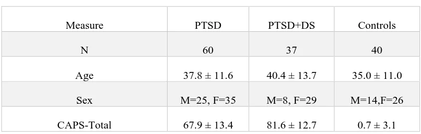
Table 2.1 Clinical and Demographic Information
Measure PTSD PTSD+DS Controls
N 60 37 40
Age 37.8 ± 11.6 40.4 ± 13.7 35.0 ± 11.0
Sex M=25, F=35 M=8, F=29 M=14,F=26
CTQ – Total 56.3 ± 24.7 68.2 ± 19.1 31.6 ± 8.6
BDI 22.8 ± 7.5 33.0 ± 10.3 1.2 ± 2.1
MDI – Total 54.1 ± 15.2 77.2 ± 22.0 33.7 ± 3.4
MDI – Depersonalization 6.6 ± 2.7 12.0 ± 5.2 5.2 ± 0.6
MDI – Derealization 8.6 ± 3.4 12.7 ± 4.0 5.2 ± 0.5
MDD n=11(24) n=23(9) -
Panic Disorder/Agoraphobia n=10(6) n=9(6) -
Social Phobia n=2(2) n=6(0) -
OCD n=3(2) n=0(2) -
GAD n=1(0) n=0(0) -
Age, sex, CAPS and self-report questionnaires (CTQ, MDI, BDI) are reported as mean ± SD. Psychiatric disorders assessed via SCID-I (MDD, Panic Disorder/Agoraphobia, Social Phobia, OCD and GAD) are reported in frequencies, as n = current(past) cases.
Abbreviations: PTSD, non-dissociative posttraumatic stress disorder patients; PTSD+DS, dissociative posttraumatic stress disorder patients; M, Males; F, Females; CAPS,
Clinician-Administered PTSD Scale; CTQ, Child Trauma Questionnaire; BDI, Beck Depression Inventory; MDI, Multiscale Dissociation Inventory; MDD, Major Depression Disorder; OCD, Obsessive Compulsive Disorder; GAD, Generalized Anxiety Disorder.
2.2.2
Data Acquisition
Whole-brain fMRI (functional magnetic resonance) data was obtained using a 3.0T scanner (Magnetom Tim Trio, Siemens Medical Solutions, Erlangen, Germany) with a 32-channel phased array head coil where the participant’s head was supported with foam padding. BOLD (blood-oxygen level dependent) fMRI data was collected using a