Interrogating the Structural Landscape of Malaria
Biomarkers with Epitope Targeted Peptide Capture Agents
Thesis by
JingXin Liang
In Partial Fulfillment of the Requirements for the degree of
Doctor of Philosophy
CALIFORNIA INSTITUTE OF TECHNOLOGY Pasadena, California
2018
ã 2018
JingXin Liang
Acknowledgements
There’s a famous proverb that says, “It takes a village to raise a child.” In my opinion, it takes no less than that – in actuality, even more – to teach, nurture, and grow a scientist. The journey towards becoming a scientist is an arduous one, filled with twists and turns, experimentation, learning on the fly, and displaying tenacity in the face of adversity. Completing a doctoral program is as much an intellectual feat as it is an exercise of persistence. As my doctoral advisor would say, “You can’t do science in a vacuum,” and he is absolutely right. There are people, both within and outside of the scientific community, who have helped me, shaped me, and contributed along my journey.
Voilà! Here is my village.
First and foremost, I thank my parents, H.X. Liang and C.W. Leung, as well as my brother, J.Y. Leung. We are a family of first-generation immigrants to America. My parents are the most selfless and brave people I know – it is through their sacrifices that their children had better lives, educations, and opportunities. I cannot even begin to fathom the amount of strength, persistence, and courage it took for them to create new lives on foreign shores, to build something from nearly nothing. From humble beginnings, they created possibilities for their children and our family. When I struggle, I think of the bravery they displayed to overcome even greater obstacles, and it gives me the perspective to push on and persevere. I would not be here without my family. My deepest gratitude and love goes out to them.
multiple disciplines. When I am at a scientific crossroads, I tend to think, “What would Jim do?” I appreciate his dedication to science, his willingness to help when I had a scientific problem, his insight and constructive criticism that helped me grow the independence to captain my own ship. I am privileged to have had the opportunity to learn from him and grow under his mentorship.
I also express gratitude for the team of scientists who are my committee members: my chair, Professor Douglas C. Rees; my secondary advisor, Professor William A. Goddard, III; and Professor Shu-ou Shan. They provided time, questions, and valuable insight during meetings and exams. I thank Professor Mitchio Okumura and Agnes Tong (Caltech Y) for the meaningful contributions they made during my first year at Caltech. I must acknowledge and thank Professor Spiridoula Matsika, who was most formative in my nascent years of scientific research at Temple University.
I thank my partner, Mark Nesbit, for his love, companionship, patience, and support. Thank you for enriching my life, for being the kind of partner with whom I can be comfortable around all the time, and a steady ship. We all have to sail through rough seas from time to time – the journey is infinitely more pleasant with you on board. I love you and I’m so proud of you. Through Mark, I’ve also had the blessing of growing my family: Dorothy and Skip, who have always treated me like their own; Katherine, my marine biologist sister; Veronica, my warrior sister. I love you all.
Kozak, Professor Zhen Lu, Professor Kurt A. Kistler, and Catherine Triandafillou, who all played significant parts leading up to today.
“A smooth sea never made a skilled sailor.”
- Franklin D. Roosevelt
Good timber does not grow with ease:
The stronger wind, the stronger trees;
The further sky, the greater length;
The more the storm, the more the strength.
By sun and cold, by rain and snow,
In trees and men good timbers grow.
- Douglas Malloch, “Good Timber”
Abstract
Antibodies have conventionally been used as molecular recognition agents against epitopes, or antigenic regions, for protein capture and detection. The ability of monoclonal and polyclonal antibodies to selectively bind their targets with high affinities makes them excellent agents for specific protein recognition. However, as large proteins themselves (~150 kDa), antibodies are susceptible to changes in pH, temperature, and biochemical environment, particularly proteolytic cleavage. Additionally, epitope binding on antibodies is reliant on their rigid tertiary structure to position key functional groups that facilitation antigen recognition. Retaining the integrity of the protein structure creates rigid limitations against chemical modifications of antibodies to suit unique needs.
Protein-catalyzed capture agents (PCCs) developed within the Heath group at Caltech address the limitation of antibodies as affinity agents. Using epitope-targeted in situ click screening methodology, the Heath group has developed peptidomimetic molecules that offer an alternative solution to antibodies. These PCCs exhibit high affinity and selectivity for their protein targets. As peptide-based molecules, PCCs can be engineered to be biochemically stable and resistant to changes in their chemical environment. Their peptide-based structures are readily amenable to chemical modifications and allow for adaptation to a range of applications.
unique challenges for protein capture. The LDH biomarker is homologous across malaria species, whereas HRP2 is highly polymorphic and lacks distinct secondary structure. The variation in sensitivity of HRP2 detection by antibody-based tests has been attributed to the genetic polymorphism of the biomarker.
In Chapter 1, we describe the development of high affinity PCCs that bind selectively to the LDH biomarker. We targeted an epitope that was highly homologous across LDH species. This chapter also details the expansion of mono-valent PCC agents into bivalent ligands using the protein architecture to select secondary ligands for binding improvement. For the HRP2 biomarker, we developed a multiple epitope targeting strategy to address protein polymorphism. We targeted for epitopes in HRP2 and developed PCCs that bind in the range of monoclonal antibodies.
Chapter 2 details the expansion of PCC agents developed against HRP2 into multivalent molecules for improved binding. The development of bivalent ligands from combinatorial screening of linker libraries is presented. The optimal linker lengths determined by the screens are described.
In Chapter 3, a general strategy for targeting the protein landscape to inhibit formation of a protein and biomolecule complex with PCCs against HRP2 is demonstrated. Specifically, the inhibition of heme sequestration by HRP2 is shown. A bivalent ligand that targets two epitopes on HRP2 is shown to have enhanced inhibitory potency over any single or cocktail combination of PCCs.
Published Content and Contributions
1. *S. Das, *A. Nag, J.X. Liang, D.N. Bunck, A. Umeda, B. Farrow, M.B. Coppock, D.A. Sarkes, A.S. Finch, H.D. Agnew, S. Pitram, B. Lai, M.B. Yu, A.K. Museth, K.M. Deyle, B. Lepe, F.P. Rodriguez-Rivera, A. McCarthy, B. Alvarez-Villalonga, A. Chen, J. Heath, D.N. Stratis-Cullum, J.R. Heath Angewandte Chemie International Edition, 2015, 54(45), 13219-13224. DOI: 10.1002/anie.201505243
- J.X. Liang conceived of the strategy, executed, and tested capture agents developed against PfHRP2. J.X. Liang also participated in the screening, synthesis, and validation of capture agents and epitopes for affinity agents against PfLDH.
2. J. R. Heath, H. Agnew, B. Farrow, D. Bunck, J.X. Liang, A. Nag, S. Das, B.T. Lai, S.M. Pitram “Il-17f-specific capture agents, compositions, and methods of using and making” US15211759, Patent Pending, Published February 23, 2017.
- J.X. Liang innovated a bivalent capture agent against PfHRP2 that demonstrated inhibition of protein function.
3. J.X. Liang, D.N. Bunck, A. Mishra, M. Idso, J.R. Heath, “Inhibition of heme
sequestration of Histidine-Rich Protein 2 using multiple epitope-targeted peptides”
Submitted 2018
Table of Contents
Acknowledgements………...iii
Abstract ………...viii
Published Content and Contributions...………...x
Table of Contents………...xi
List of Figures and Tables ………...………..xiii
Abbreviations………...…………..….…xvi
Chapter 1: An Introduction to Protein-Catalyzed Capture Agents ...1
1.1 Peptidomimetics as Antibody Alternatives in RDTs ... ...1
1.2 Peptidomimetics as Antibody Alternatives in RDTs ... 4
1.3 Engineering PCCs with High Affinity and Target Selectivity ... 6
1.4 Chapter Summaries ... 10
1.5 References ... 12
Chapter 2: Rapid Discovery of Capture and Detection Agents for Plasmodium falciparum Lactate Dehydrogenase ... 16
2.1 Introduction ... 16
2.2 Materials and Methods ... 19
2.3 Results and Discussion ... 27
2.3.1 Epitope Selection for Targeting Plasmodium LDH ... 27
2.3.2 Discovery of Capture and Detection PCCs Against PfLDH ... 29
2.3.3 Improving Affinity of the PfLDH Capture PCC Through Olefin Metathesis. . 33
2.3.4 Expansion of Anchor PCC Agents into Bivalent Ligands ... 34
2.4 Conclusion ... 41
2.5 Acknowledgements ... 42
2.6 References ... 43
Chapter 3: A Cocktail of Multi-Epitope Targeted Protein-Catalyzed Capture Agents Against Plasmodium falciparum Histidine-Rich Protein 2 ... 47
3.1 Introduction ... 47
3.2 Materials and Methods ... 50
3.3.1 A Multi-Epitope Targeting Strategy ... 50
3.3.2 Development of a Capture Agent Against PfHRP2 ... 52
3.3.3 Development of a Capture and Detection PCCs Against PfHRP2 ... 53
3.3.4 A Limitation to Epitope Targeting ... 60
3.3.5 Assessment of a Multi-Epitope Targeted Cocktail of PCCs ... 61
3.4 Conclusion ... 63
3.5 Acknowledgements ... 64
Chapter 4: A Linker Screen for Bivalent Ligands in an Unstructured Protein Landscape Using In Situ Click Chemistry ... 68
4.1 Introduction ... 68
4.2 Materials and Methods ... 70
4.3 Results and Discussion ... 72
4.3.1 Target-Guided Linker Screens... 72
4.3.2 Comparison of PfHRP2 Biligands to Anchor PCCs ... 74
4.3.3 Comparison of Dual PCC Cocktails to Biligands ... 76
4.4 Conclusion ... 79
4.5 Acknowledgements ... 79
4.6 References ... 79
Chapter 5: Inhibition of heme sequestration of Histidine-Rich Protein 2 using multiple epitope-targeted peptides ... 82
5.1 Introduction ... 82
5.2 Materials and Methods ... 86
5.3 Results and Discussion ... 91
5.3.1 Development of Macrocylic Peptide Ligands ... 91
5.3.2 Heme Binding Assays ... 92
5.3.3 Heme Binding By Native HRP2 ... 99
5.3.4 The Potency of a Bivalent Ligand ... 101
5.4 Conclusion ... 104
5.5 Acknowledgements ... 106
5.6 References ... 106
List of Figures and Tables
Chapter 1
Figure 1-1. Schematic of a lateral flow assay...2
Figure 1-2. A comparison of antibodies to PCCs...3
Figure 1-3. OBOC Library Architectures...7
Figure 1-4. Principle of SynEps...8
Figure 1-5. Schematic of a high throughput screen...9
Chapter 2 Figure 2-1. Schematic of an RDT for P. falciparum infection...18
Figure 2-2. Targeted epitopes in PfLDH...28
Figure 2-3. Structure and binding of hevwh...29
Table 2-1. Hit peptides for PfLDH Capture...30
Figure 2-4. Sandwich ELISA of capture PCC candidate...31
Figure 2-5. Structure and assays of cyHWSAN...32
Figure 2-6. Structure and assays of GHWSANRCM...34
Scheme 2-1. Schematic of target guided in situ click screening...36
Figure 2-7. Structure of GHWSANRCM anchors for screening...37
Table 2-2. Secondary ligand hits for capture PCC...37
Figure 2-8 Structure of capture biligands...38
Figure 2-9. Binding assays of capture biligands...39
Figure 2-10. Structure and assays detection biligand...40
Chapter 3 Table 3-1. Repeat motifs in PfHRP2...49
Figure 3-1. Schematic of multiple epitope targeting against PfHRP2...51
Figure 3-2. Capture PCC hits against PfHRP2...52
Figure 3-3. Assays of GHWSANRCM...53
Table 3-2. Hits against Type 2 SynEp...54
Figure 3-5. Sandwich ELISA of cyYYYKV optimizations...55
Figure 3-6. Structure of cyYYYKV improvements...56
Figure 3-7. Binding and performance of cyY4FFYRV...56
Figure 3-8. Structure of cyYKYYR monomer and intramolecular dimerization...57
Figure 3-9. Bidning of cyYKYYR monomer/dimer and performance...58
Table 3-3. Hits against N-terminal SynEp...59
Figure 3-10. Binding and performance of cyRYKHY and variants...60
Figure 3-11. Structure of Type 6 epitope and hits...60
Figure 3-12. Sandwich ELISA comparing performance of cyPWEVH to other detection PCCs...61
Figure 3-13. Sandwich ELISA of cocktails versus standalone PCCs...62
Chapter 4 Scheme 4-1. OtBu protection of Fmoc-L-propargylglycine-OH...71
Figure 4-1. Schematic of PfHRP2-guided linker screen...73
Figure 4-2. Amino acids comprising the linker region of OBO library...73
Figure 4-3. Sequenced hits from linker screen...74
Figure 4-4. General schematic of biligand structures...75
Figure 4-5. Sandwich ELISA comparing biligands to constituents...76
Figure 4-6. Structure of peglyated cyYKYYR biligand...77
Figure 4-7. Sandwich ELISA comparing performances of combinations, biligands, and constituent PCCs...78
Figure 4-8. Structure of cyYKYYR-Peg1-cyY4FFYRV...78
Chapter 5 Figure 5-1. Sequence of PfHRP2...84
Figure 5-2. Heme-binding targeting strategy...85
Figure 5-3. Electronic absoroption spectra of heme binding controls...93
Figure 5-4. Heme-binding inhibition of single PCCs...94
Table 5-1. Quantitative assessment of inhibition...95
Figure 5-6. Chemical structure of bivalent ligand...97
Figure 5-7. Comparison of heme-binding inhibition at full and half dosage...99
Figure 5-8. Heme-binding capacity of PfHRP2 (native and with fusion GST tag)...100
Figure 5-9. ∆A415 at 7 µM of heme with and without inhibitors...101
Figure 5-10. Circular dichroism spectra of PfHRP2 titrated with biligand...102
Figure 5-S1. UV-Vis absorption spectra of heme binding assay with GST control.112 Figure 5-S2. Heme binding assay with NYRWL control...112
Figure 5-S3. Heme binding assay with 10 µM CQ...113
Figure 5-S4. Heme binding assay with 10 µM L1 (cyRYKHY)...113
Figure 5-S5. Heme binding assay with 10 µM L2 (cyYKYYR)...114
Figure 5-S6. Heme binding assay with 10 µM L3 (cyY4FFYRV)...114
Figure 5-S7. Heme binding assay with 10 µM L2-P1-L3...115
Figure 5-S8. EC50 binding curves for L1, L2, L3, and L2-P1-L3...115
Figure 5-S9. UV-Vis Spectra of CQ interactions with heme...116
Figure 5-S10. UV-Vis absorption spectra of L1 with heme...117
Figure 5-S11. UV-Vis absorption spectra of L2 Interaction with heme...117
Figure 5-S12. UV-Vis absorption spectra of L3 Interaction with heme...117
Figure 5-S13. UV-Vis absorption spectra of L2-P1-L3 Interaction with Heme...118
Figure 5-S14. Absorption spectra of geme and ligands...119
Figure 5-S15. Difference absorption spectra of heme and ligands...120
Figure 5-S16. Quantification of ligand interactions with heme...121
Figure 5-S17. HPLC chromatogram for L1 purification...121
Figure 5-S18. MALDI-TOF mass spectrum of L1 (cyRYKHY)...122
Figure 5-S19. HPLC chromatogram for L2 purification...122
Figure 5-S20. MALDI-TOF Mass Spectrum of L2 (cyYKYYR)...123
Figure 5-S21. HPLC chromatograms for L3 purification...123
Figure 5-S22. MALDI-TOF Mass Spectrum of L3 (cyY4FFYRV)...124
Figure 5-S23. HPLC chromatogram for L2-P1-L3 Purification...124
Figure 5-S24. MALDI-TOF Mass Spectrum for L2-P1-L3...125
Figure 5-S25. FPLC traces of HRP2-GST and GST...125
Abbrevations
Aib (aminoisobutyric acid)
AP (alkaline phosphatase)
Az4 (Fmoc-Lys(N3)-OH, azide click handle)
BCIP/NBT (5-bromo-4-chloro-3'-indolyphosphate, nitro-blue tetrazolium) BSA (bovine serum albumin)
CuACC (Copper(I)-catalyzed azide-alkyne cycloaddition)
cy (denotes triazole cyclization, prefix*)
DCM (dichloromethane)
DIEA (N,N-Diisoproprylethylamine)
DMSO (dimethylsulfoxide)
EC50(half-maximal effective concentration)
ELISA (enzyme linked immunoassay)
FPLC (fast protein liquid chromatography)
GST (glutathione S-transferase)
HATU (2-(7-Aza-1H-benzotriazole-1-yl)-1,1,3,3-tetramethylammonium
hexafluorophosphate)
HPLC (high-performance liquid chromatography)
HRP (horseradish peroxidase)
KD (dissociation constant)
MALDI-TOF (matrix assisted laser desorption ionization-time-of-flight)
NMP (N-methylpyrrolidone)
PCC (protein-catalyzed capture agent)
PEG (polyethylene glycol)
Pra (Fmoc-L-propargylglycine-OH, alkyne click handle)
PfHRP2 (Plasmodium falciparum histidine-rich protein 2)
PfLDH (Plasmodium falciparum lactate dehydrogenase)
PvLDH (Plasmodium vivax lactate dehydrogenase) RCM (ring-closing metathesis)
RDT (rapid diagnostic test)
SynEp (synthetic epitope)
TBS (tris buffered saline)
TES (triethylsilane)
Chapter 1
An Introduction to Protein-Catalyzed Capture Agents
1.1 Peptidomimetics as Antibody Alternatives in RDTs
Conventional methods of protein capture and detection rely on the use of antibodies
as agents of molecular recognition.1 Monoclonal and polyclonal antibodies are large
proteins on the scale of ~150 kDa which exhibit high affinity and selectivity for their targets.
One application of antibodies for molecular recognition is the antibody-based rapid
diagnostic test (RDT), which is essentially a sandwich immunoassay employed as a fast
screening platform for myriad health conditions. A typical antibody-based RDT is a lateral
flow device, or dipstick, that is used to measure biomarkers in samples such as urine and
blood (Figure 1-1A).2 A capture antibody is immobilized on the surface, which is typically
nitrocellulose, that pulls down the target protein out of the biological sample by
recognizing a particular region on the antigen. A second antibody binds to an orthogonal
region of the antigen and is typically tethered to a colored particle that provides a
colorimetric readout to indicate a positive or negative confirmation. RDTs require no
trained personal and can provide a diagnostic answer in the time frame of minutes. They
are easy to use, require no trained personal or external resources such facilities, and can be
easily transported and distributed.3 As such, antibody-based RDTs have utility for disease
detection where diagnoses of large populations are required, such as global infections like
However, while RDTs provide a means for rapid disease detection, there are
drawbacks to these antibody-based platforms. Antibodies are expensive to produce, exhibit
batch-to-batch variability, and like all proteins, require storage and use in controlled pH,
temperature, and chemical environments. They are susceptible to biochemical processes
such as proteolytic cleavage and their sensitivity to abrupt changes in their environments
limitss their widespread use outside of laboratory settings. The ideal affinity agent would
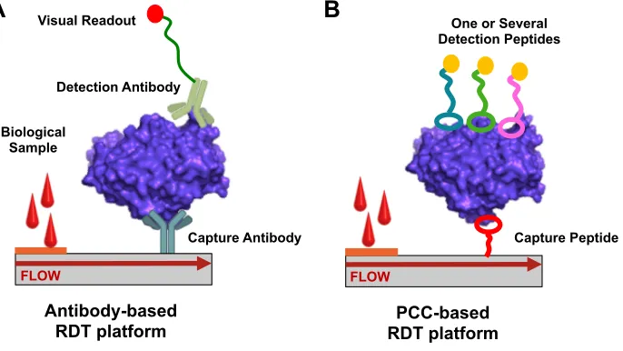
[image:19.612.159.502.273.466.2]be one that is cost-effective, easy to synthesize, and stable against biochemical fluctuations.
Figure 1-1. A typical rapid diagnostic test (RDT) is a lateral flow assay. (A) An antibody-based RDT relies on proteins to capture and detect the antigen. (B) An alternative RDT where the antibodies are replaced by PCCs.
Protein-catalyzed capture agents (PCCs), developed in collaboration between
Heath and Sharpless labs, offers an alternative to antibodies as agents of molecular
recognition.36–9,9,10 These synthetic PCCs are developed from high throughput screening
of peptide-based one-bead-one-compound (OBOC) libraries. The goal of PCC
development for diagnostics is to create pairs of molecular recognition agents that can
replace antibodies in a lateral flow assay (Figure 1-1B). Using high throughput screening
Antibody-based RDT platform
FLOW FLOW
Capture Antibody Detection Antibody
Visual Readout
Biological Sample
Capture Peptide One or Several
Detection Peptides
PCC-based RDT platform
methodology allows for the rapid discovery of new ligands with precision targeting and
engineering of affinities. Like antibodies, PCCs developed within the Heath group are
built from amino acid chains (peptides), but these peptidomimetic molecules have multiple
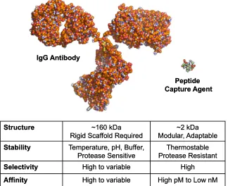
[image:20.612.164.488.191.455.2]advantages over their protein counterparts (Figure 1-2).
Figure 1-2. A comparison of antibodies to peptide-based affinity agents (PCCs) for protein capture and detection. (PDB: 1IGT)
First, PCCs have high affinity and selectivity for their protein targets, similar to
antibodies, but are developed synthetically. Peptides are cost-effective to produce relative
to antibodies and display excellent thermostability against high temperatures.11 Second,
the synthesis of small peptidomimetic molecules from amino acid building blocks allows
for increased chemical flexibility in design that cannot be accomplished with antibodies.
For example, unnatural D-amino acid buildings blocks can be used to generate PCCs that
Structure ~160 kDa
Rigid Scaffold Required
~2 kDa Modular, Adaptable
Stability Temperature, pH, Buffer,
Protease Sensitive Protease ResistantThermostable
Selectivity High to variable High
Affinity High to variable High pM to Low nM
IgG Antibody
Peptide Capture Agent
Structure ~160 kDa
Rigid Scaffold Required
~2 kDa Modular, Adaptable Stability Temperature, pH, Buffer,
Protease Sensitive Protease ResistantThermostable
Selectivity High to variable High
Affinity High to variable High pM to Low nM IgG Antibody
are resistant to proteases.9 Cyclization of the peptide backbone increases stability against
degradation and allows for chemical flexibility of affinity agent architectures. In addition,
the synthesis of PCCs on solid support allows for modular and adaptable synthesis of the
peptidomimetic scaffold. The synthetic flexibility of PCCs allows for the generation of
molecules ranging from ~1 to ~5kDa. The synthesis of PCCs on large scale can be
accomplished via automated techniques, which makes their production cost effective, and
the simplicity of their structures relative to antibodies significantly reduces batch-to-batch
variability.
1.2 PCCs as Antibody-Alternatives in Malaria Diagnostics
This thesis describes the development of PCCs as alternatives to antibodies in
lateral flow assays for malaria diagnostics. Malaria, a mosquito-borne infectious disease
caused by the protozoan Plasmodium, persists in subtropical and tropical regions of the
world. Despite widespread measures to treat and prevent the disease, malaria persists as a
global health epidemic. The disease infects over 200 million people annually and claims
over 600,000 lives.12 The most lethal species of the disease is caused by Plasmodium
falciparum, which contributes to the majority of malarial deaths. The gold standard of
disease detection is through blood smear microscopy, but its widespread use is hindered by
the need for dedicated facilities and personnel. In malaria endemic regions, RDTs are the
most cost-effective and efficient means to rapidly diagnose infection in large populations.
Currently, malaria RDTs exist that detect the antigens P. falciparum lactate
dehydrogenase (PfLDH) and histidine-rich protein II (PfHRP2) for the diagnosis of lethal
detection in instances where parasitemia, or parasite load, is low (<200 parasites/µL). This
variation in sensitivity is especially a concern for tests that detect PfHRP2, which is highly
polymorphic.4,14 The inaccuracy of diagnoses can lead to false negatives, which
contributes to untreated infections and potential death. Thus, an ideal RDT must reliably
detect antigens that are unique to P. falciparum and have the sensitivity to capture
biomarkers at low concentrations.
There are several factors that make the diagnosis of malaria infection through
traditional antibody-based RDTs problematic. First, antibodies are sensitive to heat and
humidity, which are less than ideal for the climates in which malaria persists. Second,
PfHRP2 antigen detection tests have been shown to exhibit variable performance, which
has been attributed to the genetic variations of the protein across regions of the world.4,14–
16 In total, over 400 isolates of PfHRP2 exist globally.4 These isolates have variations in
repeat sequences of the proteins or deleted sections,94,14 making antigen detection
problematic if monoclonal antibodies in RDTs are targeted against such regions. Another
issue is the existence of P. falciparum isolates lacking the pfhrp2 or pfhrp3 genes which
encode for PfHRP2 and its homolog, PfHRP3.17 The complete absence of PfHRP2 antigen
would result in false positives, which underscores the importance of RDTs that reliably
detect PfLDH protein. However, PfLDH antigen tests also exhibit issues of their own
which include low specificity.13 The variability in detection of malaria infection results in
under- and over-diagnosis of the disease. Poorly implemented treatment programs that
result from misdiagnosis can contribute to antimalarial drug resistance, which also
increases malaria-related fatalities.18,19 In order to treat and eradicate malaria, the
management, disease surveillance, and properly implemented drug treatment programs. In
this thesis, we leverage and expand upon existent technology in the Heath group to innovate
PCCs that address the shortcomings on antibodies with applications towards future RDTs
for malaria.
1.3 Engineering PCCs with High Affinity and Target Selectivity
Within the Heath group, PCCs have been developed through combinatorial
screening of combinatorial OBOC peptide libraries of varying architectures (Figure 1-3).
Earlier screening methodology utilized randomized 5-mer linear peptide libraries.7,9,10,20
These libraries were synthesized on bead via split-and-mix synthesis using amino acids of
L- or D-stereochemistry and yielded affinity agents that bound in micromolar to nanomolar
affinities (Figure 1-3A).7,9,20 Later PCC methodology utilized libraries that were cyclized
through clicking between azide and alkyne or ring closing metathesis, the latter of which
afforded a larger, more flexible ring structure (Figure 1-3B). Cyclization of the 5-mer
libraries restricts the numbers of conformations accessible to PCCS. Since linear PCCs
sample more conformations than cyclic PCCs, they exhibit lower binding affinities.
Conformational restriction through cyclization drastically limits the conformation
accessible through bond rotation, which can be thought of as prepaying the entropic cost
of PCC:target binding. In later work within the Heath group, cyclic PCCs have
demonstrated affinities in the picomolar regime.7,21 The OBOC libraries are
comprehensive in 18 of naturally occurring amino acid side chains with cysteine and
methionine eliminated for chemical stability which yields 185, or roughly 2 million, diverse
with an azide or alkyne click handle, also denoted as Az4 or Pra, respectively. Linear
libraries require no cyclization and are immediately appended with a handle. The libraries
are validated for confirmation of cyclization and successful synthesis by sequencing on an
Edman instrument.
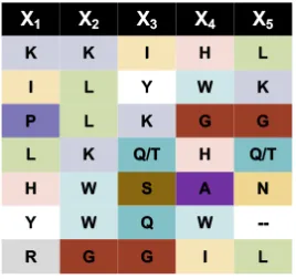
Figure 1-3. Architectures of OBOC libraries used for high throughput screening. The
libraries are comprehensive in 18 amino acids, can be constructed with L- or D- stereocenters, and bear alkyne/Pra or azide/Az4 click handles.
Standard development of PCCs within the Heath group first begins with epitope
targeting.7,8,11,20,21 Epitopes are antigenic determinants, also known as amino acid
sequences which are recognized by antibodies. After identification of the antigen, one or
several epitopes are selected after inspection of the primary protein sequence. Ideally, this
epitope is a minimum of ~10 amino acids in length, and is unique to the target, thus
engineering selectivity into the final PCC. It is also beneficial to select an epitope that is
accessible to an affinity agent, i.e., not buried within the protein core, and that has some
secondary structure that might be maintained in a synthetic peptide. However, defined H2N
O N3 N H O H N O N H O H N O N H O X1 X2 X3 X4 X5 NH
X = Arg, His, Lys, Asp, Glu, Ser, Thr, Asn, Gln, Gly, Pro, Ala, Val, Ile, Leu, Phe, Tyr, Trp, 4FPhe
NH NH HN H N HN NH NH O X1 O X2 O X3 O X4 O X5 O N O HN NN O NH2 N3 A B
Linear 5-mer peptide library with click handle
Cyclic 5-mer peptide library with click handle
Triazole Cyclization Ring Closing Metathesis i, i + 7 Cyclization
secondary structure is not a necessarily a prerequisite for generating high-binding hits in
screens. Following selection, the synthetic epitope (SynEp) is constructed via standard
solid phase peptide synthesis, fitted with an azide or alkyne click handle that is
complementary to the one on the OBOC library, and appended with a biotin tag for
detection. (Figure 1-4).
Figure 1-4. A targeted peptide sequence (epitope) is selected from a protein and synthesized (SynEp). The SynEp is affixed with an azide/Az4 (shown) or alkyne/Pra click handle and appended with a biotin tag for detection by an antibody. (PDB: 1LDG)
A general protocol of pre-clearing followed by screening of the OBOC library is
followed (Figure 1-5) which includes non-specific binding to detection antibodies, such as
anti-biotin or streptavidin, and human serum to yield a focused library. For increased
selectivity in the final PCC, the library can be pre-screened against SynEps from off-target
proteins to weed out binding to homologous biomolecules or even adjacent sites on the
target. This focused library is then incubated with the SynEp from the target of interest.7
Library elements that bind strongly to the SynEp position the complementary alkyne and
azide click handles together to form a covalent triazole product.
12 amino acid SynEp
Click Handle
Biotin Tag (Detection) Target
The classic Copper(I)-catalyzed azide-alkyne cycloaddition (CuACC) reaction
forms a covalent heterocyclic triazole product.22 The Copper(I) catalyst increases the rate
of reaction between azide and alkyne with complete conversion. In the absence of the
Copper(I) catalyst in our screens, the triazole click product is low-yielding. Covalent
linkage is entirely dependent on the molecular recognition between the SynEp and the
library elements to hold the azide and alkyne fragments in close enough proximity to
facilitate triazole formation.6 Thus, it is largely the strength, or affinity, of binding between
the SynEp and library elements that promote the click product in the absence of Copper(I).
The affinity-driven catalysis of the click reaction allows for the development of PCC agents
[image:26.612.155.492.350.572.2]with nanomolar affinities in a single-generation screen.7
Figure 1-5. Schematic of a high throughout screen using epitope targeting, in situ click chemistry, and OBOC library methodologies. False-positives such as binders to detection antibodies, off-target epitopes, and human serum are removed from the OBOC library in a pre-clear. The pre-cleared library is incubated with the SynEp that bears a click handle complementary to the one on bead. Strong library binders promote formation of the covalent click product.
Pre-Screen
Library of ~500,000 beads
1. Detection Ab 2. Off-Target Epitopes 3. Human Serum
✗ Strip Library Screen Target Epitope(s) BIOTIN BIOTIN Tz Tz
X1X 2X3X4X5
1.4 Chapter Summaries
This thesis presents the application of the aforementioned PCC technology for the
development of peptidomimetics in malaria RDTs that detect lethal P. falciparum infection.
In Chapter 2, the development of monovalent and bivalent ligands against the PfLDH and
Pan-Plasmodium LDH antigens is described. The use of cyclic OBOC libraries over linear
libraries demonstrates the superiority of conformational restriction in generating peptide
binders with nanomolar affinities. The expansion of PCCs into bivalent ligands through
secondary ligand screens with the target antigen is also described within this chapter. In
the instance of a linear peptide, we find that expansion into a bivalent structure can increase
affinity.
Chapter 3 describes the development of cocktail combination of PCCs to bind the
PfHRP2 antigen. Unlike PfLDH, PfHRP2 is a highly patterned and polymorphic protein
that lacks defined secondary structure. The primary amino acid sequence of this antigen
differs across P. falciparum isolates and has been suggested to contribute to the variations
in sensitivity observed in RDTs that detect PfHRP2.4,14–16 To address PfHRP2
polymorphism, we devised a strategy where we target four unique epitopes within the
protein. By developing a cocktail of affinity agents that target conserved and variant
regions of PfHRP2, we can account for variations or deletions in repeats through
simultaneously binding multiple regions in a single protein for built in sensitivity
amplification. In total, four regions of the PfHRP2 antigen are targeted.
Chapter 4 describes the expansion of PfHRP2 monovalent ligands into bivalent
ligands through OBOC linker screens. A small library of peptide-based linkers was
Chapter 3. Even without defined secondary structure in the target antigen, we found that
the linker screens selected for a defined ligation length though the hits generated were
nonspecific. Knowledge obtained from this work was applied towards development of a
bivalent ligand that demonstrated inhibitory properties against PfHRP2 function as
described in Chapter 5.
In addition to its diagnostic utility, PfHRP2 has long been implicated as a target for
the sequestration of cytotoxic free heme in the malaria parasite. In Chapter 5, the PCCs
developed in Chapter 3 are demonstrated to inhibit the sequestration of PfHRP2 binding to
heme through targeting heme-binding epitope motifs. Through ligation of two PCCs that
target internal regions of PfHRP2, we find that inhibition of heme sequestration is
significant and improved over using monovalent peptides.
Altogether, this thesis describes the interrogation of the structural landscape of two
malarial proteins with unique diagnostic and scientific challenges. Firstly, PCCs were
developed against PfLDH, a highly homologous biomarker with a defined protein
architecture for the generation of mono- and bivalent affinity agents. Secondly, PCCs were
developed that could simultaneously target multiple regions in a single and unstructured
biomarker with the added ability to inhibit protein function. Through the use of epitope
targeting and in situ chemistry, PCCs were developed that bind to their P. falciparum
targets with high affinity and selectivity. In addition, we demonstrate that the PCCs have
utility outside of diagnostics and can inhibit protein and biomolecule interactions, showing
References
(1) Borrebaeck, C. A. . Antibodies in Diagnostics – from Immunoassays to Protein
Chips. Immunol. Today2000, 21 (8), 379–382.
(2) Koczula, K. M.; Gallotta, A. Lateral Flow Assays. Essays Biochem.2016, 60 (1),
111–120.
(3) Mouatcho, J. C.; Goldring, J. P. D. Malaria Rapid Diagnostic Tests: Challenges
and Prospects. J. Med. Microbiol.2013, 62 (10), 1491–1505.
(4) Baker, J.; Ho, M.-F.; Pelecanos, A.; Gatton, M.; Chen, N.; Abdullah, S.; Albertini,
A.; Ariey, F.; Barnwell, J.; Bell, D.; et al. Global Sequence Variation in the
Histidine-Rich Proteins 2 and 3 of Plasmodium Falciparum: Implications for the
Performance of Malaria Rapid Diagnostic Tests. Malar. J.2010, 9 (1), 129.
(5) Birku, Y.; Welday, D.; Ayele, D.; Shepherd, A. Rapid Diagnosis of Severe Malaria
Based on the Detection of PfHRP-2 Antigen. Ethiop Med J1999, 37.
(6) Agnew, H. D.; Rohde, R. D.; Millward, S. W.; Nag, A.; Yeo, W.-S.; Hein, J. E.;
Pitram, S. M.; Tariq, A. A.; Burns, V. M.; Krom, R. J.; et al. Iterative In Situ Click
Chemistry Creates Antibody-like Protein-Capture Agents. Angew. Chem. Int. Ed
Engl.2009, 48 (27), 4944–4948.
(7) Das, S.; Nag, A.; Liang, J.; Bunck, D. N.; Umeda, A.; Farrow, B.; Coppock, M. B.;
Sarkes, D. A.; Finch, A. S.; Agnew, H. D.; et al. A General Synthetic Approach for
Designing Epitope Targeted Macrocyclic Peptide Ligands. Angew. Chem. Int. Ed
(8) Lai, B. T.; Wilson, J. A.; Malette Loredo, J.; Pitram, S. M.; LaBerge, N. A.; Heath,
J. R.; Agnew, H. Epitope Targeted Macrocyclic Peptide Ligand with Picomolar
Cooperative Binding to Interleukin-17F. Chem. – Eur. J. n/a-n/a.
(9) Deyle, K. M.; Farrow, B.; Qiao Hee, Y.; Work, J.; Wong, M.; Lai, B.; Umeda, A.;
Millward, S. W.; Nag, A.; Das, S.; et al. A Protein-Targeting Strategy Used to
Develop a Selective Inhibitor of the E17K Point Mutation in the PH Domain of
Akt1. 2015, 7, 455.
(10) Millward, S. W.; Henning, R. K.; Kwong, G. A.; Pitram, S.; Agnew, H. D.; Deyle,
K. M.; Nag, A.; Hein, J.; Lee, S. S.; Lim, J.; et al. Iterative in Situ Click Chemistry
Assembles a Branched Capture Agent and Allosteric Inhibitor for Akt1. J. Am.
Chem. Soc.2011, 133 (45), 18280–18288.
(11) Pfeilsticker, J. A.; Umeda, A.; Farrow, B.; Hsueh, C. L.; Deyle, K. M.; Kim, J. T.;
Lai, B. T.; Heath, J. R. A Cocktail of Thermally Stable, Chemically Synthesized
Capture Agents for the Efficient Detection of Anti-Gp41 Antibodies from Human
Sera. PLOS ONE2013, 8 (10), e76224.
(12) WHO. World Malaria Report 2016. World Health Organization December 2016.
(13) Houzé, S.; Boly, M. D.; Le Bras, J.; Deloron, P.; Faucher, J.-F. PfHRP2 and
PfLDH Antigen Detection for Monitoring the Efficacy of Artemisinin-Based
Combination Therapy (ACT) in the Treatment of Uncomplicated Falciparum
Malaria. Malar. J.2009, 8, 211–211.
(14) Baker, J.; McCarthy, J.; Gatton, M.; Kyle, D. E.; Belizario, V.; Luchavez, J.; Bell,
2 (PfHRP2) and Its Effect on the Performance of PfHRP2-Based Rapid Diagnostic
Tests. J Infect Dis2005, 192.
(15) Lee, N.; Baker, J.; Andrews, K. T.; Gatton, M. L.; Bell, D.; Cheng, Q.; McCarthy,
J. Effect of Sequence Variation in Plasmodium Falciparum Histidine Rich Protein
2 on Binding of Specific Monoclonal Antibodies: Implications for Rapid
Diagnostic Tests for Malaria. J Clin Microbiol2006, 44.
(16) Kumar, N.; Singh, J. P.; Pande, V.; Mishra, N.; Srivastava, B.; Kapoor, R.;
Valecha, N.; Anvikar, A. R. Genetic Variation in Histidine Rich Proteins among
Indian Plasmodium Falciparum Population: Possible Cause of Variable Sensitivity
of Malaria Rapid Diagnostic Tests. Malar. J.2012, 11, 298–298.
(17) Menegon, M.; L’Episcopia, M.; Nurahmed, A. M.; Talha, A. A.; Nour, B. Y. M.;
Severini, C. Identification of Plasmodium Falciparum Isolates Lacking
Histidine-Rich Protein 2 and 3 in Eritrea. Infect. Genet. Evol.2017, 55, 131–134.
(18) Le Bras, J.; Durand, R. The Mechanisms of Resistance to Antimalarial Drugs in
Plasmodium Falciparum. Fundam. Clin. Pharmacol.2003, 17 (2), 147–153.
(19) White, N. J. Antimalarial Drug Resistance. J. Clin. Invest.2004, 113 (8), 1084–
1092.
(20) Nag, A.; Das, S.; Yu, M. B.; Deyle, K. M.; Millward, S. W.; Heath, J. R. A
Chemical Epitope-Targeting Strategy for Protein Capture Agents: The Serine 474
Epitope of the Kinase Akt2. Angew. Chem. Int. Ed.2013, 52 (52), 13975–13979.
(21) Farrow, B.; Wong, M.; Malette, J.; Lai, B.; Deyle, K. M.; Das, S.; Nag, A.; Agnew,
Target-Guided Synthesis of a Potent In-Cell Inhibitor of Botulinum Neurotoxin. Angew.
Chem. Int. Ed.2015, 54 (24), 7114–7119.
(22) Kolb, H. C.; Sharpless, K. B. The Growing Impact of Click Chemistry on Drug
Chapter 2
Rapid Discovery of Capture and Detection Agents for
Plasmodium
falciparum
Lactate Dehydrogenase
Reproduced in part with permission from:
*S. Das, *A. Nag, J.X. Liang, D.N. Bunck, A. Umeda, B. Farrow, M.B. Coppock, D.A.
Sarkes, A.S. Finch, H.D. Agnew, S. Pitram, B. Lai, M.B. Yu, A.K. Museth, K.M. Deyle,
B. Lepe, F.P. Rodriguez-Rivera, A. McCarthy, B. Alvarez-Villalonga, A. Chen, J. Heath,
D.N. Stratis-Cullum, J.R. Heath
Angewandte Chemie International Edition, 2015, 54(45), 13219-13224.
DOI: 10.1002/anie.201505243
2.1 Introduction
The mosquito-borne disease malaria is caused by the protozoan parasite,
Plasmodium, and infects over 200 million human hosts annually in tropical and subtropical
regions of the world.1 The P. falciparum species is the most lethal and contributes to nearly
all malaria related deaths. Eradication of this global epidemic requires the rapid and
accurate diagnosis of the large human populations affected. Lateral flow
immunochromatographic assays, also known as rapid diagnostic tests (RDTs), provide a
means of rapidly diagnosing large populations with results in minutes, which allows for
sandwich assays where a protein bioarker is captured by one antibody and detected by a
second antibody, and the colorimetric readouts provide a positive or negative result.7
Though the gold standard of malarial disease detection is microscopy,4,5 RDTs are
advantageous because they do not require dedicated facilities or personnel for examination
of patient samples. Relative to other techniques, RDTs are relatively low-cost and provide
results within 5 to 15 minutes.
There are commercially available RDTs for the P. falciparum histidine-rich protein
2 (PfHRP2) and lactate dehydrogenase (PfLDH) biomarkers, the latter of which is the focus
of this chapter.2,3,6 PfLDH is key enzyme utilized by the parasite for glycolysis and energy
production and interconverts lactate and pyruvate while simultaneously converting
NADH/NAD+.8 The LDH enzymes exists in other malarial species such as P. vivax, with
which it is nearly 75% genetically identical with 90% residue similarity.9,10 Since P.
falciparum infection is so severe, it is imperative that malarial LDHs can be differentially
detected in a single test. In addition to diagnostics, the selective detection of PfLDH
antigen by RDT is useful for assessing parasite burden. Unlike another P. falciparum
-specific antigen, histidine-rich protein 2 (PfHRP2), which is detected in malaria RDTS and
persists even after treatment, the PLDH antigen clears from the blood within 24 hours after
infection clearance, which holds prognostic utility.11–14 The rapid clearance of the antigen
is imperative for patient case management, determining the success of drug treatments such
as artemisinin combination therapy, and for identifying
recurrent malaria infections.11,15
Currently, PLDH RDTs are available that can detect P. falciparum-specific or
(schematic provided in Figure 2-1).5 However, whilst malaria RDTs provide a means of
rapidly diagnosing patients infected with P. falciparum for the administration of drug
treatments, they have limitations. At high parasitemia, or high parasite densities, greater
than 90% of sensitivities are achieved for P. falciparum specific RDTs. However, at low
parasitemia where parasite density varies from 100 – 500 parasites/µL, sensitivities of these
antibody-based RDTs can drop below 80%.15 Antibody-based RDTS, particularly for
PfLDH detection, have been shown to be sensitive to temperature fluctuations and generate
false negatives under heat.2 The degradation in performance at higher temperatures is
especially problematic for diagnostics in the tropical and subtropical climates in which
malaria is prevalent.2 PLDH-based RDTs are also less sensitive than their PfHRP assay
counterparts and cannot always detect clinical infection.12
Figure 2-1. Schematic of an RDT for P. falciparum-specific and pan-Plasmodium
infection. Representative results for (A) P. falciparum and possible mixed infection, (B) non-P. falciparum-specific infection, and (C) a negative result for malarial infection are shown. Figure adapted from the literature.5
This chapter focuses on the development of reagents that address the limitations of
targeting PfLDH antigen in antibody-based RDTs.2,5,14 An ideal RDT (schematic provided Capture
Detection
Control
pLDH
PfLDH
+ / + + / - /
in Figure 2-1) for malaria should be able to distinguish between species infection, generate
true positive/negative results, withstand temperature fluctuations for use in endemic
regions of the world, be cost-effective, and be sensitive in low to high parasitemia. For
prompt treatment of lethal malaria, it is especially important to distinguish P. falciparum
from other Plasmodium species from an RDT.
In this chapter, we describe the development of peptidomimetic protein-catalyzed
capture agents (PCCs) as synthetic, easily synthesizable, and cost-effective antibody
alternatives for PfLDH RDTs. We use epitope-targeted in situ click chemistry screening
methodology,16–18,18–20 which allows for the rapid discovery of peptide-based ligands from
high throughput screening of one-bead-one-compound (OBOC) to develop affinity agents
against PfLDH. We use an epitope targeting strategy to engineer PCCs with specificity for
PfLDH over PvLDH and other off-target proteins. A comparison of PCCs developed from
linear and cyclic OBOC libraries is provided that demonstrates the superior utility of
entropy restricted structures for affinity agents. We also explore using the structured
landscape of PfLDH antigen to screen for secondary ligands to develop bivalent PCCs.
2.2 Materials and Methods
Materials. Epitope and peptide syntheses were accomplished using standard Fmoc amino
acids with acid-labile side chain protecting groups, which were purchased from Anaspec,
Chempep, Chem-Impex International, and Aapptec. Specialty amino acids such as
Fmoc-NH-Pegn-CH2CH2CO2H, (S)-N-Fmoc-2-(4’-pentenyl)alanine, and
(R)-N-Fmoc-2-(7’-octenyl)alanine were purchased from Chempep and Sigma Aldrich, respectively. Grubbs
Aldrich. All peptide syntheses were completed on solid support with standard
methodology using Biotin NovaTagTM resin (Millipore Sigma) for biotinylated peptides or
Rink Amide MBHA resin (Aapptec). The reagents N-Methyl-2-pyrrolidone (NMP, BDH
Chemicals), N,N-Diisoproprylethylamine (DIEA, Sigma Aldrich),
2-(7-Aza-1H-benzotriazole-1-yl)-1,1,3,3-tetramethylammonium hexafluorophosphate (HATU,
Chempep), and piperidine (Alfa Aesar) were used in synthesis. Trifluoroacetic acid (TFA,
Chem-Impex International) and 2.5% triethylsilane (TES, Sigma Aldrich) were used for
removal of peptides from solid support. TentaGel S-NH2 resin (Rapp Polymere) was used
for OBOC library synthesis. Copper (I) iodine (CuI) and L-Ascorbic Acid were purchased
from Sigma Aldrich. Sodium diethyldithiocarbamate was purchased from Chem-Impex
International. Dimethyl sulfoxide (DMSO) was purchased from EMD Millipore.
Recombinant PfLDH and PfHRP2 antigens were purchased from CTK Biotech.
Human LDH was obtained form Abnova. Anti-biotin alkaline phosphatase antibody was
purchased from Sigma-Aldrich. Anti-GST antibody conjugated to HRP was purchased
from Abcam. For screening, 5-bromo-4-chloro-3’-indolylphosphate p-toludine
salt/nitro-blue tetrazolium chloride (BCIP/NBT Color Development Substrate) was purchased from
Promega.
General Preparation of OBOC Libraries. Linear OBOC libraries were synthesized on
90 µm TentaGel resin using standard split-and-mix synthesis on a 5g scale to generate a
combinatorial mixture of pentameric peptides. The 18 standard L-amino acids,
Fmoc-X-OH, where X= Ala, Val, Leu, Ile, Pro, Phe, Trp(Boc), Gly, Ser(tBu), Thr(tBu), Tyr(tBu),
with methionine and cysteine omitted for chemical stability. D-libraries were generated
using the enantiomers of the standard amino acids. All syntheses were accomplished using
standard solid phase techniques from the C- to N-terminus on a Titan 357 Peptide
Synthesizer (Aapptec) with 0.2 M amino acid, 0.2 M HATU, 2 M DIEA solutions, and
20% piperidine/NMP solutions. An azide or alkyne click handle was appended to the
N-termini of each library after synthesis of the pentameric region. Cyclic OBOC libraries
were generated in the same manner with the addition of azide and alkyne amino acids
(alkyne-X1X2X3X4X5-azide). The libraries were cyclized overnight by treatement with 2
equivalents of CuI and 5 equivalents of ascorbic acid in 20% piperidine/NMP. The copper
was removed by washing with 2% sodium diethyldithiocarbamate (w/v) and 2% DIEA (v/v)
in NMP. An azide of alkyne click handle was then appended to the N-termini of the
cyclized libraries. The side chain protecting groups were removed by treatment with 95%
TFA, 2.5% TES, and 2.5% deionized water for 2 hours. The libraries were validated for
cyclization and completion by sequencing via Edman degradation on a 494 CLC Procise
Sequencer (Life Technologies). All reactions were performed at ambient temperature.
General Peptide Synthesis. All resin was preswelled in NMP for a minimum of 2 hours
prior to synthesis. Fmoc groups were removed by treatment of 2 x 20 minutes in 20%
piperidine/NMP. Couplings were accomplished using excesses of 4 equivalents of amino
acid, 4 equivalents of HATU, and 12 equivalents of DIEA in NMP. Washes were
performed between all steps with NMP. Peptides were cleaved off the resin by treatment
with 95% TFA, 2.5% TES, and 2.5% deionized water for 2 hours with agitation. The
centrifugation and resuspended in minimal DMSO for purification. All reactions were
performed at ambient temperature.
Protocols for Cyclization. For click cyclization, peptides bearing the azide and alkyne
handles were subjected to 2 equivalents of CuI and 5 equivalents of ascorbic acid overnight
at ambient temperature. Following the overnight reaction, the peptides were washed with
2% sodium diethyldithiocarbamate (w/v) and 2% DIEA (v/v) in NMP, followed by
rigorous rinsing with NMP. Peptides cyclized by ring closing metathesis (RCM) had the
specialty peptides, (S)-N-Fmoc-2-(4’-pentenyl)alanine, and
(R)-N-Fmoc-2-(7’-octenyl)alanine, in place of the azide and alkyne click handles. The resin was dried down
in DCM before resuspension in anhydrous DCE. The resin was placed under inert
atmosphere and subjected to 8 mM Grubbs catalyst prepared in anhydrous DCE. The
reaction was allowed to stir under argon for 6 hours, at which time all solution was drained
from the flask, and fresh 8mM Grubbs catalyst solution was added. The reaction was
allowed to proceed overnight under inert atmosphere at room temperature. Following the
RCM procedure, the resin was washed rigorously with 10% sodium diethyldithiocarbamate
(w/v) and 5% DIEA (v/v) in dimethyl formamide (DMF). Synthesis, cleavage, and
characterization were accomplished as described.
Purification and Characterization. Synthetic epitopes and peptide hits were purified via
reversed phase high-performance liquid chromatography on either a Beckman Coulter
HPLC instrument with a Luna 10 µm C18(2) 100A column. Gradients were composed of
Peptides were characterized after purification by matrix assisted laser desorption ionization
time-of-flight (MALDI-TOF) mass spectrometry. Product fractions were collected,
lyophilized, and reconstituted for quantification prior to assays. Peptides were quantified
by determination of extinction coefficients and measurement of absorbance at 280 nm on
a ThermoFisher Nanodrop 2000c UV-Vis spectrophotometer.
Screening Protocols. All screening protocols were conducted using binding buffer (20
mM Tris, 150 mM NaCl, 0.1% bovine serum albumin, 0.05% Tween 20, pH 7.5) unless
otherwise denoted. High salt buffer (25 mM Tris, 750 mM NaCl, 10 mM MgCl2, pH 7.5)
and BCIP buffer (100 mM Tris, 10 mM NaCl, 1 mM MgCl2, pH 9) were also used. All
steps were performed at ambient temperature unless otherwise denoted. All screens were
conducted on 300 to 500 mg of library beads bearing the combinatorial pentamers.
General Library Preclear and Epitope Antiscreen. Step 1: The library beads were swelled
in buffer with 0.05% TWEEN20 for 6 hours at 4°C. Step 2: The library was incubated with
7.5 µM of scrambled epitope (where applicable) for 6 hrs. After incubation, the library
beads were washed for 3 x 5 minutes buffer, 3 x 5 minutes in TBST (TBS + 0.05%
TWEEN20), and 3 x 5 minutes in TBS (standard wash). Non-covalent binders were
removed by a 2 hour wash with guanidinium HCl solution (pH 2). Step 3: The library was
treated with anti-biotin alkaline phosphatase at 1/10000 dilution and developed with
BCIP/NBT for 15 to 25 minutes. The blue/purple beads (nonspecific hits) were discarded
and the clear beads were retained for subsequent screens. Step 4: The clear beads were
They were then agitated in guanidine hydrochloride for 3 hours followed by 10 x 5 minutes
washes in deionized water.
General Human Antiscreen. Step 1: Library beads were swelled in binding buffer until
homogenous. Step 2: Beads were blocked in 5% milk in buffer overnight. Step 3: The
beads were washed 3 x 5 minutes in buffer followed by incubation with 1% filtered human
serum in 0.5% milk for 1 hour. Step 4: The beads were washed 3 x 5 minutes in buffered
followed by incubation with 1/10000 rabbit anti-whole human serum polyclonal antibody
in 0.5% milk in buffer for 1 hour. Step 5: The beads were washed 3 x 5 minutes in buffer
before incubation with 1/10000 goat anti-rabbit IgG polyclonal antibody in 0.5% milk in
TBS for 1 hour. Step 6: The beads were washed 3 x 5 minutes in high salt TBS followed
with an additional 1 hour wash. The beads were washed with BCIP buffer, pH 9. Step 7:
The beads were developed with BCIP/NBT as described above.
General Product Screen (Single Ligand). Step 1: The beads retained from the
antiscreen/preclear were swelled as described followed by incubation with 5 to 10 µM of
target SynEP. Step 2: Beads were washed 3 x 5 minutes with buffer. Non-covalent binders
were removed by washing in guanidinium hydrochloride for 2.5 hours. The beads were
washed 10 x 5 minutes with deionized water. Step 3: The beads were swelled in buffered
and blocked in 5% milk overnight at 4°C. Step 4: The beads were incubated with
streptavidin or anti-biotin AP (1/10000) for 1 hour. The beads were washed for 3 x 5
in BCIP buffer and developed as described above. Step 6: The colored/hit beads were
isolated, decolorized, and retained for sequencing via Edman degradation.
Secondary Ligand Antiscreen and Preclear (Applicable to PfLDH Only for alkyne bearing
library). Step 1: The library beads were swelled in buffer. A solution of 100 µM of the
best anchor peptide hit with a C-terminal azide click handle, 200 mM human LDH, and
500 nM of GST was incubated for 30 minutes. The solution was subsequently incubated
with the library for 90 minutes followed by 3 x 5 minutes washes in buffer. Step 2: The
library beads were incubated with anti-biotin AP (1/10000) for 1 hour, washed in 3 x 5
minutes in buffer, and washed again in BCIP buffer. Step 3: The library was developed
with BCIP/NBT. Hits (binders to the anchor ligand, hLDH, GST, and the detection
antibody) were detected and removed. The library was stripped and prepped as usual for
the next screening step.
Secondary Ligand Target Screen (PfLDH – screen for noncovalent binding). Step 1:
Following the antiscreen/preclear, the library beads were swelled in buffer overnight at
4°C. Step 2: A solution of 90 µM of anchor ligand with an N-terminal azide click handle
was incubated with 200 nM of PfLDH-GSTfor 30 minutes. The anchor + protein solution
was then incubated with the library for 90 minutes. Step 3: The library beads were washed
3 x 5 minutes in buffer and incubated with anti-GST (1/2000) antibody conjugated for 1
hour. Step 4: The library was then washed in BCIP buffer and developed with BCIP/NBT.
Secondary Ligand Product Screen. Step 1: The library beads retained from the target
screen were incubated for 1 hour with anti-biotin AP antibody in TBST at 4°C. Step 2:
The colored hit beads were washed for 5 minutes in BCIP buffer and developed with BCIP
only. The darkest beads were isolated, stripped of protein, decolorized, and washed
vigorously with water. The hit peptide sequences were identified by Edman degradation.
General Enzyme-Linked Immunosorbent Assay (ELISA) Protocols. The solution
conditions of ELISAs were designed to mimic those in the screens. The same TBS binding
buffer was used for all assays in general unless otherwise specified. All sandwich ELISAs
were performed on NeutrAvidin or Streptavidin Coated Microtiter Plates (Pierce). All
assay steps were performed at ambient temperature unless otherwise specified.
General Sandwich ELISA Protocol. Step 1: The plate was washed 3X with buffer, 200 µL
per well at RT. The plate was then incubated with blanks or biotinylated ligands at 1 – 2
µM solutions for 2 hours. Lower concentrations can be used to reduce surface coverage
for binding assays. Step 2: The plate was washed 3X and blocked with 3% BSA or 5%
milk in TBST for two hours at RT or longer at 4°C. Step 3: The plate was washed 3X and
incubated with either a single protein concentration or a dilution series when constructing
a binding curve. The incubations were performed at ambient temperature if incubation
time was less than 3 hours or at 4°C for longer times. Step 4: The plate was washed 3X
and incubated with anti-GST-HRP (Abcam) antibody at 1:2000 dilution for 1 hour. Step
5: The place was then washed 3X with buffer, 1X with TBS, and developed with TMB
allowed to proceed for no longer than 10 minutes, at which time the reaction was quenched
with 1M sulfuric acid. Absorbance measurements were taken at 450 nm on a plate reader.
The data was worked up with blanks or control ligands removed as necessary for
background subtraction. Measurements were taken in duplicate to triplicate, contigent on
reagent availability.
2.3 Results and Discussion
2.3.1 Epitope Selection for Targeting Plasmodium LDH
The development of a sandwich pair of PCCs to target PfHRP2 in an RDT requires
two orthogonal epitopes for capture and detection (see Figure 1-1). A selective capture
agent must be able to target PfLDH over its homologs which are ~75% identical. 9,10 In
line with a general sandwich assay, we targeted two orthogonal regions in PfLDH so that
affinity agents would not cross-react during simultaneous binding events. To engineer a
capture PCC specific to PfLDH, we targeted an epitope specific to the antigen. With the
rational in mind that the capture PCC would solely pull down PfLDH and thus eliminate
concerns of cross-reactivity, we targeted a second epitope that is common to all
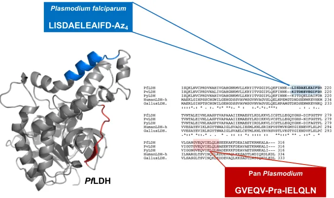
Figure 2-2. An epitope (blue) that is specific to PfLDH was targeted for capture and fitted with an azide (Az4) click handle. A pan-species epitope (red) was selected for detection and fixed with an alkyne (Pra) click handle. The general schematic of the capture and detection PCC pair in a sandwich assay format is shown in the upper left-hand corner. (PDB: 1LDG, figure partially adapted.8)
For capture of PfLDH, the amino acid sequence from 208-219, LISDAELEAIFD,
was selected for epitope synthesis as illustrated in Figure 2-2. It is important to note the
high homology of the LISDAELEAIFD epitope to the homogolous epitopes in other
malaria species which requires that the final PCC be able to distinguish between small
changes in sequence identity. For example, amino acids 208-219 differ only by four amino
acids between P. falciparum and P. vivax LDH antigens.14 For the PCC screening process,
the LISDAELEAIFD epitope was synthesized with an alkyne click handle, on the
C-terminus to yield LISDAELEAIFD-Az4.18 A scrambled version of this epitope,
DDIAILIEEFALS-Az4, was also synthesized for prescreening. Inclusion of a scrambled
epitope in a prescreen helps to eliminate hits that might generate false positives from lack
of sequence specificity.18 A pan-species epitope was selected for the development of a Plasmodium falciparum
LISDAELEAIFD-Az4
Pan Plasmodium
GVEQV-Pra-IELQLN
PfLDH
Pan
Plasmodium
detection PCC. The amino acids 297-308 of LDH, GVEQVIELQLN, are common to at
least P. falciparum, P. vivax, and P. yoeli species of malaria.14 The SynEp for PxLDH
detection was synthesized with a Pra in the center to yield GVEQV-Pra-VIELQLN.18
2.3.2 Discovery of Capture and Detection PCCs Against PfLDH
The primary detection PCC agent against GVEQVIELQLN was developed using
older screening methodology from the Heath group that utilized in situ click screening with
OBOC linear peptide libraries.16,16 The screen was conducted against a OBOC
D-stereochemistry library fitted with an Az4 click handle that was prescreened to remove
false-positives. This screen yielded a linear 5-mer D-peptide with the sequence hevwh.18
The use of D-amino acids in a linear library is to provide increased proteolytic stability in
the final peptide sequences. The EC50 of hevwh as determined by enzyme-linked
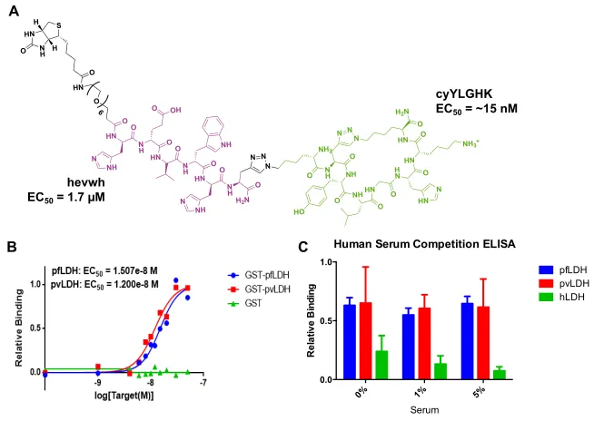
immunosorbent assay (ELISA) was found to be 1.7 µM (Figure 2-3).18 The EC
50 value
offers an upper limit to the true binding affinity, or KD, of a ligand to substrate, and the
affinity of this PCC is considered modest. Further work detailed in this chapter will
describe the expansion of this primary ligand into a bivalent molecule for increased target
binding.
Figure 2-3. (A) Structure of hevwh. (B) Sandwich ELISA of the binding affinity of