Applications and Extensions of Living Ring-Opening Metathesis
Polymerization
Thesis by
John B. Matson
In partial fulfillment of the requirements
for the degree of
doctor of philosophy
California Institute of Technology
Pasadena, CA
2010
©
2010
John B. Matson
Acknowledgments
I suppose the most chronologically accurate place to begin my thesis acknowledgements is with my Mom and Dad, who long, long ago bought several educational placemats for the kitchen table. I ate breakfast staring at a periodic table at the age of ten, routinely asking my parents what atomic numbers and masses were. The atmosphere in our house growing up taught me to ask questions and created a love of learning that my siblings and I continue to enjoy. Thank you Mom, Dad, Tim, and Abby for teaching me how to learn and how to teach.
While my family may have set the stage, the one who opened the curtain was my high school chemistry teacher, Carolyn Morse. I remember coming home from school and telling Mom and Dad about the polymer demonstrations that she did one day. Remembering my fascination from high school, I decided to take Karen Wooley’s polymer chemistry class at WashU during my sophomore year. This led to undergraduate research in her lab, where I was trained by Brooke Van Horn and Maisie Joralemon. I owe Brooke and Maisie a lot for teaching, encouraging, and mostly dealing with all the mistakes I’m sure I made during that time.
Grubbs family and the confidence to investigate important problems in science. Bob has also been a great friend. Whether it was rock climbing, drinking at the Ath, shooting hoops in the gym, or just chatting, Bob was fun to be around. It was during these times that I learned other important things, such as when to use a double-fisherman’s knot (almost always), who not to get in a drinking contest with (anyone from Ireland), and what to do when you don’t know the answer (mumble). I look forward to more beers and more stories with Bob in the future.
My committee members, Professors Dave Tirrell, Jim Heath, and Mark Davis, were also valuable resources during my time here. Dave was my committee chair and kept everything focused and running smoothly. Jim is great fun to discuss science with because his excitement is contagious and inspiring. Mark was an outstanding resource due to his interest in nanoparticles in cancer therapy. I especially thank him for his comments on my proposals and his friendly, laid-back attitude.
I am also indebted to Scott Virgil, director of the Center for Catalysis and Chemical Synthesis. Scott ran the robot that was a vital component of Chapter 6 of this thesis, but he did so much more than that. Scott took the time to understand the broader goals of my project and contributed valuable ideas that turned into components of the finished product. Additionally, he took care of the meticulous details that I overlooked, as far down as the shape of stirbars and the withdrawal rates of syringes. I am sure that Scott will be an important resource in the department for many students to come.
have space to work until February. At that point I worked in Ron Walker’s hood because he was writing his candidacy report. My desk was a flammables cabinet. Ron and Masao Yanagawa helped me hit the ground running, and Ron taught me nearly everything I know about GPC. Erin Guidry and Jason Jordan were also very helpful in those early days. Mike Page came along a little later, working on an offshoot of my project. He was a great guy to discuss chemistry with and a fantastic teacher, and we had a lot of fun traveling to various conferences. The rest of the polymer subgroup was also very helpful along the way. This includes Yan Xia, Rosemary Conrad, Andy Hejl, Irina Gorodetskaya, AJ Boydston, Paul Clark, Grace Chan, Erin Guidry, Jeremiah Johnson, Jasim Uddin, Bahar Bingöl, and several others. Other important folks in the lab were my various baymates: Donde Anderson, Connie Hou, Chris Daeffler (Man Bay), and Masao Yanagwa. Labmates who helped me along the way in various other ways were Jean Li (softball organizer, paper proofreader, back-walker, and group baker), Chris Douglas (organic synthesis czar and Irish car bomb expert), Rosemary Conrad (organic synthesis wisdom and travel buddy), Ian Stewart and Kevin Kuhn (general metathesis knowledge), Cheol Chung (knows everything), AJ Boydston (best guy to read your paper and get you the basketball on the block), Keith Keitz (helping clean up chromium), Matthew Van Wingerden (solvent column apprentice), and Chris Daeffler (country music). A special acknowledgement goes to the 5th Year Braintrust, which includes Kevin Kuhn, Matt Whited, and me. I am both a founding member and the last remaining member of the 5th
Year Braintrust, and it was a great honor to be a part of this exclusive organization.
He was a dedicated and enthusiastic student who contributed quite a bit to a chapter in this thesis. He also provided a lot of entertainment in the Man Bay with his music videos and undying love of New Jersey.
My thanks to people outside of the Grubbs group must start with the NanoSystems Biology Cancer Center (NSBCC). I began with the NSBCC when it started in early 2006, and I’ve learned a lot about biology and cancer from all the people involved in the center. Important people from this group include Jim Heath (NSBCC director), Mike Phelps (director of my project and director of the Crump Institute for Molecular Imaging at UCLA), and Mark Davis (nanoparticle advice). I also would like to pay special thanks to Nagichettiar Satyamurthy (Saty), Phillip Marchis, and Arkadij Elizarov for running radiofluorination reactions on my samples. In addition, thanks go to several others at Caltech who helped along the way, including Raymond Archer and Chris Alabi (DLS training), Peigen Cao (AFM training and tireless help), Udi Vermesh and Ke Xu (SEM), Pat Keon (TEM), Young In Oh, Dave Montgomery and John Phillips (HPLC), Dan Harki (fluorine-18 discussions), and Chris Henry (proposal reading). Michelle Chen at Wyatt Technology was also very helpful with all GPC-related things.
has taught me while working on the solvent columns. His indefatigable working style, attention to detail, and creative genius would have served him well had he been a Caltech graduate student.
Sports provided a great way to let out the frustrations of lab work. I played softball for several years with the Grubbs group team Imperial Palace, where we won several games and a few championships, both in the Caltech A league and the JPL B league. I also played basketball with a Grubbs group team of rotating players with team names including Brokeback Cowgirls, Black Thunder, and Def Halen Van Journey Snake. We won a few games and usually contended for last place. I was a founding member and am again the last remaining member of the retired WashU swimmers swim team, Pasadena chapter. Once the chapter was reduced to one person, I began swimming with the Caltech Masters Swim Team. Both of these groups provided a nice avenue to swim and socialize.
Metz, Mark Ambrosi, and Nick Graham were friends from WashU that were in the area. We had a lot of great times partying in Hollywood, Hermosa, Westwood, Huntington Beach, and once all around town in a Hummer Limo. I thank all of you for making my time out of lab fun and worthwhile. I’d also like to thank Mariann Ramirez for being on Colorado Boulevard on New Years 2008 and the Ramirez family for treating me like one of their own.
I owe the Athenaeum crew a proverbial round for always being there for a Friday-afternoon, gluten-free outing. The regulars over my years here were Tim Funk, Andy Hejl, Tobias Ritter, Nick Graham, Kate Ashton, Jacob Berlin, Daryl Allen, Matt Whited, Charlotte Whited, Patricio Romero, Ian Stewart, Kevin Kuhn, Rosemary Conrad, Vince Lavallo, Paul Clark, and Keith Keitz. Tim and Andy first invited me, and then I quickly became a regular downstairs or on the lawn in the summertime. Jerry Rodriguez, head of food and beverage, liked to make me the girliest, pinkest drinks he could come up with, and Mike Echevarria, my favorite bartender ever, gave us entirely too many free pitchers. They were also just nice guys to chat with.
Abstract
Living ring-opening metathesis polymerization (ROMP) is a polymerization method that has recently become popular in the synthesis of complex polymers due to advances in olefin metathesis catalyst design. The unrivaled degree of functional group tolerance of the method, coupled with a high level of control and synthetic ease, make living ROMP a valuable tool in the assembly of complex nanostructures and functional polymers. Work in this thesis details methods for applying living ROMP in the assembly of complex nanostructures and extending the uses of living ROMP to end-functionalized polymers and to polymers synthesized in a catalyst economical manner.
Chapter 2 describes the synthesis and radiofluorination of fluorine-18 functionalized nanoparticles assembled from polynorbornene block copolymers synthesized via living ROMP. The block copolymers include a hydrophobic photo-crosslinkable block made from a novel cinnamate-containing norbornene, as well as a hydrophilic block made from a PEGylated norbornene. Chapter 3 illustrates another application of ROMP-based nanoparticles in which polynorbornene block copolymers are assembled into Janus (hemispherical) nanoparticles.
A thorough study of pulsed-addition ROMP (PA-ROMP) performed using a Symyx robotic system is presented in Chapter 6. Extending the end-capping methodology described in Chapters 4 and 5 to the synthesis of additional polymer chains led to a homo- and block copolymerization strategy that can produce more than one polymer chain per molecule of metal initiatior. The PA-ROMP strategy reduces catalyst consumption as much as sevenfold in the synthesis of polynorbornenes.
Table
of
Contents
Biological Applications of ROMP ... 8
Telechelic ROMP polymers ... 8
Nanoparticles as in vivo Molecular Imaging Agents ... 10
Thesis Research ... 12
References ... 14
Chapter 2. Synthesis of Fluorine-18 Functionalized Nanoparticles for use as in vivo Molecular Imaging Agents ... 20
Abstract ... 21
Introduction ... 21
Results and Discussion ... 25
Monomer Syntheses and Evaluations ... 25
Polymer Syntheses ... 30
Micelle Formation and Crosslinking ... 33
Radiofluorination ... 38
Conclusions ... 39
Acknowledgement ... 40
Experimental Section ... 40
References ... 51
Chapter 3. A Living Ring-Opening Metathesis Polymerization Route to Janus Nanoparticles ... 54
Abstract ... 55
Introduction ... 55
Results and Discussion ... 56
Monomer Syntheses ... 57
Polymer Syntheses ... 59
Nanoparticle assembly and staining ... 62
Conclusions ... 65
Acknowledgement ... 66
Experimental Section ... 66
Chapter 4. ROMP-ATRP Block Copolymers Prepared from Monotelechelic
Poly(oxa)norbornenes using a Difunctional Terminating Agent... 76
Abstract ... 77
Introduction ... 77
Results and Discussion ... 81
Conclusions ... 89
Acknowledgement ... 89
Experimental Section ... 90
References ... 98
Chapter 5. Monotelechelic Poly(oxa)norbornenes by Ring-Opening Metathesis Polymerization using Direct End-Capping and Cross Metathesis ... 100
Abstract ... 101
Introduction ... 101
Results and Discussion ... 104
TA Syntheses ... 106
End-functionalization by direct end-capping ... 108
End-functionalization using CM ... 112
Conclusions ... 117
Acknowledgment ... 117
Experimental Section ... 118
References ... 131
Chapter 6. Pulsed-Addition Ring-Opening Metathesis Polymerization: Catalyst-Economical Syntheses of Homopolymers and Block Copolymers ... 134
Abstract ... 135
Introduction ... 136
Results and Discussion ... 139
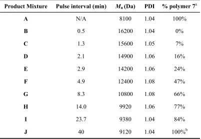
Pulse Interval Optimization ... 141
Homopolymers by PA-ROMP ... 146
Block Copolymers by PA-ROMP ... 160
Acknowledgement ... 166
Conclusions ... 167
Experimental Section ... 168
References ... 177
Appendix 1. Additional Monomers and Polymers Synthesized ... 179
Introduction ... 180
Results and Discussion ... 180
Acknowledgement ... 183
Experimental Section ... 183
Appendix 2. Theoretical PA-ROMP Molecular Weight Data and
Determination of Catalyst Death Rates ... 191
Introduction ... 192
Results and Discussion ... 192
Theoretical PA-ROMP Molecular Weight Data ... 192
Determination of Catalyst Death Rates... 194
List
of
Figures
Chapter 1.
Figure 1.1. Olefin metathesis catalysts. ... 4
Figure 1.2. Comparison of activity and functional group tolerance in olefin metathesis catalysts. ... 5
Figure 1.3. Monomers typically used in ROMP. ... 7
Chapter 2. Figure 2.1. GPC of homopolymer of monomer 3 ... 27
Figure 2.2. Norbornenes designed to test leaving group reactivity ... 29
Figure 2.3. GPC traces of block copolymers. ... 33
Figure 2.4. AFM images of micelles and crosslinked nanoparticles ... 35
Figure 2.5. Nanoparticle core crosslinking percentage dependence on irradiation time. ... 36
Figure 2.6. AFM images of micelles made from block copolymers with values of the PEG chain length. ... 38
Figure 2.7. RadioTLC of radiofluorinated nanoparticles ... 39
Chapter 3. Figure 3.1. Ruthenium metathesis catalyst (1) and photocrosslinking monomer (2) used in this study. ... 57
Figure 3.2. AFM image of control nanoparticles on silicon. ... 64
Figure 3.3. SEM images of Janus nanoparticles and control nanoparticles ... 65
Chapter 4. Figure 4.1. Monomers and metathesis catalyst used in ROMP reactions ... 82
Figure 4.2. GPC traces of P(tBENI) homopolymer and P(tBENI-b-S) block copolymer ... 84
Figure 4.3. GPC traces of P(tBENI) homopolymer, P(tBENI-b-S) block copolymer and P(tBENI-b-tBA) block copolymer ... 88
Chapter 5. Figure 5.1. Ruthenium olefin metathesis catalysts used in this study ... 105
Figure 5.2. Monomers and previously reported TAs used in this study ... 106
Chapter 6. Figure 6.1. Ruthenium olefin metathesis catalysts and monomers used in PA-ROMP reactions ... 140
Figure 6.2. GPC traces of products A, B, F, and J from pulse interval optimization experiment ... 146
Figure 6.3. GPC traces of P(tBENI) product mixtures K−T from one to ten cycles of PA-ROMP ... 149
Figure 6.5. GPC trace of PA-ROMP product in single-vial experiment ... 154 Figure 6.6. GPC traces of P(tBENI) product mixtures U−DD from one to ten
cycles of PA-ROMP ... 155 Figure 6.7. Dependence of Mn and PDI on number of cycles of PA-ROMP for
P(tBENI) homopolymers with M/C = 100 ... 157 Figure 6.8. GPC traces of P(tBENI) product mixtures EE−NN from one to ten
cycles of PA-ROMP ... 158 Figure 6.9. Dependence of Mn and PDI on number of cycles of PA-ROMP for
P(tBENI) homopolymers with M/C = 25 at 20 ºC with 60 min pulse interval. ... 160 Figure 6.10. GPC traces of block copolymer product mixtures OO−XX from one
to ten cycles of PA-ROMP. ... 163 Figure 6.11. Dependence of Mn and PDI on number of cycles of PA-ROMP for
P(tBENI-b-NMONI) block copolymers. ... 165 Appendix 2.
Figure A2.1. Dependence of Mn (total) on molecular weight on the number of
List
of
Schemes
Chapter 1.
Scheme 1.1. Mechanism of olefin metathesis. ... 2
Scheme 2.1. Monomer syntheses ... 26
Chapter 2. Scheme 2.2. Homopolymerization of monomer 3 ... 27
Scheme 2.3. Fluorination of test substrates ... 29
Scheme 2.4. Synthesis of block copolymers. ... 31
Scheme 2.5. Fluorinated Nanoparticle Synthesis. ... 34
Chapter 3. Scheme 3.1. Syntheses of complementary hydrogen-bonding monomers. ... 58
Scheme 3.2. Syntheses of acid-containing monomer and PEGylated monomer. ... 59
Scheme 3.3. Synthesis of amphiphilic block copolymer containing ADA H-bonding group ... 60
Scheme 3.4. Synthesis of amphiphilic block copolymer (12) containing DAD hydrogen-bonding group ... 61
Scheme 3.5. Synthesis of control amphiphilic block copolymer (13). ... 62
Scheme 3.6. Assembly of Janus nanoparticles. ... 63
Scheme 3.7. Assembly of control nanoparticles. ... 63
Chapter 4. Scheme 4.1. Synthesis of Vinyl Ether. ... 81
Scheme 4.2. ROMP and chain termination using vinyl ether followed by ATRP of styrene. ... 83
Scheme 4.3. Preparation of terminating agent. ... 85
Scheme 4.4. Synthesis of monotelechelic poly(oxa)norbornenes and ROMP-ATRP block copolymers. ... 86
Chapter 5. Scheme 5.1. Synthesis of Several Functionalized Internal cis-Olefins for Poly(oxa)norbornene End-Functionalization. ... 107
Scheme 5.2. Synthesis of NHBoc, FITC, and Biotin-Containing TAs. ... 108
Scheme 5.3. End-functionalization of P(tBENI) or P(NMONI) using the direct end-capping method. ... 109
Scheme 5.4. Removal of Boc and tert-Butyl Ester Groups to Afford Amine-Terminated Polynorbornene... 111
Scheme 5.5. Synthesis of methylene-capped P(tBENI) ... 113
Chapter 6.
Scheme 6.1. Mechanism of PA-ROMP ... 138
Scheme 6.2. Experiment designed to determine the optimal pulse interval required for PA-ROMP ... 143
Scheme 6.3. Synthesis of polynorbornenes by PA-ROMP. ... 147
Scheme 6.4. Synthesis of a single batch of low polydispersity homopolymer by PA-ROMP. ... 152
Scheme 6.5. Synthesis of P(tBENI-b-NMONI) block copolymers by PA-ROMP. ... 162
Appendix 1. Scheme A1.1 Synthesis of DOTA-containing norbornene monomer. ... 180
Scheme A1.2. ROMP of monomers 3 and 5. ... 181
Scheme A1.3. Synthesis of galactose-containing norbornene monomer. ... 182
List
of
Tables
Chapter 2.
Table 2.1. Products from fluorination of representative monomers. ... 30
Table 2.2. GPC Characterization of Block Copolymers ... 32
Table 2.3. Characterization of Nanoparticles with varied block lengths ... 37
Table 2.4. Characterization of Nanoparticles with varied PEG chain lengths ... 38
Chapter 4. Table 4.1. Polymer characterization data for monotelechelic poly(oxa)norbornenes and ROMP-ATRP block copolymers ... 87
Chapter 5. Table 5.1. End-capping Efficiency of P(tBENI) or P(NMONI) Polymers using the Direct End-Capping Method. ... 110
Table 5.2. End-capping Efficiency of P(tBENI) or P(NMONI) Polymers using Cross Metathesis ... 115
Chapter 6. Table 6.1. Characterization of the products formed in the pulse interval optimization experiment ... 145
Table 6.2. Characterization of P(tBENI) homopolymer products by GPC from 10 cycles of PA-ROMP at 25 ºC with M/C = 25 and a pulse interval of 30 min ... 150
Table 6.3. Addition volumes of monomer solution assuming 8.5% catalyst death rate for each of ten cycles of PA-ROMP ... 153
Table 6.4. Characterization of P(tBENI) homopolymer products by GPC from 10 cycles of PA-ROMP at 25 ºC with M/C = 100 and a pulse interval of 30 min ... 156
Table 6.5. Characterization of P(tBENI) homopolymer products by GPC from ten cycles of PA-ROMP at 20 ºC with M/C = 25 and a pulse time of 60 min ... 159
Table 6.6. Characterization of P(tBENI-b-NMONI) block copolymer products by GPC from ten cycles of PA-ROMP at 25 ºC with M/C = 25 and a pulse interval of 30 min. ... 164
Appendix 2. Table A2.1. Theoretical molecular weight trend over 40 cycles of PA-ROMP ... 193
Table A2.2. Determination of catalyst death rate for P(tBENI) homopolymers with M/C = 25 and a pulse time of 30 min in PA-ROMP ... 195
Table A2.3. Determination of catalyst death rate for P(tBENI) homopolymers with M/C = 25 and a pulse time of 30 min in PA-ROMP ... 196
Table A2.4. Determination of catalyst death rate for P(tBENI) homopolymers with M/C = 25, a temperature of 20 ºC and a pulse time of 60 min in PA-ROMP ... 197
Chapter
1
Olefin
Metathesis
General Aspects
The olefin metathesis reaction has become a valuable and versatile tool in organic and polymer synthesis over the past few decades.1 Although several mechanisms for olefin metathesis had been proposed, the now accepted mechanism for the reaction was first described by Chauvin in 19712 and later confirmed by Grubbs in 19753 and Katz in 1976.4 Mediated by a metal carbene species, olefin metathesis in its most basic form is capable of taking two different, symmetrical olefins and forming a new, unsymmetrical olefin (Scheme 1.1). The reaction proceeds via a four-membered metallacyclobutane intermediate and is often reversible and therefore under thermodynamic control.
Scheme 1.1. Mechanism of olefin metathesis.
In its early years, olefin metathesis was catalyzed by ill-defined, multi-component systems, usually made up of transition metal salts such as WCl6 and MoCl5 and
alkylating agents such as SnR4 and AlCl2R.12 These early catalysts were highly active,
but they suffered from lack of air, moisture, and functional group tolerance, and their heterogeneous nature made them difficult to study. A single-component, well-defined olefin metathesis catalyst (1) was developed by Grubbs in the 1980s13 derived from the Tebbe reagent (Figure 1.1).14 Titanocyclobutane catalyst 1 was the first catalyst capable of mediating the living ROMP of norbornene, affording low polydispersity polymers with control over molecular weight. However, catalyst 1, unable to tolerate polar monomers, was limited in utility to hydrocarbons. Innovations in molybdenum and tungsten carbenes, such as Schrock’s family of catalysts (NAr)(OR)2MCHR’ (2),15 as well as late
Figure 1.1. Olefin metathesis catalysts.
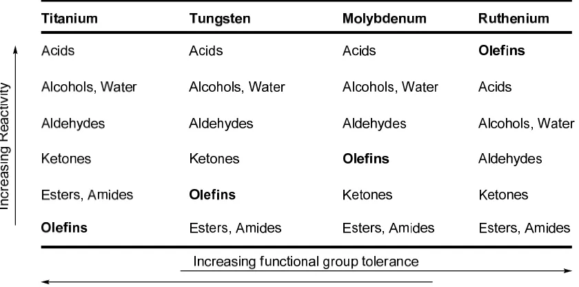
(Figure 1.2). However, the catalyst was only active for the ROMP of norbornene. Replacement of the triphenyl phosphine ligands with the bulkier and more electron-donating tricyclohexyl phosphine ligands and changing the vinylidene component to a benzylidene led to the more active and more stable catalyst 4, first reported by Grubbs in 1996.21 Catalyst 4 has the functional group tolerance of catalyst 3 but has an increased substrate scope for CM and RCM and can mediate ROMP of low-strain monomers.22
Figure 1.2. Comparison of activity and functional group tolerance in olefin metathesis catalysts (adapted from reference 22).
Recent Developments
room temperature in just a few seconds.25 Additionally, electron-poor alkenes can undergo metathesis with catalyst 5, allowing for inclusion of acrylates, trisubstituted olefins, vinyl phosponates and many other functional groups into the olefin metathesis substrate table.26 Chelating isopropoxy-styrene containing catalysts, such as 6, have been found to be more stable than their phosphine-containing counterparts.27 Catalysts such as 6 are very useful in RCM and CM, but their utility in ROMP is limited due to their
generally slow initiation kinetics. Water-soluble, NHC-containing, ruthenium metathesis catalysts,28 as well as catalysts capable of performing asymmetric metathesis29 and catalysts that can effect RCM of tetrasubstituted olefins30 have also been developed in recent years.
ROMP
General Aspects
Figure 1.3. Monomers typically used in ROMP.
Living ROMP
Other techniques, such as adding phosphine or running reactions at reduced temperatures, can be used to prevent secondary metathesis reactions of less bulky monomers.33 Pyridine-containing catalysts 7a and 7b have been shown to initiate ROMP rapidly, resulting in the formation of low polydispersity polymers with low PDIs.34
Biological Applications of ROMP
Highly functional, low polydispersity ROMP polymers have been made for biological applications using various olefin metathesis catalysts.35 ROMP is an ideal polymerization strategy for the synthesis of biologically-relevant polymers because of the functional group and steric tolerance of ruthenium olefin metathesis catalysts. Sugar-containing monomers,36 peptidic monomers,37 and charged monomers38 have all been polymerized with high efficiency using ROMP. There also exist two reports on nanoparticle imaging agents made by ROMP, using magnetic resonance imaging (MRI)39 and near-IR optical imaging.40 Chapter 2 of this thesis concerns the synthesis and
self-assembly of ROMP block copolymers into nanoparticles for use as in vivo molecular imaging agents using positron emission tomography.
Telechelic ROMP polymers
which acts as a chain transfer agent (CTA).41 Because thermodynamic equilibrium needs to be reached to ensure complete end-functionalization, these reactions are typically run at elevated temperature for 12-24 h. Additionally, the amount of catalyst in such a reaction needs to be as small as possible because the benzylidene fragment of the catalyst competes with CTA in the capping of polymer chains. This process results in polymers with a theoretical PDI of 2.0. High PDIs are acceptable for some purposes, but low PDIs are required for many applications, such as when specific morphologies of block copolymers are desired.
carbonates.46 More recently the sacrificial monomer method has been developed to produce highly functional, low polydispersity polynorbornenes via ROMP.47 This method consists of the ROMP of dioxepine (or another readily degradable monomer) on the end of the desired homopolymer. Subsequent degradation of the poly(dioxipene) block yields an alcohol terminated polymer. This method is limited to a few functional groups, all of which must be further derivatized after polymerization to add any additional functionality. A portion of this thesis addresses the synthetic limitations of end-functionalized ROMP polymers, presenting methodology for the production of mono- and ditelechelic ROMP polymers with high end-capping efficiency.
Nanoparticles
as
in
vivo
Molecular
Imaging
Agents
Nanoparticles have recently received a vast amount of interest as imaging and therapeutic agents in cancer.48 Much of this interest stems from Maeda’s seminal report in 1986 detailing what is now known as the enhanced permeability and retention (EPR) effect.49 The EPR effect describes the observation that particles in the range of approximately 10-150 nm are able to selectively penetrate and remain in tumor tissue, while surrounding healthy tissue is left undisturbed.50 The EPR effect occurs as a result of hypervascularization, enhanced vasculature permeability, and poor lymphatic system development in tumor tissue.
drug delivery and imaging agents, including a size range of 10-150 nm, near neutral surface charge and a “stealth” surface to minimize opsonization and subsequent removal by the rectoendothelial system.56 Adherence to these guidelines appears to be vital for achieving effective nanoparticle drug delivery and imaging agents.
Thesis
Research
Increasing applications for functionally complex polymers and nanostructures in biomedical fields will need to be met by an analogous increase in synthetic methodology to incorporate functionality into macromolecules. ROMP provides a highly functional group tolerant polymerization strategy with which to make functional polymers and nanostructures, yet its uses in biomedical applications remain limited. Work in this thesis details methods for applying living ROMP in the assembly of complex nanostructures and extending the uses of living ROMP to end-functionalized polymers and to polymers synthesized in a catalyst economical manner.
Chapter 2 describes the synthesis and radiofluorination of fluorine-18 functionalized nanoparticles. The nanoparticles are assembled from polynorbornene block copolymers synthesized via living ROMP. The block copolymers include a hydrophobic photo-crosslinkable block made from a novel cinnamate-containing norbornene, as well as a hydrophilic block made from a PEGylated norbornene.
Chapter 3 illustrates another application of ROMP-based nanoparticles in which polynorbornene block copolymers are assembled into Janus (hemispherical) nanoparticles. One hemisphere of the particles was labeled with small gold nanoparticles, and scanning electron microscopy (SEM) was used to image the Janus nanoparticles.
to confirm complete end-functionalization and synthesize mechanistically incompatible block copolymers.
Chapter 5 is an extension of Chapter 4, extending the polymer end-functionalization approach described in Chapter 4 to additional functional groups, including alcohols, bromides, thioacetates, fluorescent compounds, biotin, and others.
A thorough study of pulsed-addition ROMP (PA-ROMP) performed using a Symyx robotic system is presented in Chapter 6. Extending the end-capping methodology described in Chapters 4 and 5 to the synthesis of additional polymer chains led to a homo- and block copolymerization strategy that can produce more than one polymer chain per molecule of metal initiatior. The PA-ROMP strategy reduces catalyst consumption and ruthenium contamination in the polymer products.
References
(1) Grubbs, R. H. Handbook of Metathesis; Wiley-VCH: Weinheim, 2003. (2) Herrison, J. L.; Chauvin, Y. Makromol. Chem. 1971, 141, 161. Handbook of Metathesis; Wiley-VCH: Weinheim, 2003, Vol. 3. (e) Bielawski, C. W.; Grubbs, R. H. Prog. Polym. Sci. 2007, 32, 1–29. Saucier, P. C.; Wasserman, E. P. Organometallics 2004, 23, 2027–2047.
(11) (a) Bielawski, C. W.; Benitez, D.; Grubbs, R. H. Science 2002, 297, 2041. (b) Bielawski, C. W.; Benitez, D.; Grubbs, R. H. J. Am. Chem. Soc. 2003, 125, 8424. (c) Boydston, A. J.; Xia, Y.; Kornfield, J. A.; Gorodetskaya, I. A.; Grubbs, R. H. J. Am. Chem. Soc. 2008, 130, 12775–12782. (d) Xia, Y.; Boydston, A. J.; Yao, Y.; Kornfield, J. A.; Gorodetskaya, I. A.; Spiess, H. W.; Grubbs, R. H. J. Am. Chem. Soc. 2009, 131, 2670–2677.
(13) (a) Howard, T. R.; Lee, J. B.; Grubbs, R. H. J. Am. Chem. Soc. 1980, 102, 6876.
Chem. Soc. 2006, 128, 3508–3509. (e) Jordan, J. P.; Grubbs, R. H. Angew. Chem., Int. Ed. 2007, 46, 5152–5155.
(29) (a) Fujimura, O.; Grubbs, R. H. J. Am. Chem. Soc. 1996, 118, 2499–2500. (b) Fujimura, O.; de la Mata, F. J.; Grubbs, R. H. Organometallics 1996, 15, 1865– 1871. (c) Van Veldhuizen, J. J.; Gillingham, D. G.; Garber, S. B.; Kataoka, O.; Hoveyda, A. H. J. Am. Chem. Soc. 2003, 125, 12502–12508. (d) Van Veldhuizen,
(32) Matyjaszewski, K.; Mueller, A. H. E. ACS, Macromolecular Nomenclature Note No. 12; http://www.polyacs.org/main/ nomenclature.shtml.
(33) (a) Myers, S. B.; Register, R. A. Macromolecules 2008, 41, 6773–6779. (b) Walker, R.; Conrad, R. M.; Grubbs, R. H. Macromolecules 2009, 42, 599–605. (34) Choi, T. L.; Grubbs, R. H. Angew. Chem., Int. Ed. 2004, 42, 1743–1746.
(35) For recent reviews see: (a) Ladmiral, V.; Melia, E.; Haddleton, D.M. Eur. Polym. J. 2004, 40, 431–449. (b) Lee, Y.; Sampson, N. S. Curr. Opin. Struct. Biol. 2006, 16, 544–550. (c) Smith, D.; Pentzer, E. B.; Nguyen, S. T. Polym. Rev. 2007, 47, 419–459.
(36) (a) Mortell, K. H.; Gingras, M.; Kiessling, L. L. J. Am. Chem. Soc. 1994, 116, 12053–12054. (b) Manning, D. D.; Hu, X.; Beck, P.; Kiessling, L. L. J. Am. Chem. Soc. 1997, 119, 3161–3162. (c) Gestwicki, J. E.; Strong, L. E.; Kiessling, L. L. Chem. Biol. 2000, 7, 583–591. (d) Puffer, E. B.; Pontrello, J. K.; Hollenbeck, J. J.; Kink, J. A.; Kiessling, L. L. ACS Chem. Biol. 2007, 2, 252–262. (e) Fraser, C.; Grubbs, R. H. Macromolecules 1995, 28, 7248–7255. (f) Nomura, K.; Schrock, R. R. Macromolecules 1996, 29, 540–545. (g) Camm, K. D.; Castro, N. M.; Liu, Y.; Czechura, P.; Snelgrove, J. L.; Fogg, D. E. J. Am. Chem. Soc. 2007, 129, 4168–4169. (h) Rawat, M.; Gama, C. I.; Matson, J. B.; Hsieh-Wilson, L. C. J. Am. Chem. Soc. 2008, 130, 2959–2961.
North, M. Polymer 1998, 39, 1007–1014. (c) Biagini, S. C. G.; Davies, R. G.; Gibson, V. C.; Giles, M. R.; Marshall, E. L.; North, M.; Robson, D. A. Chem. Commun. 1999, 235–236. (d) Maynard, H. D.; Grubbs, R. H. Macromolecules 1999, 32, 6917–6924. (e) Maynard, H. D.; Okada, S. Y.; Grubbs, R. H. Macromolecules, 2000, 33, 6239–6248. (f) Maynard, H. D.; Okada, S. Y.; Grubbs, R. H. J. Am. Chem. Soc. 2001, 23, 1275–1279. (g) Breitenkamp, R. B.; Ou, Z.; Breitenkamp, K.; Muthukumar, M.; Emrick, T. Macromolecules 2007, 40, 7617–7624. (h) Biagini, S. C. G.; Parry, A. L. J. Polym. Sci., Part A: Polym. Chem. 2007, 45, 3178–3190. (i) Sutthasupa, S.; Sandra, F.; Masuda, T. Macromolecules 2009, 42, 1519–1525.
(38) (a) Han, H. J.; Chen, F. X.; Yu, J. H.; Dang, J. Y.; Ma, Z.; Zang, Y. Q.; Xie, M. R. Maughon, B. R.; Morita, T.; Bielawski, C. W.; Grubbs, R. H. Macromolecules 2000, 33, 1929–1935.
(42) (a) Bielawski, C. W.; Louie, J.; Grubbs, R. H. J. Am. Chem. Soc. 2000, 122, 12872–12873. (b) Burtscher, D.; Saf, R.; Slugovc, C. J. Polym. Sci., Part A: Polym. Chem. 2006, 44, 6136–6145.
(47) (a) Hilf, S.; Berger-Nicoletti, E.; Grubbs, R. H.; Kilbinger, A. F. M. Angew. Chem., Int. Ed. 2006, 45, 8045–8048. (b) Hilf, S.; Kilbinger, A. F. M. Macromol. Rapid Comm. 2007, 28, 1225–1230. (c) Hilf, S.; Hanik, N.; Kilbinger, A. F. M. J. Poly. Sci., Part A: Polym. Chem. 2008, 46, 2913–2921. (d) Hilf, S.; Kilbinger, A. F. M. Macromolecules 2009, 42, 1099–1106.
(48) For recent reviews see: (a) Brigger, I.; Dubernet, C.; Couvreur, P. Adv. Drug. Del. Rev. 2002, 54, 631–651. (b) Ferrari, M. Nat. Rev. Cancer 2005, 5, 161–171. (c) Chem. Soc. 2007, 129, 15096–15097. (b) Torchilin, V. P. Nat. Rev. Drug Discovery 2005, 4, 145–160.
Chapter
2
Synthesis
of
Fluorine
18
Functionalized
Nanoparticles
for
use
as
in
vivo
Molecular
Imaging
Agents
Portions of the text in this chapter have been reproduced with permission from: Matson, J. B.; Grubbs, R. H. J. Am. Chem. Soc. 2008, 130, 6731-6733.
Copyright 2008 American Chemical Society
Abstract
Nanoparticles containing fluorine-18 were prepared from block copolymers made by ring-opening metathesis polymerization (ROMP). Using the fast initiating ruthenium metathesis catalyst (H2IMes)(pyr)2(Cl)2RuCHPh, low polydispersity amphiphilic block
copolymers were prepared from a cinnamoyl-containing hydrophobic norbornene monomer and a mesyl-terminated, PEG-containing hydrophilic norbornene monomer. Self-assembly into micelles and subsequent crosslinking of the micelle cores by light-activated dimerization of the cinnamoyl groups yielded stable nanoparticles. Incorporation of fluorine-18 was achieved by nucleophilic displacement of the mesylates by the radioactive fluoride ion with 31% incorporation of radioactivity. The resulting positron-emitting nanoparticles are to be used as in vivo molecular imaging agents for use in tumor imaging.
Introduction
The EPR effect was first observed in the 1980s when Maeda and coworkers reported that large molecules (> 50 kDa) showed a tendency to enter and remain in cancer cells.8 They attributed these results to four characteristics of tumor vascularization: a) hypervascularization; b) enhanced vascular permeability; c) little recovery of macromolecules via the blood vessels; and d) little recovery of macromolecules from the lymphatic system. Since then the existence of the EPR effect across many types of tumors has been studied,9 and the phenomenon was later found to be consistent across all solid tumors. Interestingly, the maximum size of a molecule that can cross into tumor tissue varies widely, from 100 nm to 2 μm, depending on the tumor cell line.10 Currently there are no reports that seek to optimize nanoparticle size to deliver the maximum amount of chemotherapeutic to tumors.
Imaging of tumors using nanostructures designed to exploit the EPR effect has been accomplished using several in vivo imaging techniques, including magnetic resonance (MR),11 near-IR fluorescence (NIR),12 and positron emission tomography (PET).13 PET is a specific, highly sensitive and versatile three-dimensional molecular imaging technique, and PET is the most sensitive and accurate method of measuring the temporal pattern in the biodistribution of labeled compounds. The most widespread radionuclide used in PET imaging is fluorine-18, which is the positron-emitting isotope in the commonly used PET tracer 18-fluorodeoxyglucose. Its relatively long half-life (t1/2
= 109 min) makes fluorine-18-containing radiotracers more synthetically accessible than radiotracers containing other small, positron-emitting nuclides, such as carbon-11 (t1/2 =
radiotracer syntheses has led to a dramatic increase in recent years in the production of fluorine-18, which is produced by the proton bombardment of [18O]H2O in a cyclotron.
While nanoparticles incorporating positron-emitting metals such as copper-64 (t1/2
= 12.7 h)13a-c have been synthesized, rapid and efficient incorporation of fluorine-18 into nanoparticles remains elusive.14 Incorporation of fluorine-18 into nanoparticles is expected to pave the way for precise and accurate in vivo PET imaging using nanostructured materials.
One of the most common imaging agents in PET scanning is 18-fluorodeoxyglucose (FDG).15 A hydrogen atom in a glucose molecule is replaced by a radioactive fluorine atom, and the positron-emitting compound is injected into the body where it is preferentially consumed by a growing tumor. A problem with FDG, as well as other small molecule imaging agents, is that approximately one out of every thousand FDG molecules has the radioactive label—the rest are unlabeled 19-fluorodeoxyglucose molecules and therefore cannot be visualized using a PET scan. Sites in the body that cannot absorb over one thousand glucose molecules can become saturated with these small molecule imaging agents, and the tumor cells are not imaged.16 To solve this
problem, either more 18F-containing molecules relative to 19F-containing molecules are needed (higher specific activity), or the number of possible fluorination sites per molecule needs to be increased. While significantly increasing the percent of radioactive fluorine is currently both difficult and unsafe, organic nanoparticles can be used to increase the possible fluorination sites per molecule and thus improve tumor imaging.
at the appropriate concentration in a solvent that is selective for only one of the blocks, the less soluble block will form a tight core, while the soluble block will form a loose shell or corona. Indeed, polymeric micelles have found use in medical diagnostic imaging17 and drug delivery;18 however, since there are no covalent bonds holding the micelles together, there is the possibility that they can dissociate into individual polymer chains upon dilution in the bloodstream. Crosslinking either the core or the corona avoids this problem by covalently linking all of the chains, turning the micelles into nanoparticles. Since polymeric micelles are typically comprised of a few dozen to several hundred individual polymer chains, there can be thousands of sites available to incorporate functionality in each nanoparticle. In the case where each nanoparticle possesses thousands of potential radioactive sites, the chances of having a particle without a radioactive label are very low, and the oversaturation problem is avoided.
post-polymerization functionalization steps. Additionally, many of the steps performed after polymerization require lengthy purification procedures such as dialysis. ROMP can be used to avoid these steps, which are time-consuming and not atom economical, by allowing for the direct polymerization of a variety of functional monomers.21 Living ROMP using substituted norbornenes also produces polymers whose degrees of polymerization can be easily and precisely controlled by adjusting the monomer to catalyst ratio.22 When substituted norbornenes are used as the monomers, ROMP is free of chain-transfer and termination events. Reactions are therefore typically run to complete conversion, allowing for extremely precise control over polymer molecular weight by modifying the monomer to catalyst ratio. Factors affecting nanoparticle size and shape, such as the length and relative ratio of the hydrophilic and hydrophobic blocks, can be easily modified to quickly produce a wide variety of nanoparticle architectures using ROMP. We describe here the synthesis of organic nanoparticles that can be easily synthesized and efficiently functionalized with fluorine-18 using amphiphilic block copolymers made by ROMP.
Results
and
Discussion
Monomer Syntheses and Evaluations
because the condensation of exo-anhydride 1 with functionalized amines is a versatile reaction capable of forming a variety of monomers.
Scheme 2.1. Monomer syntheses. Reaction conditions: i) NEt3, toluene, reflux, DS-trap.
ii) EDC, DMAP, CH2Cl2, rt. iii) C6H6, DS-trap, reflux. iv) MsCl, NEt3, -30 ºC.
Hydrophobic monomer 3 was synthesized by reaction of exo-norbornene anhydride 1 with aminoethanol to produce norbornene-imide 2, followed by coupling with trans-cinnamic acid using EDC (Scheme 2.1). The cinnamoyl group has recently become popular as a photo-crosslinking group in nanoparticle synthesis since its development by Liu in the mid 1990s.23 Irradiation with ultraviolet light causes the trans-olefin to undergo a [2+2] dimerization, affording a tetrasubstituted cyclobutane ring. The olefin present in the cinnamoyl group of 3 was not expected to participate in metathesis due to its electron deficiency. Homopolymerization of monomer 3 (Scheme 2.2) was carried out in CH2Cl2 using catalyst 10 (vide infra) and found to afford a
The PEGylated norbornene imide 4 was synthesized by reaction of a previously reported mono-aminated poly(ethylene glycol) (PEG) chain with exo-norbornene anhydride 1, followed by installment of a mesylate using mesyl chloride to produce hydrophilic monomer 5 (Scheme 2.1). PEG was chosen because it is well-known to be non-immunogenic and non-toxic, and both linear and grafted PEG chains have been shown to provide a stealth coating for nanoparticles in the bloodstream.24 Many lengths of PEG chains were examined, but PEG600 was found to provide the desired solubility while retaining high activity during ROMP.
(trifluoromethyl sulfonate ester) leaving group was also synthesized, but it was found to decompose during purification by column chromatography.
Figure 2.2. Norbornenes designed to test leaving group reactivity.
A standard test reaction was used to approximate the relative reactivities of monomers 6-9 (Scheme 2.3). In this reaction, the desired substrate (0.1 M in CD3CN)
was reacted with KF (3 equiv) in the presence of K2CO3 (3.5 equiv) and kryptofix 222 (2
equiv) for 20 min at room temperature.
Scheme 2.3. Fluorination of test substrates.
8 was expected to be faster than monomer 7, but only 21% conversion to the desired
product was observed. The reason for this slower than expected reactivity is unknown. Monomer 9 was indeed the fastest of the group with complete consumption of starting material, but 19% of the starting material was converted to an unknown side product.
Table 2.1. Products from fluorination of representative monomers.
Monomer % desired product % elimination product % other products
6 22 55 0
7 28 0 0
8 21 N/A 0
9 81 0 19
After examining the results of this study, the mesylate group was chosen as the leaving group for displacement by radioactive fluorine-18. Although the nosylate group was faster, the formation of side products was unacceptable. Additionally, the hydrophobic nature of the nosylate group prevented micellization of the block copolymers formed from this monomer.
Polymer Syntheses
(H2IMes)(pyr)2(Cl)2RuCHPh (10) was selected as the initiator due to its ability to
produce extremely low polydispersity polymers. Recently, pyridine-containing, fast-initiating ruthenium catalysts such as 10 have shown remarkable reactivity as initiators for living ROMP.22,27 The rate of dissociation of the two pyridine ligands from catalyst 6 has been shown to be over five orders of magnitude faster than the rate of phosphine dissociation of the parent complex (H2IMes)(PCy3)(Cl)2RuCHPh,28 leading to polymer
syntheses that can be completed in less than a minute.29 Sequential copolymerization of the two monomers was carried out on the benchtop under argon in THF. After polymerization of the first monomer had reached completion (1-2 min), the second monomer was added to the reaction mixture. All reactions reached completion in 30 min. Quenching with ethyl vinyl either, stirring for 10 min and precipitation into ether/hexanes (1:1) afforded the desired products in excellent yields.
Scheme 2.4. Synthesis of block copolymers.
Table 2.2. GPC Characterization of Block Copolymers.
Entry m n Mn (theo) Mn (GPC) PDI
1 50 150 140,400 133,200 1.01 2 100 300 280,500 280,000 1.03 3 200 600 560,100 544,000 1.18 4 400 1200 1,124,000 1,222,000 1.73
Scheme 2.5. Fluorinated Nanoparticle Synthesis. Black lines represent the polymer backbone; blue lines and red lines represent pendent PEG and cinnamoyl groups, respectively. Purple balls represent mesylate groups and green balls represent fluorine atoms. Conditions: (i) dialysis against H2O, 24 h. (ii) hν, 3 min. (iii) (1) K18F,
kryptofix 222, K2CO3, BHT, MeCN, 120 ºC, 60 min; (2) K19F, kryptofix 222, MeCN,
80 ºC, 30 min.
evidence that significant crosslinking occurs while the micelle solution is standing in incident light. The extent of the reaction was kept between 15% and 25%, as longer reaction times caused the nanoparticles to become insoluble.
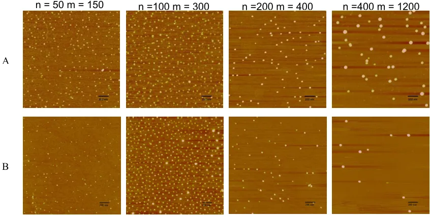
Figure 2.4. A) AFM images of micelles. B) AFM images of crosslinked nanoparticles. The nanoparticle diameters are observed to increase with increasing molecular weight of the constituent block copolymers.
n =100 m = 300 n =200 m = 400 n =400 m = 1200
A
B
Figure 2.5. Crosslinking percentage dependence on irradiation time.
Characterization of the nanoparticles (Table 2.3) was accomplished in the solid state by AFM, as shown in Figure 2.4, and in solution by DLS. Similar to the micelles, the expected trend of increasing nanoparticle diameter with increasing polymer molecular weight is observed, with nanoparticle diameters ranging from 12.7 nm to 39.7 nm by AFM and 47.4 nm to 142.5 nm by DLS. These data are comparable to the values observed for the micelles. The apparent diameter of the nanoparticles is 2-3 times larger when measured using DLS than when measured using AFM. This effect is likely due to the swelling of the polymer chains in solution as well as the hydration sphere surrounding the particles in aqueous environments. Because AFM measures dehydrated particles on a substrate, hydrodynamic diameter measured by DLS is a better indicator of the particle size in vivo. DLS measurements show that the particles fall into the desired range to effectively probe the limits of the EPR effect.1d
Table 2.3. Characterization of Nanoparticles with varied block lengths.
Entry Polymer Mn Diameter (AFM) Diameter (DLS)
1 133,200 12.7 ± 2.6 nm 47.4 ± 7.5 nm 2 280,000 16.4 ± 4.5 nm 58.1 ± 1.8 nm 3 544,000 21.1 ± 3.9 nm 79.7 ± 9.7 nm 4 1,222,000 39.7 ± 4.0 nm 142.5 ± 6.8 nm
remaining after radiofluorination were displaced with additional fluorine-19 to avoid undesired reactions in vivo. The radiofluorinated particles were isolated by diluting the reaction mixture with water and passing this solution through neutral alumina and strongly-acidic cation exchange resin. These conditions effectively removed all of the kryptofix and most of the unreacted fluoride. The extent of reaction was established by measurement of the radioactivity of the nanoparticles, as well as radioTLC (Figure 2.7), which showed that 31% of the fluorine was incorporated into the nanoparticles. The product was recovered in 61% radiochemical purity.
Figure 2.7. RadioTLC of radiofluorinated nanoparticles.
Conclusions
as a hydrophilic block. Sequential ROMP of the two monomers followed by dissolution in water yielded aqueous micelles. Crosslinking of the micelles using ultraviolet light yielded discrete nanoparticles that exhibited hydrodynamic diameters from 47 nm to 142 nm. Standard nucleophilic fluorination chemistry was employed to incorporate fluorine-18 into the nanoparticles in 61% radiochemical purity. Ongoing in vivo studies in mice will establish the optimal size range of nanoparticles for exploitation of the EPR effect.
Acknowledgement
The author thanks Materia for catalyst as well as Peigen Cao for assistance with AFM and for providing a program for measuring the size and size distribution of the nanoparticles. The group of Mark Davis, especially Raymond Archer and Chris Alabi, assisted in the DLS measurements. Radiofluorination reactions were performed by Arkadij Elizarov and Phillip Marchis. The author also thanks Nagichettiar Satyamurthy for helpful discussions regarding radiofluorination.
Experimental
Section
General Information
NMR spectra were measured in CDCl3 or DMSO-d6 on Varian Mercury 300
MHz spectrometers unless otherwise noted. 1H and 13C NMR chemical shifts are reported in ppm relative to CDCl3. Flash column chromatography of organic compounds
(Holtsville, NY) system, including a model BI-200SM goniometer, a model BI-9000AT digital correlator, a model HC120-08 photomultiplier, and a laser operated at 659 nm. Measurements were made at 25 °C. Prior to analysis, solutions were centrifuged in a Beckman model TJ-6 centrifuge at 2000 rpm for 5 min to sediment dust particles. Scattered light was collected at a fixed angle of 90°. The digital correlator was operated with 250 ratio spaced channels, an initial delay of 5 μs, a final delay of 50 ms, and a duration of 10 min. The calculations of the particle size distributions and distribution averages were performed with the ISDA software package (Brookhaven Instruments Company), using CONTIN particle size distribution analysis routines. All measurements were made in triplicate. AFM images were taken using a Nanoscope IV Scanning Probe Microscope Controller (Digital Instruments, Veeco Metrology Group) in tapping mode in air at room temperature using Veeco model TESP tips (spring constant = 20-80 N/m, resonance frequency = 297-335 kHz). The samples were prepared by drop coating onto silicon 111 surfaces that had been prepared by immersion for 5 min at 55 ºC in a solution of H2O/NH4OH (30% in H2O)/H2O2 (50% in H2O) (5:1:1) followed by washing with DI
dn/dc values were obtained for each injection by assuming 100% mass elution from the columns. UV-Vis spectra were taken on a Beckman DU 7400 spectrophotometer.
Materials
CH2Cl2 and THF were purified by passage through solvent purification
columns.30 (H2IMes)(pyr)2(Cl)2RuCHPh (10) was prepared from
(H2IMes)(PCy3)(Cl)2RuCHPh, which was obtained from Materia, according to a
literature procedure.28 Aminohydroxy(polyethylene glycol) was made according to a literature procedure.31 cis-5-Norbornene-endo-2,3-dicarboxylic anhydride was purchased from Acros Organics. All other commercially available materials were obtained from Aldrich Chemical Company and used as received unless otherwise noted.
cis-5-Norbornene-exo-2,3-dicarboxylic anhydride (1). A round-bottom flask
N-(hydroxyethyl)-cis-5-norbornene-exo-2,3-dicarboximide (2). A
round-bottom flask was charged with anhydride 1 (2.07g, 1 equiv). To the flask was added 15 mL toluene, followed by 2-aminoethanol (800μL, 1.05 equiv) and triethylamine (200μL, 0.11 equiv). Stirring caused insoluble clumps to form. A Dean-Stark trap was attached to the flask, and the reaction mixture was heated at reflux for 8 h. Once complete consumption of 1 was observed by TLC, the reaction mixture was concentrated in vacuo to yield an off-white solid. This residue was dissolved in 40 mL CH2Cl2 and washed
with 0.1N HCl (10 mL) and brine (10 mL). The organic layer was dried over MgSO4 and
concentrated in vacuo to yield 2 as a white solid in 93% yield. 1H NMR: δ 1.34 (d, J = 9.9 Hz, 1H), 1.51 (dt, J = 9.9, 1.4 Hz, 1H), 2.3 (s, 1H), 2.71 (d, J = 1.5 Hz, 2H), 3.28 (t, J = 1.5 Hz, 2H), 3.67-3.71 (m, 2H), 3.75-3.79 (m, 2H), 6.29 (t, J =1.8 Hz, 2H). 13C NMR: δ 178.95, 138.00, 60.56, 48.07, 45.45, 42.97, 41.49. HRMS: calculated 208.0974, found
208.0984.
N-(cinnamoyl ethyl)-cis-5-norbornene-exo-2,dicarboximide (3). To a
3-necked, round-bottom flask, equipped with a stirbar, a septum, a stopper, and gas inlet, was added alcohol 2 (1.022 g, 1 eq) under argon flow. 10 mL CH2Cl2 was added,
followed by N-(3-dimethylaminopropyl)-N’-ethylcarbodiimide hydrochloride (1.411 g, 1.49 eq) and 4-dimethylaminopyridine (60 mg, 0.10 eq). trans-Cinnamic acid (886 mg, 1.21 eq) was added as a solution in 10 mL CH2Cl2 via syringe. The reaction mixture was
chromatography (3:2 hexanes/EtOAc) to yield 3 as a clear oil, which solidified into a white solid over several days, in 91% yield. 1H NMR: δ 1.32 (d, J = 9.9 Hz, 1H), 1.49 (dt, J = 9.9, 1.5 Hz, 1H), 2.72 (d, J = 1.2, 2H), 3.28 (t, J = 1.8 Hz, 2H), 3.83-3.87 (m, 2H), 4.34-4.38 (m, 2H), 6.28 (t, J = 1.8 Hz, 2H), 6.36 (d, J = 16.2 Hz, 1H), 7.37-7.50 (m, 5H), 7.64 (d, J = 15.9 Hz, 1H). 13C NMR: δ 178.00, 166.61, 151.02, 145.69, 137.98, 134.40, 130.63, 129.08, 128.35, 117.49, 61.08, 48.02, 45.44, 42.85, 37.76. HRMS: calculated 338.1392, found 338.1381.
N-(hydroxy poly(ethylene glycol))-cis-5-norbornene-exo-2,3-dicarboximide
(4). A round-bottom flask was charged with aminohydroxyPEG (1.428 g, 1 equiv) and
30 mL toluene, followed by anhydride 1 (exo anyhydride) (367 mg, 1.07 equiv) and triethylamine (40 μL, 0.1 equiv). A Dean-Stark trap was attached, and the reaction mixture was heated at reflux for 19 h, at which point complete consumption of aminohydroxyPEG was observed by TLC. The reaction mixture was concentrated in vacuo, and the residue was taken up in CH2Cl2, washed with 0.1 N HCl and brine, and
dried over MgSO4. The crude product was purfied by silica gel chromatography (5%
MeOH in CH2Cl2) to yield 4 as a clear oil in 80% yield. 1H NMR: δ 1.35 (d, J = 9.9 Hz,
1H), 1.49 (dt, J = 9.9 Hz, 1.5 Hz, 1H), 2.67 (s, 2H), 3.26 (m, 2H), 3.56-3.73 (m, 50H), 6.27 (t, J = 1.8 Hz, 2H). 13C NMR: δ 178.21, 138.00, 130.92, 70.72, 70.44, 70.01, 67.05, 61.87, 47.99, 45.44, 42.89, 37.88.
N-(mesyl poly(ethylene glycol))-cis-5-norbornene-exo-2,3-dicarboximide (5).
mixture was cooled to –35 ºC. MsCl (73 μL, 1.5 equiv) as a solution in 2 mL CH2Cl2
was added dropwise using a syringe pump at a rate of 0.05 mL/min. After complete addition, the reaction mixture was allowed to slowly warm to room temp. After 6 h complete consumption of 4 was observed by TLC, and the reaction mixture was diluted with CH2Cl2 (20 mL) and washed with 0.1 N HCl (10 mL) and brine (10 mL), then dried
over MgSO4. The crude product was purified by silica gel chromatography (5% MeOH
in CH2Cl2) to yield 5 as a clear oil in 83% yield. 1H NMR: δ 1.34 (d, J = 9.9 Hz, 1H),
1.49 (dt, J = 9.9 Hz, 1.5 Hz, 1H), 2.68 (s, 2H), 3.08 (s, 3H), 3.26 (m, 2H), 3.56-3.72 (m, 50H), 3.73 (m, 2H), 4.38 (m, 2H) 6.28 (t, J = 1.8 Hz, 2H). 13C NMR: δ 178.19, 138.01, 70.74, 70.03, 69.49, 69.20, 67.06, 47.99, 45.45, 42.90, 37.92.
N-(ethylmesyl)-cis-5-norbornene-exo-2,3-dicarboximide (6). A round bottom
flask under argon was charged with alcohol 2 (100 mg, 1 equiv) and CH2Cl2 (1 mL). The
solution was cooled to 0º C, then NEt3 (150 μL, 2.2 equiv) was added via syringe.
Lastly, MsCl (60 μL, 1.6 equiv) was added dropwise via syringe. The reaction mixture immediately turned dark yellow. After 2 h the reaction was quenched by addition of H2O
(2 mL) to the reaction mixture. The mixture was transferred to a separatory funnel, and the organic layer was removed. The organic layer was then washed with 0.01 N HCl (10 mL) and brine (10 mL) and dried over MgSO4. The yellow solid was purified on a plug
of silica, eluting with 5% MeOH in CH2Cl2 to yield a white powder (94 mg) in 68%
yield. 1H NMR: δ 1.30 (d, J = 9.9, 1H), 1.63 – 1.48 (m, 2H), 2.77 (t, J = 7.4, 2H), 3.03
N-(ethoxy-ethyl-2-(mesyl))-cis-5-norbornene-exo-2,3-dicarboximide (7).
N-(ethoxy-2-ethoxy)-cis-5-norbornene-exo-2,3-dicarboximide was first prepared by reaction of anhydride 1 (96 mg, 1 equiv) with H2N(CH2)2O(CH2)2OH (70 μL, 1.2 equiv) and NEt3
(10 μL, 0.1 equiv) in 1 mL toluene. The reaction mixture was heated at reflux with a Dean-Stark trap for 2 h. The solvent was removed by rotary evaporation, and the residue was taken up in CH2Cl2. This solution was washed with 0.1 N HCl and brine then dried
over MgSO4. A clear oil was recovered (114 mg) and used without further purification in
the next step. To this oil in 1 mL dry CH2Cl2 was added NEt3 (120 μL, 2.0 equiv). After
cooling to 0º C, MsCl (50 μL, 1.5 equiv) was added dropwise. After 12 h, the reaction was quenched by adding H2O (2 mL). The mixture was transferred to a separatory
funnel, and the organic layer was removed. The organic layer was then washed with 0.1 N HCl (10 mL) and brine (10 mL) and dried over MgSO4. The crude yellow oil was
purified by silica gel chromatography (5% MeOH in CH2Cl2, I2 vis) to yield a clear oil
(119 mg) in 83% yield. 1H NMR: δ 1.30 (d, J = 9.9, 1H) 1.63 – 1.48 (m, 2H), 2.71 (d, J = 1.1, 2H), 3.04 (s, 3H), 3.30 – 3.22 (m, 2H), 3.79 – 3.62 (m, 6H), 4.36 – 4.24 (m, 2H), 6.30 (t, J = 1.7, 2H). 13C NMR: δ 178.29, 138.02, 69.18, 68.45, 67.39, 48.06, 45.50,
42.90, 37.89, 37.76.
N-(ethoxy-ethyl-2-(mesyl)acetate)-cis-5-norbornene-exo-2,3-dicarboximide
(8). Mesyl-glycolic acid was first prepared from a modified literature procedure.32 Briefly, silver mesylate (520 mg, 1.1 equiv) was added to a 2-necked round-bottom flask under argon flow followed by dry CH3CN (2 mL). Iodoacetic acid (437 mg, 1 equiv) was
and CH3CN was added. This solution was transferred via cannula into the reaction flask,
and the flask was covered with aluminum foil. After 20 h, the yellow AgI precipitate was removed by filtering the reaction mixture through Celite. The filtrate was rotovapped to yield a clear oil. The product was extracted from the oil with Et2O. Concentration of the
Et2O solution yielded a white powder, which was recrystallized from CHCl3/actone/pet
ether (1:1:5) to yield white crystals (105 mg) in 29% yield. 1H NMR (CD3COCD3): δ
3.22 (s, 3H), 4.85 (s, 2H). The pure mesyl-glycolic acid (95 mg, 1 equiv) was then added to a 2-necked round-bottom flask under argon with CH2Cl2 (1 mL). EDC (185 mg, 1.9
equiv) and DPTS (15 mg, 0.1 equiv) were added to the reaction mixture, and the reaction mixture became cloudy yellow over several minutes. N-(ethoxy-2-ethoxy)-cis -5-norbornene-exo-2,3-dicarboximide, prepared as described previously, was added to the reaction mixture in 1 mL CH2Cl2. The reaction mixture became clear within 1 min.
After 3.5 h, the reaction was quenched by addition of H2O (3 mL). The CH2Cl2 layer was
separated off, washed with H2O and brine, and dried over MgSO4 to yield a pale yellow
oil. The product was purified by column chromatography to yield a clear oil (24 mg) in 12% yield. 1H NMR: δ 1.31 (d, J = 9.8, 1H), 1.48 (d, J = 9.8, 1H), 2.69 (s, 2H), 3.34 – 3.16 (m, 5H), 3.78 – 3.59 (m, 6H), 4.38 – 4.23 (m, 2H), 4.80 (s, 2H), 6.30 (t, J = 1.7, 2H).
13C NMR: δ 178.19, 166.99, 137.97, 68.00, 67.14, 64.89, 47.99, 45.43, 42.82, 39.29,
37.65.
N-(ethoxy-ethyl-2-(nosyl))-cis-5-norbornene-exo-2,3-dicarboximide (9).
(165 mg, 1.4 equiv). CH2Cl2 (1 mL) was added, and the flask was cooled to 0 ºC. NEt3
(110 μL, 1.5 equiv) was then added dropwise. After 15 h, the reaction was quenched by adding H2O (3 mL). The mixture was transferred to a separatory funnel, and the organic
layer was removed. The organic layer was then washed with 0.1 N HCl (10 mL) and brine (10 mL) and dried over MgSO4. The crude orange oil was purified by silica gel
chromatography (5% MeOH in CH2Cl2, I2 vis) to yield an orange oil (48 mg) in 20%
yield. 1H NMR: δ 1.29 (d, J = 9.9, 1H) , 1.55 – 1.41 (m, 1H), 2.68 (d, J = 1.2, 2H), 3.31 – 3.19 (m, 2H), 3.73 – 3.54 (m, 6H), 4.27 – 4.15 (m, 2H), 6.28 (t, J = 1.7, 2H), 8.19 – 8.06 (m, 2H), 8.50 – 8.37 (m, 2H). 13C NMR: δ 178.23, 138.00, 128.51, 124.71, 70.41, 67.90, 67.30, 48.02, 45.47, 42.88, 37.73.
Cold Fluorination Procedure. In an N2-filled glovebox, KF (11 mg, 3.0 equiv),
K2CO3 (30 mg, 3.5 equiv), and kryptofix 222 (45 mg, 2.0 equiv) were massed into a
2-necked, round-bottom flask. The flask was capped with a septum and a greased stopper. In a separate vial, the substrate to be fluorinated (1 equiv) was dissolved in 0.75 mL CH3CN. The vial and the flask were brought out of the box, and the CH3CN solution was
transferred via cannula to the reaction flask. After stirring for 20 min at room temp, the reaction was quenched by addition of a small amount of H2O. The reaction mixture was
concentrated, and the product distribution was evaluated by 1H NMR spectroscopy.
Polymerization Procedure. In a typical polymerization, a vial was charged with
mg/mL) was then injected into the vial. The reaction was allowed to proceed at room temperature under argon flow for 1-2 min, then hydrophilic norbornene 5 (21.2 mg) was added as a solution in THF (0.2 mL). All reactions were quenched by addition of ethyl vinyl ether (0.2 mL) that had been purified by passage through a short column of silica gel. The reaction mixture was allowed to stir for an additional 15 min before precipitation into 20 mL Et2O/hexanes (1:1). The products were recovered in 87-99%
yield by decanting off the supernatant and scraping the gooey solids off of the sides of the beaker. 1H NMR δ 1.30-1.70 (br s), 1.95 (s), 1.85-2.55 (br s), 2.90-3.05 (br s), 3.07 (s), 3.63 (br s), 3.75 (m), 4.36 (m), 5.38-5.80 (br m), 6.32-6.44 (br d), 7.30-7.55 (br d), 7.58-7.70 (m).
Micelle Formation Procedure. Block copolymer (10 mg) was dissolved in
filtered (0.02 μm) THF (5 mL). Once homogeneous, filtered (0.02 μm) DI water (5 mL) was added dropwise over 10 min. The micelle solution was then transferred to dialysis tubing (8000 MWCO) and dialyzed against water for 24-48 h while protected from light.
Radiofluorination Procedure. A 5 mL reaction vessel in a fume hood with a leaded glass sash was charged with hydrated K18F (361 mCi). K2CO3 (1 mg) and
kryptofix 222 (10 mg) in 1 mL CH3CN/H2O (94:4) were added to the vessel. The solvent
was boiled off by submersing the vessel in an oil bath at 120 ºC while bubbling N2
through the reaction mixture. BHT in dry CH3CN (1 mL) was then added to the vessel,
followed by solvent removal in the same way. Two more additions and evaporations of dry CH3CN were performed to ensure complete removal of H2O. A lyophilized
nanoparticle sample (5.7 mg) was dissolved in 3 mL dry CH3CN and added to the vessel.
The vessel was sealed and heated at 120 ºC for 60 min. In order to displace the excess mesylates, K19F (1.5 mg) and kryptofix 222 (6.5 mg) in 0.5 mL CH3CN were added to
the vessel. The vessel was sealed and heated for an additional 30 min at 80 ºC. At this point the reaction mixture was diluted with water and passed through a column containing Dowex strongly acidic macroreticular ion exchange resin, followed by a short plug of alumina. RadioTLC was used to analyze the radiochemical purity of the product.
References
Mater. 2006, 5, 971–976. (c) Mulder, W. J. M.; Koole, R.; Brandwijk, R. J.; Storm, G.; Chin, P. T. K.; Strijkers, G. J.; Donega, C. D.; Nicolay, K.; Griffioen, A. W. Nano Lett. 2006, 6, 1–6. (d) Kobayashi, H.; Brechbiel, M. W. Adv. Drug 104, 15549–15554. (c) Fukukawa, K.; Rossin, R.; Hagooly, A.; Pressly, E. D.; Hunt, J. N.; Messmore, B. W.; Wooley, K. L.; Welch, M. J.; Hawker, C. J. Biomacromolecules 2008, 9, 1329–1339. (d) Schipper, M. L.; Cheng, Z.; Lee, S. W.; Bentolila, L. A.; Iyer, G.; Rao, J.; Chen, X.; Wu, A. M.; Weiss, S.; Gambhir, S. S. J. Nucl. Med. 2007, 48, 1511–1518.
(14) Since the publication of this work, two reports concerning the synthesis of fluorine-18-containing nanoparticles have been published: (a) Devaraj, N. K.; Keliher, E. J.; Thurber, G. M.; Nahrendorf, M.; Weissleder, R. Bioconjugate Chem. 2009, 20, 397–401. (b) Herth, M. M.; Barz, M.; Moderegger, D.; Allmeroth, M.; Jahn, M.; Thews, O.; Zentel, R.; Rösch, F. Biomacromolecules 2009, 10, 1697–1703. (19) Stubenrauch, K.; Moitzi, C.; Fritz, G.; Glatter, O.; Trimmel, G.; Stelzer, F.
Macromolecules 2006, 39, 5865–5874.
(20) Bielawski, C. W.; Grubbs, R. H. Prog. Polym. Sci. 2007, 32, 1–29.
(22) Choi, T. L.; Grubbs, R. H. Angew. Chem. Int. Ed. 2003, 42, 1743–1746.
(23) (a) Liu, G. J.; Hu, N. X.; Xu, X. Q.; Yao, H. Macromolecules 1994, 27, 3892– 3895. (b) Guo, A.; Liu, G. J.; Tao, J. Macromolecules 1996, 29, 2487–2493. (c)Henselwood, F.; Liu, G. J. Macromolecules 1997, 30, 488–493.
(24) Gref, R.; Minamitake, Y.; Peracchia, M. T.; Trubetskoy, V.; Torchilin, V. P.; Langer, R. Science 1994, 263, 1600–1603.
(25) Cai, L.; Lu, S.; Pike, V. W. Eur. J. Org. Chem. 2008, 17, 2853–2873. (26) Noyce, D. S.; Virgilio, J. A. J. Org. Chem. 1972, 37, 2643–2647.
(27) (a) Camm, K. D.; Castro, N. M.; Liu, Y. W.; Czechura, P.; Snelgrove, J. L.; Fogg, D. E. J. Am. Chem. Soc. 2007, 129, 4168–4169. (b) Slugovc, C.; Demel, S.; Stelzer, F. Chem. Commun. 2002, 21, 2572–2573.
(28) Love, J. A.; Morgan, J. P.; Trnka, T. M.; Grubbs, R. H. Angew. Chem., Int. Ed. Engl. 2002, 41, 4035–4037.
(29) Bielawski, C. W.; Benitez, D.; Morita, T.; Grubbs, R. H. Macromolecules 2001, 34, 8610–8618.
(