ABSTRACT
LOOMIS, KARI DANIELLE. C/EBPα is an Epithelial Tumor Suppressor Gene, and Mitogenic Stimulation Reciprocally Regulates C/EBPα and C/EBPβ. (Under the direction of Dr. Robert C. Smart.)
incidence, multiplicity, growth rate and the rate of malignant progression. Mice hemizygous for C/EBPα displayed an intermediate enhanced tumor phenotype. Our results suggest decreased expression of C/EBPα contributes to deregulation of tumor cell proliferation. C/EBPα had been proposed to block cell cycle progression through inhibition of E2F activity. We observed that C/EBPα blocked Ras-induced and epidermal growth factor–induced E2F activity in keratinocytes and also blocked Ras-induced cell transformation and cell cycle progression.
C/EBP
α
is an Epithelial Tumor Suppressor Gene, and Mitogenic
Stimulation Reciprocally Regulates C/EBP
α
and C/EBP
β
by
Kari Danielle Loomis
A dissertation submitted to the Graduate Faculty of North Carolina State University
in partial fulfillment of the requirements for the Degree of
Doctor of Philosophy
Functional Genomics
Raleigh, North Carolina 2008
APPROVED BY:
__________________________ __________________________
David M. Bird Barbara Sherry
__________________________ __________________________ Marcelo Rodriguez-Puebla Robert C. Smart
DEDICATION
I dedicate this thesis to my grandfather, Orval Lucier. Orval was a true lifelong learner who took classes at the University of Illinois, Urbana/Champagne every semester until he passed away in 1996 at age 81. I will always strive to maintain that kind of enthusiasm for life and learning.
BIOGRAPHY
Kari Danielle Loomis was born on February 5, 1976 in Massachusetts where she graduated from Duxbury High School in 1994. She then attended Clemson University in South Carolina where she earned a BS in Biology Education in 1998. Kari then taught middle school science and math in Brewster, NY, followed by three years of teaching high school biology at Bethel High School in Connecticut. Within this time, Kari worked to update the curriculum, designed a new course in the evolution of animal behavior, and coordinated internships with Boehringer Ingelheim Pharmaceuticals, a local pharmaceutical company. Kari sought out summer internships with this company, which became the launching pad for her research career. In 2003, Kari began her doctoral studies at North Carolina State University under the direction of Dr. Robert Smart. Kari was awarded the Graduate Assistance for Areas of National Need (GAANN) biotechnology fellowship, and taught a biotechnology PCR course. Kari was an active participant in the Functional Genomics graduate student association where she served as both vice president and president. Kari presented her research at a number of local and national meetings, and she was awarded first and second place for poster presentations at the North Carolina Society of Toxicology and North Carolina State University Graduate Student research symposia, respectively. Kari has accepted a tenure track assistant professor position in Cellular and Molecular Biology at Mars Hill College, in Mars Hill, NC.
ACKNOWLEDGEMENTS
I would first and foremost like to thank my advisor, Dr. Robert C. Smart for the rigorous and thorough training I have received in his lab. I would also like to thank my committee members, Dr. David Bird, Dr. Marcelo Rodriguez-Puebla, and Dr. Barbara Sherry for their time and advice throughout my doctoral studies. I would especially like to thank Dr. Sherry as she has been a great support and inspiration to me from the moment I came for my admission interview to my doctoral defense. I thank my colleagues in the Smart lab for their help and support, including Dr. Songyun Zhu, Dr. Kyungsil Yoon, Dr. Sarah Ewing, Kristina Powers, Jeanne Burr, Elizabeth Thompson, Rakesh Ranjan, and John House. I would like to thank my family and friends for supporting me in my quest, as well as my dogs Jewels and Oscar who have had to sacrifice many nice hikes in the woods for my work. Lastly, I would like to thank my fiancé, Matthew T. Dziedzic. Matt’s support and belief in me have given me the strength I needed to complete this work.
TABLE OF CONTENTS
page
LIST OF FIGURES………... vii
GENERAL INTRODUCTION……….. 1
CHAPTER 1 Genetic ablation of CCAAT/enhancer binding protein α in epidermis reveals its role in suppression of epithelial tumorigenesis……... 34
Abstract……….. 35
Introduction……….... 37
Results……… 40
Discussion……….. 49
Materials and Methods………. 56
Acknowledgements……….. 60
References………. 61
CHAPTER 2 Stimulation with EGF induces the activation and reciprocal . regulation of C/EBPα and C/EBPβ to enhance E2F-driven cell cycle progression……... 76
Abstract……….. 77
Introduction……… 78
Results……… 81
Discussion……….. 89
Materials and Methods………. 93
Acknowledgements……….. 97
References……… 98
GENERAL DISCUSSION……….. 110
GENERAL REFERENCES……….... 116
APPENDIX... 133
C/EBPβ-/- keratinocytes have diminished expression of E2F target genes... 134
Materials and Methods... 136
LIST OF FIGURES
page
INTRODUCTION
Figure 1 Cyclin/CDK complexes... 23
Figure 2 Cyclin expression throughout the cell cycle... 24
Figure 3 Gene transcription control by E2F and Rb... 25
Figure 4 Overview of cancer process... 27
Figure 5 Oncogene activation... 28
Figure 6 Monoclonal Nature of Cancer... 29
CHAPTER 1 Figure 1 C/EBPα is not expressed in the epidermis, hair follicle, and . sebaceous gland of K5Cre/C/EBPαfl/fl mice... 71
Figure 2 C/EBPα is dispensable for normal epidermal homeostasis... 72
Figure 3 K5Cre;C/EBPαfl/fl mice are more susceptible to carcinogen -induced skin tumor development involving oncogenic . Ras... 73
Figure 4 K5Cre;C/EBPαfl/fl mice display a significant increase in tumor . growth rate and the rate of malignant progression.... 74
Figure 5 C/EBPα inhibits Ras-induced E2F transcription activity, . transformation, and cell cycle progression... 75
CHAPTER 2 Figure 1 C/EBP activity is stimulated by EGF in keratinocytes... 104
Figure 2 C/EBPα and C/EBPβ mRNA levels, protein levels, and DNA .
binding are reciprocally regulated by EGF... 105 Figure 3 C/EBPβ expression levels and transcriptional activity are .
upregulated through an EGFR/Ras/MEK/ERK pathway... 106 Figure 4 EGF signaling upregulates and activates C/EBPβ to promote .
cycle entry... 108 Figure 5 C/EBPβ-/- keratinocytes have diminished expression of E2F
target genes, and C/EBPβ can enhance EGF and Ras-
induced E2F transcription activity ... 109 GENERAL DISCUSSION
Figure 1 Proposed mechanistic role for C/EBPα and C/EBPβ in the regulation of E2F transcription and cell cycle progression... 110 APPENDIX
Figure 1 C/EBPβ-/- keratinocytes have diminished expression of
E2F target genes... 137
GENERAL INTRODUCTION
Extracellular stimuli induce a multitude of cellular responses through the specific activation of transcriptional apparatus to induce changes in gene expression: a cascade of modifications to signal-transducing molecules direct site-specific transcription factors to activate or repress gene transcription and elicit an appropriate physiological response. Cellular responses include those involving metabolism, proliferation, apoptosis/survival, differentiation, cell adhesion, inflammatory responses, and locomotion, to name a few. This complex system of gene regulation is tightly controlled and often varies significantly between cell types and organisms. Deregulation of important signal transduction pathways can result in numerous disorders and diseases, including cancer. Here I will focus on the function and regulation of the site-specific transcription factors, CCAAT/enhancer binding proteins α and β (C/EBPα and C/EBPβ), in normal epidermal homeostasis, as well as in epidermal neoplasia formation.
Skin
flexible connective tissue layer which helps bind the entire body together. The dermis contains fibroblasts and macrophages organized in a semi-fluid matrix of collagen, elastin, and reticular fibers. This layer houses most of the skin’s specialized cells and structures including blood and lymph vessels, sweat glands, hair follicles, sebaceous glands, and nerve endings. The dermis functions to regulate temperature, maintain hydration, and supply the epidermis with nutrient-rich blood. An extracellular matrix component known as the basement membrane forms a barrier which separates the epidermis and its appendages from the dermis. The basement membrane is also rich in receptor tyrosine kinase growth factors, which provide proliferative stimuli to the innermost basal layer of the epidermis1.
Small finger-like projections known as dermal papillae indent the overlying epidermis carrying pain and touch receptors as well as capillary loops. Follicular dermal papillae are composed of specialized fibroblasts that specifically populate the base of the hair follicle bulb2. The dermal papillae contain cells involved in important epithelial-mesenchymal interactions which direct various processes including the initiation of epidermal stem cell differentiation into hair follicles or other epidermal appendages1.
Epidermis
The epidermis is a thin layer of keratinized stratified squamous epithelium and is the outermost protective shield of the body. Keratinocytes are the primary epidermal cell type, but melanocytes, Merkel cells, and Langerhans’
cells are also present. In mammals, the epidermis comprises the interfollicular epidermis as well as the adnexal structures including the hair follicle and sebaceous gland.
As a self-renewing tissue, the epidermis must balance proliferation and differentiation3, 4. Proliferative keratinocytes of the innermost interfollicular epidermal layer become post-mitotic, move up to the suprabasal layers, and undergo terminal differentiation where they become flattened and eventually lead to the production of the dead, cornified epidermal layers that provide the protective covering of the skin. In addition to its role in protection from the environment, the skin and its appendages also function in sensation, communication, respiration, locomotion, immune responses, and thermoregulation5. Epidermal keratinocytes are organized into four sub-layers: the basal layer, spinous layer, granular layer, and stratum corneum.
The basal layer of the epidermis consists of a single layer of proliferating keratinocytes that give rise to terminally differentiating post-mitotic stratified layers. Keratinocytes of the basal layer are firmly attached to the extracellular matrix-rich basement membrane through focal contacts and hemidesmosomes, and are attached to one another through adherens junctions and desmosomes6. The basal layer follows a path along the surface of the interfollicular epidermis, as well as along the outer root sheath of the hair follicle. Keratinocytes in the basal layer of the interfollicular epidermis express genes encoding integrins necessary for basement membrane attachment, growth factor receptors
(particularly epidermal growth factor receptor, or EGFR), as well as the structural keratins 5 and 14 (K5 and K14)6, 7. The basal layer contains both proliferative and post mitotic keratinocytes which move upward and leave the basal cell layer. Once cells detach from the basement membrane, they are characterized as suprabasal cells, and all of the cells become post-mitotic.
The suprabasal layer consists of the spinous layer, granular layer, and stratum corneum. In these layers, cells repress basally expressed genes and switch to expressing a set of differentiation-associated proteins, including keratins 1 and 10 (K1 and K10) in early stages of differentiation and loricrin and involucrin in the later stages of differentiation. As keratinocytes continuetheir trek upward to the stratum corneum, they further adjust their transcriptional program, all metabolic activity ceases, and cytoplasmic organelles are lost, culminating in the production of dead, flattened squames which form the stratum corneum. The resulting cornified envelope is composed of highly insoluble, cross-linked proteins withcovalently attached lipids essential for the mechanicalintegrity and water impermeability of the skin8. Cells of the stratum corneum are eventually sloughed from the skin surface as new cells moving outwardreplace them4.
Epidermal Homeostasis
The epidermis is a continuously renewing tissue derived from multipotent stem cells that self-renew and also generate progeny that undergo terminal differentiation9. The best characterized stem cells reside in a region of the hair
follicle known as the bulge region10, although stem cells also reside in the interfollicular epidermis and the potentially the sebaceous gland11. The bulge region is found in a portion of the hair follicle outer root sheath just below the sebaceous gland, and the stem cells residing there are capable of producing keratinocytes, hair follicles, and sebaceous glands11. Although any epidermal stem cell can generate all of the epidermal lineages, it can be argued that the choice of lineage is determined by the specific location or microenvironment of the cell3, 12, 13. Mesenchymal stimulation from dermal papillae cells, specifically, has been shown to induce interfollicular epidermal cells to differentiate into hair and sebocyte lineages14, 15.
Although it has been proposed that stem cells from the bulge region were largely responsible for basal keratinocyte replenishment6, recent studies show that distinct stem cell populations regenerate the hair follicle and interfollicular epidermis16,. The bulge region stem cells do however participate in interfollicular regeneration in wound repair16, 17. Keratinocytes exiting the bulge region populate the outer root sheath, and are thought to fuel the highly proliferative matrix cells that are adjacent to the mesenchymal dermal papillae and are involved in hair follicle development. β1-integrin extracellular matrix receptors are important in epidermal stem cell proliferation and maintenance 1, contribute to the ability of stem cells to bind to the extracellular matrix of the basement membrane, and are potential markers of interfollicular epidermal stem cells18. In addition to high surface expression of β1 integrins, high α6
integrin, low CD71 expression, high expression of Delta1, low expression of desmoglein 3, and low expression of the EGF receptor 1 are also markers for interfollicular epidermal stem cells19.
In the widely accepted mechanism for interfollicular epidermal homeostasis, a small number of slow cycling stem cells produce a larger number of highly proliferative transit-amplifying cells3. Transit-amplifying cells have a limited replicative potential before undergoing terminal differentiation18. The stem cells are surrounded by transit-amplifying cells to form proliferative units, or EPUs, composed of hexagonally packed cells1. In vivo pulse-chase experiments identified label retaining cells of the hair follicle as cells that cycle less frequently than those of nearby basal keratinocytes. The Cairns hypothesis argues that stem cells preserve their genetic message by using selective strand segregation to maintain a constant template strand and prevent mitotic crossing over20. Due to this strand retention, mutations in the DNA of stem cells pose a greater risk for cancer development. While a large percentage of hair follicle bulge cells are label retaining cells, only a small percentage of interfollicular cells are label retaining cells. The low rate and even dispersal of label retaining cells in the interfollicular epidermis was taken as circumstantial evidence in support of the EPU mechanism described above. It is unclear however whether basal epidermal label retaining cells represent stem cells21. Comparisons between pulse-chase label retention experiments and cellular profiling using various epidermal stem cell markers such as β1
integrin, showed far greater β1 high expressing cells than label retaining cells in human skin1.
Recent studies suggesting another possible mechanism for maintenance of epidermal homeostasis show that as basal epidermal cells divide, they can polarize and localize key regulatory proteins in the cell to create an asymmetrical partition of cellular components, thus specifying the cell fate of each daughter cell as stem cell or differentiating cell22, 23. With this second mechanism, many if not all basal cells may have the capacity for self-renewal and epidermal stratification and differentiation1. Appropriate stem cell markers and a proper definition of what constitutes a stem cell are central issues in determining the true mechanism for epidermal homeostasis.
C/EBP Family of Transcription Factors
CCAAT/enhancer binding proteins, or C/EBPs, are a family of basic leucine zipper transcription factors that act as master regulators of many cellular responses. There are currently 6 C/EBP family members represented by greek letters assigned chronologically according to each gene’s date of identification (α, β, γ, δ, ε, and ζ). In the late 1980’s, the McKnight lab first identified and cloned the founding member C/EBPα (know at the time simply as C/EBP)24, 25. The protein was named according to its role in binding the CCAAT cis-regulatory promoter sequence as well as in binding an enhancer core element common to SV40, murine sarcoma virus, and polyomavirus26, 27. Further characterization of the C/EBP genes revealed their role not only in
binding to a variety of cis-regulatory DNA sequences, but also in binding to and controlling the activity of key regulatory proteins. The promiscuous and unexpected nature of C/EBP protein and DNA binding has led McKnight to describe the name C/EBP as “A silly acronym he wears around his neck like a rotting albatross27”. The next section will give an overview of the structure, expression, and function of all C/EBP family members; with a focus on the roles of C/EBPα and C/EBPβ in the epidermis.
Structure
C/EBPα, -β, -δ, and –γ are intronless genes, whereas C/EBPε and –ζ contain two and four exons, respectively28. Multiple isoforms of C/EBPε protein are produced through alternative use of promoters and differential splicing of the RNA transcript29, 30. Despite the fact that C/EBPα and C/EBPβ are intronless genes, they have multiple isoforms due to regulation of translation initiation 29, 31, 32. C/EBP family members share greater than 90% sequence identity in their C-terminal bZIP domain, consisting of a basic amino acid rich DNA binding region followed by a dimerization motif termed the leucine zipper28. Family members form homo- and heterodimers with one another through their leucine zipper domains; C/EBP dimerization is required for DNA binding. C/EBP dimerization interactions can contribute to the differential regulation of C/EBP activity31-35. The largely conserved C/EBP DNA binding domain, with the exception of C/EBPζ, results in a common but flexible C/EBP-DNA binding recognition motif of (A/G)TTGCG(C/T)AA(C/T)36, 37.
The exact mechanism by which C/EBPs achieve specificity of function while maintaining similar binding recognition domains is not yet fully elucidated, but has been shown to involve numerous processes including: 1. protein conformational changes resulting from post-translational modification, as when phosphorylation of C/EBPα at Ser21 alters C/EBPα conformation to favor monocyte differentiation and inhibit granulopoiesis through a transcriptional mechanism38, 2. protein-protein interactions with the basal transcription machinery, other transcription factors, and chromatin remodeling complexes, as when C/EBPβ binds to specific components of the RNA-polymerase-II holoenzyme to activate or repress gene transcription39, and 3. Differential regulation of C/EBP expression, as shown with the decreased expression of C/EBPα after partial hepatectomy to promote C/EBPβ rather than C/EBPα transcription40, and in C/EBPβ knock-in mice where the temporal expression of C/EBPβ from the C/ebpα gene locus was able to substitute for C/EBPα expression and rescue a lethality phenotype41. All of these mechanisms contribute to the ability of individual family members to achieve specificity of DNA binding and transcriptional regulation.
C/EBP proteins are primarily regulated through the N-terminal transactivation domain where C/EBP family members share less than 20% sequence identity. Within the N-terminus are three semi-conserved activation domains and regulatory domains, whose presence varies between family members. These conserved domains are involved in interactions between
C/EBPs and the basal transcription apparatus (TBP/TFIID), transcriptional coactivators (CBP/p300), and in sumoylation. Exceptions include C/EBPγ which lacks any functional transactivation domain outside of the basic region, and C/EBPζ which has a shortened N-terminal and does not contain any of the conserved activation or repressive domains33, 42. Also, the smaller isoforms of C/EBPα and C/EBPβ lack the major N-terminal functional domains.
Differences in the N-terminal functional domains of specific C/EBP isoforms can result in highly varied transactivation potential between family members. Heterodimerization between a relatively more and less active C/EBP can result in the ability of the less active C/EBP to dominantly inhibit the activating C/EBP. The N-terminal domain is generally considered to be the activation domain, and proteins lacking the domain are largely considered to have little to no capacity for intrinsic activity. However, this paradigm seems to be shifting somewhat as dimerization interactions between the leucine zipper domains of specific family members have been shown to activate C/EBPs lacking the N-terminal region. For instance, it has been shown that C/EBPβ and C/EBPγ heterodimerization results in the specific activation of C/EBPγ stimulatory activity in a cell type and gene promoter specific manner35. The ability of dimerization interactions in the leucine zipper to activate C/EBPγ’s transcriptional potential also supports the idea that the leucine zipper domain plays a larger role than simply enabling dimerization35. Further, protein-protein interactions take place in both the C- and N-terminal of the protein. Gene
regulation, heterodimerization, protein-protein interactions, and post-translational modifications, all contribute to the functional versatility of individual C/EBP family members to specifically respond to their cellular environments with unique and overlapping roles.
Expression
C/EBP family members are differentially expressed in numerous cells, tissues, and organisms, and in response to a variety of stimuli. The precise expression profile for each C/EBP family member is not yet fully elucidated and there are some conflicting studies in regards to these C/EBP expression patterns. The general consensus will be presented here. C/EBPα is expressed at high levels in skin, adipose tissue, liver, intestine, lung, mammary gland, adrenal gland, peripheral-blood mononuclear cells, limbal and corneal epithelial cells, ovary, and placenta28, 43-47. In both liver and adipose tissue, C/EBPα message is highest in the most differentiated post mitotic cells48, 49. C/EBPβ, also known as NF-IL6 or LAP, is expressed in most tissues, but its steady state expression levels are greatest in the skin, liver, intestine, lung, adipose tissue, spleen, kidney, and myelomonocytic cells43, 47, 48, 50-56. C/EBPγ, or Ig/EBP-1, is characterized as ubiquitously expressed in various tissues, and has been thought to be somewhat unresponsive to extracellular stimuli, in part due to the absence of any functional N-terminal domain35, 57. More recent studies have shown a high inter-individual variation in C/EBPγ transcript abundance, and a correlation between C/EBPγ and antioxidant and DNA repair genes in normal
individuals, but not in individuals with bronchogenic carcinoma58. Although C/EBPδ, also known as NF-IL6β, is expressed at low to undetectable levels in most cell types and tissues under physiological conditions 47, 48, 59, 60, it is highly inducible by a variety of extracellular stimuli including growth hormone, insulin, INFγ, IL-1, IL-6, LPS, TNFα, dexamethasone, noradrenalin, glutamate, and luteinizing hormone60, 61. C/EBPδ induction is best characterized in adipose tissue, lung, intestine, and mammary epithelium28, 62. C/EBPε has a more limited range of expression specific to myeloid and lymphoid cells28. Finally, C/EBPζ, better known as CHOP-10/GADD153 (C/EBP Homologous Protein-10/Growth Arrest- and DNA Damage-inducible gene 153), is ubiquitously expressed at very low levels, but is highly inducible by a number of extracellular growth arrest and/or DNA damaging factors including calcium ionophores, glucose deprivation, oxidative stress, reductive stress, and endoplasmic reticulum stress/activation of theacute phase response63. Although C/EBPγ, and possibly C/EBPζ and C/EBPδ are expressed in the skin, C/EBPα and C/EBPβ are the most prevalent C/EBP family members in epidermal keratinocytes (unpublished data, and43, 44, 64, 65).
Function
C/EBP family members function as transcriptional activators or repressors of many key genes involved in cellular processes such as energy metabolism, inflammation, and liver regeneration. C/EBPs are also well known regulators of differentiation and proliferation in various cell types including
adipocytes, granulocytes, hepatocytes, and keratinocytes. Although many of the functions of C/EBPs are in line with traditional roles for transcription factors, many C/EBP functions are fully independent of DNA binding and transcriptional activation or repression28.
There is considerable crosstalk between family members, as C/EBPs can either homo- or hetero-dimerize, and heterodimerization can result in modification of function. A certain level of redundancy in C/EBP protein function is also suggested by the fact that the post-natal lethality of C/EBPα knockout mice can be rescued by expression of C/EBPβ from the C/ebpα gene locus41. C/EBPs have been shown to transcriptionally self-regulate their promoters and/or regulate the promoters of other C/EBP family members, further adding to the complex modulation of C/EBP function. As a result, studying the function of individual C/EBP family members in isolation can be somewhat difficult. A general overview of all C/EBP family members’ functions will be given here, and a more detailed look specifically at the functions of C/EBPα and C/EBPβ in the epidermis and in cancer will follow.
C/EBPα plays a role in inducing genes characteristic of the terminally differentiated state in several cell types including adipocytes, lung, liver, mammary gland, skin, neurons, and within the hematopoietic system66-68. Another primary function of C/EBPα is its ability to seemingly slow or arrest mitotic growth. Much attention has been given to the ability of C/EBPα to couple these two main features of terminal differentiation66. C/EBPα also plays
a critical role in energy metabolism, as evidenced by the fact that mice with a germline mutation of the C/EBPα gene die before or shortly after birth due to severe hypoglycemia resulting from reduced expression of glycogen synthase69. The role for C/EBPα as a tumor suppressor will be discussed in a later section.
C/EBPβ functions in differentiation, specifically of mammary epithelial cells, adipocytes, ovarian granulosa cells, neurons, keratinocytes, in the lymphoid cell compartment and in the hematopoietic system28, 68. C/EBPβ is essential for ovarian follicle development in vivo, and female C/EBPβ-/- mice are sterile70. C/EBPβ is also required for the functional differentiation of mammary epithelial cells and for normal mammary gland development71. The role of C/EBPβ in proliferation arrest is less clear however, as C/EBPβ has been shown to both induce proliferation and mitotic growth arrest, as well as to induce senescence and oncogenic transformation72. Metabolism defects are also evident in one of two strains of C/EBPβ knockout mice, where mice die soon after birth owing to hypoglycemia due to an inability to mobilize their hepatic glycogen and express PEPCK73. C/EBPβ plays an important role in the innate and acquired immune response28. C/EBPβ has also been implicated in cellular transformation, cell survival, anchorage independence, and other invasive properties. Specific roles for C/EBPβ in cancer will be discussed in a later section.
C/EBPγ has been characterized primarily as a dominant negative inhibitor of other activating C/EBP family members through heterodimerization33.
More recent studies have shown that C/EBPγ modulates other C/EBPs in a cell- and isoform-specific manner74, and have begun to elucidate more activating functions for C/EBPγ including activation of the IL-6 and IL-8 promoters35, 75. C/EBPγ has been shown to have a role in protecting lungs from oxidant damage, as C/EBPγ knockout mice begin to die shortly after birth due to emphysematous lung conditions76. Further results supporting a specific activating role for C/EBPγ include its correlation with antioxidant and DNA repair genes in bronchial epithelia58.
C/EBPδ, like C/EBPα and C/EBPβ,is also associated with differentiation, specifically in adipocytes, lung epithelial cells, and myelomonocytic cells48, 77, 78. C/EBPδ also functions in mammary gland biology. However, instead of promoting mammary gland proliferation and differentiation as C/EBPβ does, C/EBPδ is associated with mammary epithelial cell G0 growth arrest and programmed cell death79. C/EBPδ has also been shown to regulate mitotic quiescence of limbal keratinocytes of the cornea byforcing cells into the G0/G1 phase of the cell cycle and promoting the self-renewal of human limbal stem cells80. C/EBPδ has also been shown to be involved in late stages of differentiation in human epidermis, although the status of C/EBPδ expression in the epidermis is questionable (unpublished data and 80, 81). C/EBPδ knockout mice, however do not exhibit an altered gross phenotype under normal physiological conditions, or after carcinogen-induced skin tumorigenesis82. Other functions of C/EBPδ involve the regulation of the acute phase
response83, 84, as well as in learning and long-term memory in the central nervous system85, 86. Loss of C/EBPδ has been shown to promote chromosomal instability60, and it has been identified as a tumor suppressor gene in acute myeloid leukemia87.
C/EBPε has a much more limited range of expression than the other C/EBP family members, and is expressed solely in the myeloid lineage28, 88. In myelopoiesis, C/EBPε is required for normal granulocytic differentiation. This was evidenced in C/EBPε knockout mice, where mice undergo early lethality due to impaired granulopoiesis and myelodysplasia89. C/EBPε has been shown to be a critical transcription factor in acute promyelocytic leukemia cells for the TNFα-induced up-regulation of PHGPx, a selenium dependent antioxidant enzyme90. Also, the N-terminal region of C/EBPε has been shown to be critical for cell cycle arrest, apoptosis, and functional maturation during myeloid differentiation91.
C/EBPζ was first characterized as a dominant negative inhibitor of other activating C/EBPs through heterodimerization92. CHOP has however been shown to activate other, non-C/EBP, transcription factors such as AP-1, while inhibiting C/EBP DNA binding and transcriptional activation93. CHOP has been implicated in cell cycle arrest and apoptosis, wheremicroinjection of GADD153 induces 3T3 cells to arrest at the G1/S boundary94, while ectopic expression of GADD153 causes induction of apoptosis in a p53 dependent manner in myeloblastic leukemia cells95. The role for CHOP in apoptosis was further
elucidated by the fact that CHOP knockout mice exhibit reduced apoptosis in response to ER stress63, 96, 97.
As outlined above, the C/EBP transcription factors are involved in numerous cellular functions, some of which are shared while some are specific to an individual C/EBP family member. C/EBPs are differentially regulated through a complex network of signal transduction pathways in order to induce various cellular responses such as differentiation, mitosis, proliferation arrest, and apoptosis. The differential regulation of these processes by C/EBPs promotes tissue homeostasis and allows cells to respond to cellular stressors. The following section will review some of the specific signal transduction networks which interact with C/EBPs in the epidermis.
Signal Transduction Networks
Cells have developed elaborate signal transduction pathways for communication to help individual cells respond to extracellular stimuli. There are multiple components to this communication system including extracellular signal molecules which are produced by cells to signal to other cells, cell surface receptors which bind to the signal molecule, plus a variety of intracellular signaling proteins that distribute the signal to multiple parts of the cell. Target proteins that lie at the end of signal transduction pathways are modified in some way in order to induce a physiological response such as differential gene regulation, ion channel opening/closing, metabolic responses, or cytoskeletal changes to induce altered cell shape or movement. The
following section will focus on specific signal transduction pathways that C/EBPα and C/EBPβ are involved with in the epidermis and in epithelial tumorigenesis.
ErbB Family of Receptor Tyrosine Kinases
Receptors of the tyrosine kinase superfamily of proteins play pivotal roles in relaying extracellular signals to intracellular signaling networks. Members of the RTK family specifically bind peptide ligands and properly integrate extracellular messages with the appropriate intracellular signal transduction pathways to invoke a fitting cellular response98. The ErbB proteins belong to subclass I of the superfamily of RTKs. There are four members of the ErbB family, ErbB1, or epidermal growth factor receptor (EGFR), ErbB2, ErbB3, and ErbB4 (also known as HER1-4 in humans). Three of these family members are expressed in the skin: EGFR, ErbB2, and ErbB3, but not ErbB499-102.
ErbB ligands are referred to as EGF-related peptide growth factors, and each contain an EGF-like domain that is sufficient to confer binding specificity103. EGF family members expressed by epidermal keratinocytes include transforming growth factor-α (TGFα)104 and amphiregulin (AR)105, which both specifically bind EGFR103; and heparin-binding EGF-like growth factor (HB-EGF)106 and epiregulin (EPR)107, which specifically bind EGFR and ErbB4103. EGF is also a potent mitogen in epidermal keratinocytes and specifically binds to EGFR108. Induction of one EGF family member generally results in the upregulation of other family members, creating an auto- and cross-induction
mechanism. These ligands are however, differentially expressed under various cellular conditions such as proliferation and differentiation to convey distinct, non-redundant biological functions107. Although all five ligands activate EGFR, each ligand evokes a unique cellular response. ErbB ligands achieve specificity of interactions and diversity of signaling through structural features that direct the interactions of ErbB receptors with one another, such as through the promotion of specific dimerization partners103.
ErbB receptors have a common extracellular ligand-binding domain, a single membrane spanning region, and a cytoplasmic protein tyrosine kinase domain103. Ligand binding stimulates specific ErbB family members to form homo- and hetero-dimers, thereby activating the cytoplasmic tyrosine kinase domain. The homo-and heterodimerization of each family member is specific to the family member and ligand present. EGFR can form homodimers, and can heterodimerize with ErbB2. ErbB2 lacks any known ligands and therefore does not homodimerize, but only heterodimerizes with the other three family members and has no known ligands103. ErbB3 receptors lack a functional tyrosine kinase domain, and therefore only form inactive homodimers. ErbB2/ErbB3 heterodimers do however form a functional unit103.
EGFR knockout mice display defective proliferation, differentiation, and maintenance of cutaneous, gustatory and gut epithelia as well as altered patterning of cutaneous innervation109. EGFR is an important regulator of interfollicular but not follicular proliferation, and of hair follicle but not
interfollicular epithelial differentiation110. In keratinocytes, ErbB1 activation promotes proliferation/survival while ErbB2 activation promotes differentiation111. ErbB2 and ErbB3 have been shown to form heterodimers in human keratinocytes in response to the ErbB3 specific ligand, heregulin, creating a potential autocrine–paracrine system involved in epidermal homeostasis and repair, as well as in hyperproliferative pathologies112.
Activating mutations can lead to the ability of EGFR to signal to downstream effectors in the absence of an activating ligand. Cell proliferation in the absence of growth factor stimulation is a hallmark of cancer, and inappropriate EGFR or ErbB2 expression or activation is implicated in a wide range of cancers including breast cancer103, 113. Targeted disruption of EGFR in epidermal keratinocytes alters phenotypic responses to the v-Ha-ras oncogene in vitro and in vivo but does not block v-Ha-ras-induced tumor development114 Receptor Tyrosine Kinase Signaling
Receptor tyrosine kinase activation initiates many cellular signaling pathways involved in development, proliferation, and differentiation in numerous tissues of epithelial, mesenchymal, and neuronal origin. Three major signaling pathways activated by RTKs include the PI3K/AKT pathway, the Ras/MAP kinase pathway, and the JAK/STAT pathway.
Ras: From activation to cell cycle progression
Activation of the Ras/MAP kinase signaling cascade can transmit signals from activated EGFR to the cell nucleus, thereby inducing cell proliferation.
EGFR couples to the MAP kinase pathway through adaptor proteins. Adaptor proteins contain Src homology 2 (SH2) or phosphotyrosine binding (PTB) domains, and dock to phosphorylated regions of the tyrosine kinase domain103. Examples of proteins known to bind these domains include phospholipase Cγ (PLCγ), PI3K, SRC, GTPase activating protein (GAP p120), and the adaptor proteins Shc and Grb2. Adaptor proteins function to recruit additional proteins to the activated RTK. GRB2 recruits a guanine nucleotide exchange factor (GEF) known as Son of Sevenless (SOS) to the inner leaflet of the plasma membrane.
Ras is tethered to the plasma membrane, and GRB2 recruitment of SOS to the activated EGFR, brings the GEF into close proximity to Ras. Ras acts as a molecular switch, bound to GDP in its inactive state and bound to GTP in its active state. The exchange of GDP for GTP is facilitated by the guanine exchange factor SOS, tying EGFR activation and GRB2 recruitment of SOS, to Ras activation. Ras exhibits intrinsic GTPase activity which is largely stimulated by the GTPase activating protein GAPp120. This mechanism of activation and subsequent inactivation of Ras allows for a tightly controlled system of signal transduction. The active GTP-bound form of Ras undergoes a conformational change which allows it to interact with downstream effectors such as RAF, PI3K, RALGDS, and RAC.
Ras-GTP binds to and activates the serine threonine kinase RAF-1, which is free to move about the cytoplasm and carry on the signal. RAF-1 then
phosphorylates MAP kinase-extracellular signal regulated kinase, or MEK1, which consequently activates its intrinsic threonine and tyrosine kinase abilities. MEK furthers the signal by then phosphorylating and activating ERK1 and ERK2 (extracellular regulated MAP kinase), which have numerous cellular substrates and activate a number of transcription factors. Within a few minutes of mitogenic stimulation, activated ERK isoforms translocate to the nucleus thereby transmitting mitogenic signals to the cell nucleus115. One important target for MAP kinase signaling is cyclin D1.
Cyclin D1 is an important cell cycle regulator whose induction leads to the de-repression of E2F transcription through activation of Cdk4 and Cdk6 to phosphorylate pRb and release E2F. E2F transcription leads to the upregulation of genes required for progression from G1, through the restriction point, into S-phase of the cell cycle. Once cells have passed through the restriction point of the cell cycle, further cell cycle progression is growth factor independent.
Ras/MAP kinase signaling can thereby transmit proliferative signals to the nucleus and translate growth factor stimulation into cellular proliferation. It is apparent, then how deregulation or aberrant activation of proteins at any point along the MAP kinase pathway could result in uncontrolled growth. Characteristic activating Ras mutations in the 12th, 13th, or 61st codon impair Ras’s intrinsic and GAPp120-induced GTPase activity, thereby allowing Ras to remain in an active, GTP-bound state. This sort of growth factor independent
proliferation is a hallmark of cancer113, and Ras is mutated in approximately 25% of all human tumors. This is of course only one of many possible signaling pathways activated by EGFR, and there is considerable cross talk between pathways further adding to the complexity of growth factor induced cell signaling networks.
Cell Cycle
The numerous processes that must take place as cells undergo mitotic expansion characterize the three stages of cell cycle progression: G1, S, and G2/M. Another stage known as G0, is said to be a stage where non-proliferating cells perform basic cellular functions, and that cells are only
considered to be in G1 upon induction of proliferation. The G1 or gap phase 1 of the cell cycle is the longest phase of the cell cycle and is the period in which the cell increases in size and begins to prepare for cell division. The S- phase, or synthesis phase of the cell cycle is the period of time when cells replicate their DNA, and the G2 (gap-2) phase allows for final preparations before entering mitosis. There are numerous stopping points or checkpoints in the cell cycle
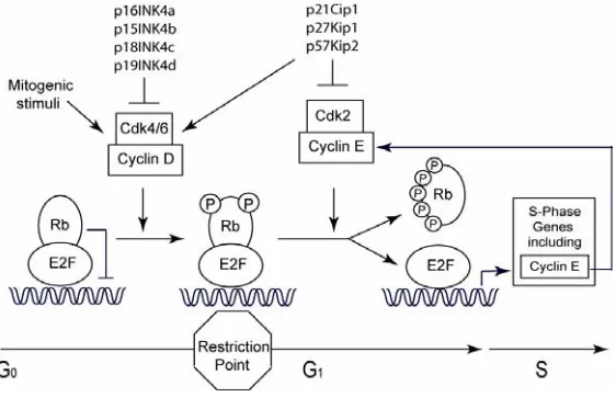
Figure 1. Cyclin/CDK complexes 116
which allow the cell to halt proliferation in response to cytogenic stressors such as UVB irradiation.
As described above, entry into the cell cycle is tightly regulated by upstream signaling pathways that culminate in the increased expression of numerous genes including cyclin D1. Cyclins bind to and activate cyclin dependent kinases (Cdks) to promote cell cycle progression. Cdk expression is constant, however its kinase activity is not stimulated until it interacts with the
Figure 2. Cyclin expression throughout the cell cycle116
appropriate cyclin. The increased expression of specific cyclins at different phases of the cell cycle is associated with activation of Cdks involved various phases of the cell cycle. As shown in figures 1 and 2, cyclin D1 levels are induced as cells enter G1, and cyclin D1 binds to and activates Cdk4 and Cdk6. The activation of Cdk4 and Cdk6 by cyclin D1 leads to phosphorylation of the tumor suppressor Rb.
The Rb/E2F cell cycle regulatory mechanism generally inhibits G1 to S-phase transition through Rb’s repression of E2F-mediated transcription, as depicted in figure 3. The repressive effects of Rb on E2F transcription are released upon sequential hyperphosphorylation of Rb by Cdk4 and Cdk6, followed by Cdk2. E2F transcription induces genes required for entry into S phase, including cyclin E, thereby further phosphorylating Rb and derepressing E2F’s transcriptional activities. As shown in figures 1-3, the point beyond which cell cycle progression continues without growth factor stimulation is known as
Figure 3. Gene transcription control by E2F and Rb116
the restriction, or R point, and it falls between mid and late G1. Further oversight of cellular proliferation is enforced through specific inhibitors of the cell cycle such as p16INK4A and p21Cip1. Replicative senescence and
differentiation of keratinocytesare associated with increased levels of p16INK4A and involucrin, which indicate irreversible exit from the cell cycle and onsetof terminal differentiation, respectively117, 118.
Cancer
Cancer is not a single disease, but is rather a group of diseases characterized by cells that have acquired a number of unifying characteristics. Hanahan and Weinberg propose that there are six hallmarks of cancer which alter cell physiology to promote malignant growth: 1. Self sufficiency in growth signals, 2. Insensitivity to growth-inhibitory signals, 3. Tissue invasion and metastasis, 4. Limitless replicative potential, 5. Sustained angiogenesis, and 6. Evading apoptosis113. They propose that cancer cells must acquire all 6 characteristic hallmarks of cancer, and that the acquisition of these traits occurs through somatic mutations of DNA. These traits could be acquired from as few as 2 to 3 mutations, or as many as 8, depending on the specific gene mutations and the interactions between that gene and other mutated genes. All of these characteristics can be regulated by signal transduction pathways, and the perturbation of these signaling pathways can result in cancer development. For instance, mutation of p53 could promote both angiogenesis and resistance to apoptosis, allowing the cell to acquire 2 characteristic hallmarks of cancer through a single mutation. Upon acquisition of each new acquired trait, the cell becomes more out of control, feeding an environment of instability and promoting further gene mutations, otherwise known as genomic instability.
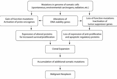
Specific genes involved in cancer have been deemed oncogenes and tumor suppressor genes. Oncogenes are genes which promote tumorigenesis, while tumor suppressor genes inhibit carcinogenesis. The interplay between oncogenes, tumor suppressor genes, and genes that promote genomic stability such as DNA repair genes drive the cancer process and result in the acquisition of the characteristic hallmarks of cancer. As shown in figure 4, Oncogenic activation can lead to altered expression of proteins to promote increased survival/proliferation, tumor suppressor inactivation results in the loss of
Figure 4. Overview of cancer process116
expression of anti-proliferative and apoptotic regulatory proteins, and faulty DNA repair genes can contribute to these changes. As stated above, once the cell has acquired a hallmark of cancer the cell starts to lose control, resulting in an increased rate of acquisition of further mutations leading towards malignant neoplasia formation.
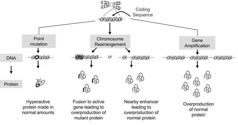
There are a number of mechanisms by which oncogenes can be activated, as shown in figure 5. A point mutation in an oncogene can lead to its activation, as with the characteristic mutations in the Ras oncogene mentioned above. Chromosome rearrangements can result in the overproduction of normal protein by altered regulation of gene expression due to translocation of a foreign sequence to the promoter of an oncogene. Translocation of a portion of a chromosome can also lead to the production of a fusion protein, where the
Coding Sequence or Chromosome Rearrangement DNA Protein Hyperactive protein made in normal amounts
Fusion to active gene leading to overproduction of mutant protein Nearby enhancer leading to overproduction of normal protein Overproduction of normal protein Point
mutation AmplificationGene
Figure 5. Oncogene activation116
mutant protein has acquired additional oncogenic characteristics. Another mechanism of oncogene activation by mutation is through gene amplification, where multiple copies of the gene become inserted into and expressed from the genome. All of these mechanisms lead to the activation/overexpression of oncoproteins to promote proliferation and survival, and further neoplastic progression.
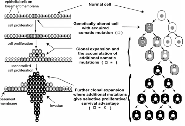
This idea of a single cell acquiring cumulative mutations as it progresses towards malignancy is known as the somatic mutation theory of clonal expansion. Figure 6 depicts how a single cell with cumulative acquired somatic mutations can clonally expand to produce a primarily clonal neoplasm. For instance, the first mutation may have inactivated a tumor suppressor gene,
Figure 6. Monoclonal Nature of Cancer. A tumor develops through successive rounds of proliferation and mutation. As a cell acquires a proliferative/survival advantage through mutation, it becomes more susceptable to further mutation. Eventually, an individual cell with successive mutations gives rise to a population of identical cells that
have the proliferative/survival capacity to form a tumor116.
resulting in unchecked growth. The cell is now replicating its DNA without any checks on proliferation making it much more susceptible to further mutation. A further mutation involving the activation of an oncogene would thereby promoting excessive proliferation. The drive for proliferation is now fully unchecked by the mutated tumor suppressor genes, thereby resulting in further mutations, including mutations of DNA repair genes. The uncontrolled proliferation of cells renders cells more prone to errors in DNA replication, but if the DNA repair genes are inactive they are unable to repair the damage. The mutated cells pass on the acquired somatic mutations, eventually leading to possible further mutation and neoplasm formation. One requirement for cancer development is that at some point cells must replicate and pass on their acquired mutations. This taken with other evidence has led to the theory that epithelial cancers are derived from highly proliferative stem cells that have a limitless replicative potential119. Further, oncogenes such as c-Myc are capable of immortalizing cells. Limitless replicative potential is in fact one of the hallmarks of cancer, thereby giving stem cells one of the hallmark characteristics of cancer even without mutation.
C/EBPs and Cancer
C/EBPα and C/EBPβ have been implicated in the cancer process through a number of studies in numerous tissues and cell types. C/EBPα has been characterized as a tumor suppressor which is inactivated by mutation in ~10% of all acute myeloid leukemia (AML)66. C/EBPα expression is also diminished in
numerous tumors of various cell types66. C/EBPα was first identified as an inhibitor of mitotic growth in fibroblasts by the McKnight lab120, and many further studies have confirmed this function in multiple cell types66. The mechanism by which C/EBPα inhibits growth arrest is not yet fully elucidated, although a number of possible mechanisms has been proposed: 1. stabilization of the Cdk inhibitor p21, 2. promotion of a shift in Rb complex constitution, from proliferative E2F-p107 complexes to resting E2F-p130 complexes, 3. direct binding to E2F and repression of E2F transcription, 4. direct binding to Cdk2 and Cdk4 and repression of their kinase activity, and 5. a requirement for interactions with the SWI/SNF chromatin remodeling complex66. These mechanisms suggest a primarily non-transcriptional role for C/EBPα in growth arrest. As stated previously, it is not yet determined which if any of these proposed mechanisms is responsible for the growth inhibitory role of C/EBPα.
The role for C/EBPβ in cancer is not quite as clear as that for C/EBPα. C/EBPβ-/- primary keratinocytes express partial resistance to calcium induced growth arrest associated with induction of differentiation and forced expression of C/EBPα or C/EBPβ inhibits the proliferation of keratinocytes in culture64. A tumor suppressor function for C/EBPβ has been suggested by its important role in Rasv12-induced senescence involving Rb/E2F complexes. Opposing roles for C/EBPα and C/EBPβ are seen in the suppression and stimulation, respectively, of ras-induced cellular transformation121, 122. Ras signaling downregulates C/EBPα but not C/EBPβ expression, and co-expression of Ras and C/EBPβ
results in the synergistic activation of a C/EBP-responsive reporter reporter121, 123. C/EBPβ is required for transition from G1 to S phase of the cell cycle in hepatocytes induced to proliferate after partial hepatectomy, and in adipocytes clonal expansion72. An important piece of evidence which truly highlights the importance of C/EBPβ in cancer is that loss of C/EBPβ renders mice completely refractory to carcinogen-induced skin tumorigenesis121. Similar oncogenic functions of C/EBPβ can be observed in macrophages, mammary epithelial cells, uterine epithelium and stroma, B lymphocytes, and human tumors with elevated cyclin D172. The oncogenic and tumor suppressive functions of C/EBPβ may allow C/EBPβ to serve as a key regulator involved in the balance between proliferation and growth inhibition.
C/EBPα and C/EBPβ may play key roles in epidermal homeostasis through the regulation of differentiation and cellular proliferation, and these functions equip C/EBPα and C/EBPβ with the ability to have both oncogenic (C/EBPβ) and tumor suppressive (C/EBPα and C/EBPβ) roles in oncogenesis.
Research Hypothesis and Objectives
C/EBPα and C/EBPβ are key regulators of cellular growth and differentiation in numerous tissues and cell types28. The anti-proliferative function of C/EBPα is widely established, but the implications for its growth inhibitory role in normal epidermal homeostasis are not yet known66. Diminished expression of C/EBPα is also associated with epithelial tumor development124. Causal or genetic evidence that C/EBPα can function as an epithelial tumor suppressor is lacking as C/EBPα mutations have not been detected in epithelial tumors and C/EBPα deficient mice die before or shortly after birth69. I therefore hypothesized that C/EBPα’s anti-proliferative function is associated with normal epidermal homeostasis and that C/EBPα acts as a tumor suppressor in epithelial tissue. C/EBPβ is involved with both the inhibition and stimulation of proliferation, and C/EBPβ-/- mice are completely refractory to carcinogen-induced skin tumorigenesis72, 121. I also hypothesized then, that C/EBPα and C/EBPβ are differentially regulated and may have opposing roles in epidermal homeostasis and tumorigenesis. My objectives were to generate an epidermal specific C/EBPα-/- mouse to determine the effects of C/EBPα loss on epidermal homoestasis and tumorigenesis. I also sought to explore the regulation of C/EBPα and C/EBPβ in the keratinocytes by mitogenic stimulation.
CHAPTER 1
Genetic Ablation of C/EBPα in Epidermis Reveals its Role in Suppression
of Epithelial Tumorigenesis
Kari D. Loomis1,2, Songyun Zhu1, Kyungsil Yoon1, Peter F. Johnson3, and Robert C. Smart1,2*
1 Cell Signaling and Cancer Group, Department of Environmental& Molecular Toxicology, 2Functional Genomics Program, North Carolina State University,
Raleigh, NC 27695-7633; 3Laboratory of Protein Dynamics and Signaling, National Cancer Institute, Frederick, MD 21702-1201
Running Title: C/EBPα is a suppressor of epithelial tumorigenesis Key Words: Tumor Suppressor, C/EBP, Keratinocytes
Financial Support: NCI Grant # CA46637, NIEHS Grant # ES12473, GAANN Fellowship
CORRESPONDING AUTHOR:
Robert C. Smart
North Carolina State University Campus Box 7633 Raleigh, NC 27695
[email protected] PUBLISHED IN:
Cancer Res 2007; 67: (14). July 15, 2007
Abstract
C/EBPα is a b-zip transcription factor that inhibits cell cycle progression and regulates differentiation in various cell types. C/EBPα is inactivated by mutation in acute myeloid leukemia (AML) and is considered a human tumor suppressor in AML. While C/EBPα mutations have not been observed in malignancies other than AML, greatly diminished expression of C/EBPα occurs in numerous human epithelial cancers including lung, liver, endometrial, skin and breast suggesting a possible tumor suppressor function. However, direct evidence for C/EBPα as an epithelial tumor suppressor is lacking due to the absence of C/EBPα mutations in epithelial tumors and the lethal effect of C/EBPα deletion in mouse model systems. To examine the function of C/EBPα in epithelial tumor development, an epidermal-specific C/EBPα knockout mouse was generated. The epidermal-specific C/EBPα knockout mice survived and displayed no detectable abnormalities in epidermal keratinocyte proliferation, differentiation or apoptosis; demonstrating that C/EBPα is dispensable for normal epidermal homeostasis. In spite of this, the epidermal-specific C/EBPα knockout mice were highly susceptible to skin tumor development involving oncogenic Ras. These mice displayed decreased tumor latency and striking increases in tumor incidence, multiplicity, growth rate and the rate of malignant progression. Mice hemizygous for C/EBPα displayed an intermediate enhanced tumor phenotype. Our results suggest decreased expression of C/EBPα contributes to deregulation of tumor cell proliferation. C/EBPα had been proposed to block
cell cycle progression through inhibition of E2F activity. We observed that C/EBPα blocked Ras- and EGF-induced E2F activity in keratinocytes and also blocked Ras-induced cell transformation and cell cycle progression. Our study demonstrates that C/EBPα is dispensable for epidermal homeostasis and provides genetic evidence that C/EBPα is a suppressor gene in epithelial tumorigenesis.
INTRODUCTION
C/EBPα is a basic leucine zipper transcription factor and has a role in energy metabolism, differentiation and mitotic growth arrest (1). Forced expression of C/EBPα results in the inhibition of cell cycle progression in most cell types including those with activated oncogenes and inactivated tumor suppressor genes (2, 3). C/EBPα has been reported to inhibit cell proliferation through mechanisms involving; i) regulation, stabilization and activation of the CDK inhibitor p21 (4, 5), ii) direct inhibition of CDK4 and CDK2 activity (6), iii) interaction with Rb family members (7, 8), iv) interaction with and repression of E2F-mediated transcription activity (9, 10) and v) interaction with a SWI/SNF complex (11). Whether all of these possible mechanisms are operative in all cells or whether certain cells utilize a specific subset of C/EBPα inhibitory mechanisms is not known (12).
C/EBPα is inactivated through specific somatic mutations in approximately 10% of acute myeloid leukemia (AML) patients (13, 14) and these studies along with work demonstrating that C/EBPα is required for granulopoiesis in C/EBPα mutant mice (15) provide compelling evidence that C/EBPα is a tumor suppressor in AML. While C/EBPα mutations have not been observed in malignancies other than AML, loss or greatly decreased expression occurs in numerous epithelial cancers, including lung (16), skin (17, 18), liver (19), endometrial (20) and breast cancer (21). Re-expression of C/EBPα in hepatoma cell lines (22), lung cancer lines (16) or skin squamous cell
carcinomas (SCC) cell lines (18) blocks cell cycle progression. Thus, it appears that diminished expression of C/EBPα is associated with epithelial tumor development (23). However, causal or genetic evidence that C/EBPα can function as an epithelial tumor suppressor is lacking as C/EBPα mutations have not been detected in epithelial tumors and C/EBPα deficient mice die before or shortly after birth, presumably from altered hepatic glucose and glycogen metabolism (24). Conditional or tissue specific knockout of C/EBPα in a tissue/organ in which tumors derived from this tissue are known to display decreased C/EBPα expression would be an ideal model system for testing whether C/EBPα has tumor suppressor function. However, this approach has also been problematic as the lung specific loss of C/EBPα in mice results in respiratory failure at birth (25).
C/EBPα is expressed in epidermal keratinocytes of human and mouse skin (17, 26, 27). Forced expression of C/EBPα in keratinocytes inhibits cell cycle progression (28). In terms of stress responses, C/EBPα is induced in keratinocytes by a variety of DNA damaging agents and has a role in the G1/S checkpoint in response to UVB-induced DNA damage (29). C/EBPα is expressed primarily in the suprabasal layers of the epidermis where post-mitotic keratinocytes undergo differentiation, and to a lesser extent, in a subpopulation of basal keratinocytes (17, 29). The location of C/EBPα expression within the epidermis suggests that it may be involved in cell cycle exit associated with stratified squamous differentiation and/or the regulation of differentiation
specific genes. However, a role for C/EBPα in squamous differentiation remains unidentified.
The mouse skin model of multistage chemical-induced carcinogenesis is a well defined in vivo model of epithelial neoplasia where oncogenic Ras mutations precede p53 and INK4A/ARF mutations during tumor development and progression (30, 31). Carcinogen-induced oncogenic Ras mutation is the initial critical event responsible for the development of the squamous papilloma (32). To examine the function of C/EBPα in epithelial tumor development as well as in epidermal homeostasis, we generated an epidermal specific C/EBPα knockout mouse using the Cre-loxP recombination system. We observed that C/EBPα is dispensable for normal epidermal homeostasis, however, in spite of this the epidermal specific C/EBPα knockout mice are highly susceptible to Ras-induced skin tumorigenesis. Either reduced or ablated expression conferred increased susceptibility to tumorigenesis. Thus, C/EBPα functions as a haploinsufficient tumor suppressor gene in epithelial tumorigenesis and our results suggest that C/EBPα suppresses Ras-mediated tumorigenesis through repression of E2F activity.
RESULTS
C/EBPα expression is ablated in epidermis, hair follicles and sebaceous
glands of K5-Cre;C/EBPαfl/fl mice
To achieve epidermal-specific ablation of C/EBPα, C/EBPαfl/fl mice (33) were crossed with K5-Cre transgenic mice in which Cre recombinase expression is directed to the epidermis and other stratified epithelia by the keratin 5 promoter (34). Immunoblot analysis of epidermal lysates prepared from wild type, C/EBPαfl/fl, K5Cre and K5Cre;C/EBPαfl/fl mice was conducted (Fig 1A) . C/EBPα protein was not detectable in the epidermal lysates prepared from K5Cre;C/EBPαf/fl mice, while it was expressed at normal levels in the three other genotypes. To document the specificity of the ablation of C/EBPα in epidermis, we examined C/EBPα protein levels in liver, lung and fat; three tissues known to express relatively high levels of C/EBPα (Fig 1A). Immunoblot analysis revealed that C/EBPα was expressed at normal levels in all three tissues of all four genotypes. To examine the efficiency and location of Cre-induced recombination within the epidermis, we conducted immunohistochemical staining. In C/EBPαfl/fl mouse skin, C/EBPα staining was observed in the nuclei of interfollicular epidermal basal and suprabasal keratinocytes (Fig 1B) as well as in hair follicles and sebaceous gland cells (Fig 1C). In contrast, C/EBPα was not detected in any of the above structures in K5Cre;C/EBPαfl/fl mice (Fig 1B and 1C), reflecting the fact that epithelial cells of the epidermis and its appendages are all derived from a common pluripotent K5
expressing stem cell (43). Epidermal stem cells are considered to be the target precursors cells for skin tumor development (44) and our results indicate that C/EBPα is ablated in these cells.
Ablation of C/EBPα in the epidermis has no effect on epidermal
homeostasis
Mice with an epidermal specific ablation of C/EBPα were born at normal Mendelian frequency. These mice did not display a visible phenotype and were grossly indistinguishable from control mice. To determine whether the loss of C/EBPα in the epidermis has any effect on epidermal homeostasis we examined epidermal keratinocyte proliferation, apoptosis and squamous differentiation. Surprisingly, K5Cre;C/EBPαfl/fl mice did not display any detectable alterations in epidermal keratinocyte proliferation as determined by epidermal thickness, number of nucleated cell layers (data not shown) and the number of BrdU positive S-phase cells (Fig 2A) when compared to control mice. Similarly there were no differences in the number of apoptotic keratinocytes between control and K5Cre;C/EBPαfl/flmice (Fig 2A). To determine whether the loss of C/EBPα expression results in alterations in epidermal stratified squamous differentiation we examined the expression of keratin 5 (K5), keratin 10 (K10), keratin 1 (K1), involucrin and loricrin. K5 is expressed in the basal layer keratinocytes while K10 and K1 are first expressed in the transition from the basal to spinous layer and involucrin and loricrin are expressed later in the differentiation program. Immunoblot analysis revealed that all of these markers
were expressed at normal levels in the absence of epidermal C/EBPα (Fig 2B). Immunohistochemical staining of the epidermis demonstrated that the spatial expression of these markers was also normal in the K5Cre;C/EBPαfl/fl mice (Fig 2C). Collectively, these results indicate that the ablation of C/EBPα in the epidermis does not alter epidermal keratinocyte proliferation, squamous differentiation or apoptosis, demonstrating that C/EBPα is dispensable for normal epidermal homeostasis.
C/EBPβ and p21 are up-regulated in C/EBPα deficient epidermis.
The lack of effect of C/EBPα deficiency on epidermal proliferation and differentiation was unexpected and could be due to the compensatory up-regulation of genes with similar functions. C/EBPβ is expressed in the epidermis and is involved in squamous differentiation (42, 45). Therefore, we examined C/EBPβ protein levels in K5Cre;C/EBPαfl/fl epidermis. As shown in Figure 2D, C/EBPβ was up-regulated ~2 fold in C/EBPα deficient epidermis compared to control epidermis. The cyclin dependent kinase inhibitor, p21, a regulator of the G1 to S phase transition in the cell cycle, was also up regulated in the K5Cre;C/EBPαfl/fl epidermis. Increased expression of C/EBPβ and p21 may compensate for the loss of C/EBPα and potentially mask the role of C/EBPα in keratinocyte differentiation and proliferation.
Loss of C/EBPα in the epidermis results in increased susceptibility to
Ras-induced skin tumorigenesis.
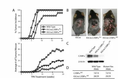
The loss of C/EBPα in the epidermis and presumably in the epidermal stem cell compartment is not sufficient in itself for skin tumor development, as untreated K5Cre;C/EBPαfl/fl mice held for one year did not develop any skin tumors. These results indicate that additional events are required for skin tumor development. The mouse skin model of multistage carcinogenesis involves treatment of mouse skin with DMBA followed by weekly TPA treatments and results in the production of squamous papillomas, the majority of which (>95%) contain an A->T182 transversion in Ha-ras (32). To determine whether K5Cre;C/EBPαfl/fl mice have an altered susceptibility to tumorigenesis involving oncogenic Ras, we subjected mice to a DMBA/TPA two-stage carcinogenesis protocol. As shown in Fig 3A, wild type mice developed their first tumor at week 7 and at week 19 developed their maximum tumor incidence of 90% with a tumor multiplicity of approximately 10 tumors/mouse. C/EBPαfl/fl and K5-Cre mice displayed similar tumor latency, incidence and multiplicity as wild type mice (data not shown). In contrast, K5Cre;C/EBPαfl/fl mice developed their first tumor at week 4, which is nearly 50% earlier than that of wild type mice. All of K5Cre;C/EBPαfl/fl mice developed papillomas by week 8 and these mice developed ~40 tumors/mouse. Dramatic differences in both tumor number and size were evident in the K5Cre;C/EBPαfl/fl mice (Fig 3B). Mice that were hemizygous for epidermal C/EBPα (K5Cre;C/EBPαfl/+) had reduced levels of C/EBPα in their epidermis (Fig 3C) and displayed an intermediate tumor phenotype between wild type mice and mice completely deficient in epidermal
C/EBPα (Fig 3A and B). Collectively, these results demonstrate that ablation or reduced expression of epidermal C/EBPα has a multifaceted effect on tumor development involving decreased tumor latency, increased tumor incidence and increased tumor multiplicity in DMBA/TPA-treated mice.
DMBA-induced mutation of Ha-Ras in epidermal stem cells is considered the critical event for skin tumor development (32, 44) and earlier studies demonstrated that forced expression of C/EBPα can override the proliferative effects of oncogenic Ras in skin SCC cell lines (18). Therefore, tumors of the epidermal-specific C/EBPα knockout and control mice were examined by mutation specific PCR to verify the presence of the DMBA-induced oncogenic Ras precursor tumor cell lesions (46). An A->T transversion in the 61st codon of H-Ras was present in all tumors isolated from K5Cre;C/EBPαfl/fl mice (Fig 3D). Collectively, these results indicate that reduced or ablated expression of epidermal C/EBPα results in increased susceptibility to Ras-induced tumorigenesis.
Premalignant tumors of K5Cre;C/EBPαfl/fl mice display increased growth
rate and an increased rate of malignant progression
During the course of the tumor experiments, it was evident that there were striking differences in the tumor growth rate as indicated by tumor size. Grossly these tumors were identified as papillomas. Measurement of tumor diameters showed that at 14 weeks only 22% of the wild type mouse tumors were greater than 2 mm in diameter; in contrast, 71% of the K5Cre;C/EBPαfl/fl tumors were