Volume 2012, Article ID 656914,8pages doi:10.1155/2012/656914
Research Article
Factorial Study of Compressive Mechanical Properties and
Primary
In Vitro
Osteoblast Response of PHBV/PLLA Scaffolds
Naznin Sultana
1, 2and Tareef Hayat Khan
31Department of Mechanical Engineering, The University of Hong Kong, Pokfulam Road, Hong Kong
2Department of Clinical Sciences, Faculty of Biosciences and Medical Engineering, Universiti Teknologi Malaysia, Johor, 81310 Johor
Bahru, Malaysia
3Department of Architecture, Faculty of Built Enviroment, Universiti Teknologi Malaysia, Johor, 81310 Johor Bahru, Malaysia
Correspondence should be addressed to Naznin Sultana,[email protected]
Received 11 September 2012; Accepted 30 October 2012
Academic Editor: Xiao-Miao Feng
Copyright © 2012 N. Sultana and T. H. Khan. This is an open access article distributed under the Creative Commons Attribution License, which permits unrestricted use, distribution, and reproduction in any medium, provided the original work is properly cited.
For bone tissue regeneration, composite scaffolds containing biodegradable polymers and nanosized osteoconductive bioceramics have been regarded as promising biomimetic systems. Polymer blends of poly(hydroxybutyrate-co-hydroxyvalerate) (PHBV) and poly(L-lactic acid) (PLLA) can be used as the polymer matrix to control the degradation rate. In order to render the scaffolds osteoconductive, nano-sized hydroxyapatite (nHA) particles can be incorporated into the polymer matrix. In the first part of this study, a factorial design approach to investigate the influence of materials on the initial compressive mechanical properties of the scaffolds was studied. In the second part, the protein adsorption behavior and the attachment and morphology of osteoblast-like cells (Saos-2) of the scaffolds in vitro were also studied. It was observed that nHA incorporated PHBV/PLLA composite scaffolds adsorbed more bovine serum albumin (BSA) protein than PHBV or PHBV/PLLA scaffolds. In vitro studies also revealed that the attachment of human osteoblastic cells (SaOS-2) was significantly higher in nHA incorporated PHBV/PLLA composite scaffolds. From the SEM micrographs of nHA incorporated PHBV/PLLA composite scaffolds seeded with SaOS-2 cells after a 7-day cell culture period, it was observed that the cells were well expanded and spread in all directions on the scaffolds.
1. Introduction
In scaffold-based tissue engineering (TE), scaffolds play
several important roles. Scaffold material and scaffold
fabrication techniques should be selected carefully as the
scaffold properties are crucial to determine the success of
a TE approach [1, 2]. One of the basic requirements for
polymer-based scaffolds is that the scaffolds should have
controllable porous architecture that can allow cell migra-tion, attachment, and growth, leading to tissue regeneration.
The degradation product(s) of scaffolds should be
non-toxic and easily taken up or excreted via metabolic
path-ways. Scaffolds should have sufficient mechanical strength
to maintain structural integrity during culture. Some of the commonly used biodegradable polymers that are used
as scaffold materials include poly(glycolic acid) (PGA),
poly(l-lactic acid) (PLLA), and their copolymer poly(lactic acid-co-glycolic acid) (PLGA) [2]. Poly(hydroxybutyrate-co-hydroxyvalerate) (PHBV), which is a natural, biodegradable polymer, possesses good biocompatibility, also being used to
made into TE scaffolds. The in vivo degradation product of
PHBV is a normal constituent of human blood [2]. PHBV
has been investigated for tissue engineering application [3–
5]. Due to the slow degradation rate, PHBV can be blended
with PLLA in order to control degradation rate and time
[6,7]. Hydroxyapatite (HA) being the mineral component
of living bones has gained much recognition as a scaffolding
material [8, 9]. Nanosized HA (nHA) and biodegradable
polymer blends containing biocomposite scaffolds can offer
a promising strategy for bone tissue regeneration.
scaffolds and controlled pore sizes [10,11] although there are many techniques available to fabricate tissue engineering
scaffolds. If freeze-drying technique is used, a number of
factors are concerned for the ultimate morphology and properties of the scaffolds. It is possible to achieve highly organized three-dimensional structures if the parameters are selected and employed properly. In the first part of the study, the factorial design was used to investigate the material
parameters to fabricate TE scaffolds based on PHBV and
PLLA polymers and nHA.
Protein adsorption on the substrate is of importance in evaluating a TE scaffold as cell adhesion and survival could
be modulated by adsorption capacity on the substrate [12].
Within seconds of implantation, protein adsorption onto the foreign surface occurs when biomaterials are implanted into animals or humans. By rapid protein adsorption, it is meant that the arriving cells at the biomaterial surface interact with the adsorbed protein layer instead of the material itself. The response between implanted biomaterial and the body depends on the initial protein adsorption onto a biomaterial surface. Thus adsorption of protein plays a key role. The three primary plasma proteins are albumin, immunoglobulin, and fibrinogen. Among these three proteins, albumin is the predominant plasma protein which makes up 60–70% of plasma. It was reported that albumin could “passivate” the surface of biomaterial and reduced the acute inflammatory response to the biomaterial. It was reported that osteoblast adhesion was significantly greater on nanophase alumina, titania and HA which was due to enhanced adsorption of protein vibronectin by
the bioceramics [12]. Protein adsorption of cerium oxide
nanoparticles as a function of zeta potential was studied and it was reported that electrostatic interactions played an important role in protein adsorption of nanoparticles [13]. The adhesion of protein on low density polyethylene (LDPE) was studied and it was described that the surface wettability and contact time had significant effects on protein adhesion to the surface of biomaterials. In the second part of this
study, the protein adsorption behavior by the scaffolds and
the response of osteoblast attachment and morphology was also investigated for the composite scaffolds.
In the present study, a two-level fractional factorial design was formulated to determine the compressive proper-ties of the scaffolds in vitro and then the scaffolds were used for protein (bovine serum albumin, BSA) adsorption study. The attachment and morphology of osteoblast-like cells (Saos-2) when cultured on the scaffolds were also studied.
2. Materials and Methods
2.1. Materials. PHBV (6% of 3-hydroxyvalerate) was
pur-chased from Sigma-Aldrich and PLLA with an inherent
viscosity 1.6 dL g−1and 1.9 dL g−1(Medisorb 100L 1A) was
purchased from Lakeshore Biomaterials (Birmingham, AL).
The nHA nanoparticles used for composite scaffolds were
produced in-house through a nanoemulsion process [14]. All
chemicals used in this investigation were analytical grade.
2.2. Methods
2.2.1. Factorial Design. To overcome the limitations of
one-factor-at-a-time method, a factorial design study was performed to investigate the effects of material parameters to produce tissue engineering scaffolds. Five processing param-eters as variables were tested in a two-level fractional factorial
design [15] and compressive properties were determined
from compressive stress-strain curves. The variables in the experimental design were (1) the polymer concentration, (2) water phase fraction, (3) blend composition of PLLA in PHBV, (4) the amount of HA as filler, and (5) molecular weight of PLLA.
2.2.2. Scaffold Fabrication. For scaffold fabrication, polymer
was weighed accurately and poured into a centrifuge tube. Then an accurately measured amount of chloroform was added to the tube to make a solution with a desired polymer concentration. After obtaining the homogeneous polymer solution, the water phase (aqueous acetic acid solution or ultra pure water) was added to make an emulsion. The emulsion was homogenized by using a homogenizer (Ultra-Turrax, T-25; IKA-WERKE). Polymer or HA/polymer
composite scaffolds were made via freeze-drying technique
[5,16].
In order to solidify the emulsion, the beaker containing the emulsion was rapidly transferred into a deep freezer at a preset temperature for overnight. The frozen emulsion was then placed into a freeze-drying vessel (LABCONCO-Freeze dry system, USA). To remove the solvent and the water phase completely, the samples were freeze-dried for at least 46 hrs.
The polymer and composite scaffolds were subsequently
obtained. The scaffolds were stored in a vacuum dessiccator
at room temperature for storage.
2.2.3. Characterization and In Vitro Mechanical Properties
of the Scaffolds. The porous structures of PHBV scaffolds
were studied through scanning electron microscopy (SEM; Stereoscan 440, Cambridge, UK). The pore diameters were calculated using SEM micrographs. Using an Instron mechanical tester (Instron 5848, USA) with a 100 N load cell and at a crosshead speed of 0.5 mm/min, the compressive mechanical properties of PHBV and PHBV-based composite
scaffolds were obtained for dry cylindrical samples with
height of 5 mm and diameter of 10 mm at room temperature The compressive yield strength and compressive modulus was calculated from the initial linear region of stress-strain curves.
2.2.4. Protein Adsorption Study. Experiments of protein
adsorption on scaffolds (100/0 PHBV/PLLA, 50/50
PHBV/-PLLA, and 10% HA in 50/50 PHBV/PLLA) were performed
by incubating scaffold samples at 37◦C in PBS containing
concentration of protein in BSA/PBS solutions containing scaffold samples and the control (without scaffold samples), respectively, under the same incubation conditions. The amount of adsorbed proteins was determined by subtracting the amount of protein which was left in the BSA/PBS solution from the amount of protein in the control.
2.2.5. Cell Culture and Cell Seeding. Human osteoblast cells
(Saos-2) were cultured with Dulbecco’s modified Eagle medium (DMEM, Gibco, UK). The medium was supple-mented with L-glutamine, 1% streptomycin, 0.04% fungi
zone, and 10% fetal calf serum (FCS) at 37◦C in a 5% (v/v)
CO2 incubator. The cells were detached using 0.25% (w/v)
trypsin-ethylenediamietetraacetic acid (EDTA) (Invitrogen, USA) when the cells reached about 80% confluence and subcultured for subsequent use. Scaffold samples were cut to the correct height (1.5 mm) and diameter (10 mm) and were prewetted in ethanol for 24 h and then washed with PBS. Then they were sterilized by 60Co γ-irradiation. 2.2×10⁵
cells/well in the wells of a 24-well plate were then seeded
separately onto scaffold samples as well as onto a tissue
culture polystyrene (TCPS) as control. Then the culture wells
were filled with 1 mL of culture medium. The cell-scaffold
constructs were maintained until 1, 7, or 14 days postseeding.
2.2.6. Cell Attachment and Morphology. The cells were
cultured for 3 h and 24 h in order to examine the cell
attachment to the scaffolds and TCPS; Triplicate samples
were used for each time. The culture medium was removed after each time point and then the wells were gently washed with PBS to remove unattached cells. The adherent cells were enzymatically released from the specimens with the aid of 0.25% trypsin-EDTA. In order to count the cells, a haemocytometer (Tiefe Depth Profounder, Marienfeld, Germany) was used.
The viability and the morphology of cells around
the scaffold specimen on the bottom of the cell culture
plate was observed through an optical microscope (Leica MZFLIII, Germany) equipped with a digital camera. For scanning electron microscopy (SEM), cells were grown and prepared for SEM by fixing with 2.5% glutaraldehyde in
cacodylate buffer and dehydrated with ethanol. Then the
samples were dried in a critical point dryer using liquid carbon dioxide as the transition fluid. The samples were sliced, sputter coated, and analyzed with a SEM (Hitachi S3400N).
2.2.7. Statistical Analysis. All quantitative data of this study
were obtained from triplicate samples and were presented as
mean±standard deviation (SD). To test the significance, an
unpaired Student’sttest (two tail) was applied. A value of
P <0.05 was considered to be statistically significant.
3. Results and Discussion
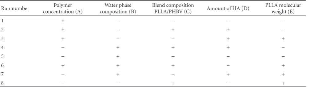
3.1. Factorial Design Study. Tables1and2show the low and
high levels of the variables which are represented by “+”
and “−” and the combinations of the variables in total of
Table 1: Low levels (−) and high levels (+) of variables in the
fractional factorial design.
Levels
Factors − +
(A) Polymer concentration (%) 7.5 10
(B) Water phase fraction 0.5 0.66
(C) Composition of PLLA in PHBV 50/50 75/25
(D) Amount of HA (%) 5 10
(E) Molecular weight of PLLA (dL g−1) 1.6 1.9
eight runs. The results of factorial design on compressive properties are given inTable 3. For the composite scaffolds, the effects of the five variables in the fractional factorial design on the compressive strength and modulus with their standard errors are given inTable 4.Figure 1is the typical
compressive stress-strain curves of a scaffold specimens
cut from one scaffolds. The compressive stress-strain curve
had three distinct regions: linear elasticity, long plateau, and densification region. The compressive modulus and compressive yield strengths were calculated from initial linear region. It can be demonstrated that the positive number indicates that the particular variable has an effect to increase the modulus as it goes from its low level to high level and a negative number indicates that the variables causes the modulus to decrease as the variable goes from its low level to its high level. As it is important to have some methods for interpreting the effects whether they are certainly real and whether they might be explained readily by chance variation, a rough rule is that effects greater than 2 or 3 times their standard error cannot be explained by chance alone [15].
In Table 4, the main effects which are not due to noise are given in italic form. It can be seen fromTable 4that the polymer concentration, amount of HA, and the molecular
weight of PLLA had the effects to increase the compressive
modulus and yield strength as they go from low levels to high levels. On the other hand, the ratio of water phase and blend
composition have the effects to decrease the compressive
modulus and yield strength when they go from low to high levels.
Yaszemski et al. investigated the effects of several com-posite material formulations and conducted a factorial study to calculate initial mechanical properties in vitro and studied the association with histological characteristics of
the resulting material in vivo [17]. It was also reported
that the synthesis of polymer allowed alterations of the
composition as well as of the physical properties to effect
resulting composites. It was also studied and reported that the polymer molecular weight, presence of a leachable salt,
and amount of cross linking monomer had strong effects
on the final strength and modulus of the composite, which were on the order of 5 MPA, an appropriate magnitude
for a temporary trabecular bone substitute [18]. In this
study, the effects of five processing parameters were studied
for the determination of the effects of these processing
Table 2: Eight-run experimental design for studying the effects of the five factors.
Run number concentration (A)Polymer composition (B)Water phase Blend compositionPLLA/PHBV (C) Amount of HA (D) PLLA molecularweight (E)
1 + − − − −
2 + − + + −
3 + − − + +
4 − + + + −
5 − + − − −
6 + + + − +
7 − + − + +
8 − − + − +
Table 3: Compressive yield strength and compressive modulus of
scaffolds. Run
number Compressive yield strength∗(MPa) Modulus∗(MPa)
1 0.25±0.07 4.0±1.2
2 0.50±0.04 11.0±3.4
3 0.80±0.06 12.0±4.1
4 0.20±0.01 1.0±0.3
5 0.11±0.01 1.2±0.4
6 0.32±0.08 0.8±0.2
7 0.23±0.04 10.0±3.1
8 0.34±0.05 2.0±0.8
∗mean±standard deviation.
to produce scaffolds by altering the combinations of these
parameters. Polymer concentration and amount of HA had the maximum effects on the scaffold compressive properties.
It was observed that the scaffolds produced from these
processing parameters which had highest effects on the
compressive properties would facilitate bone formation in
vitro [5].
3.2. Scaffold Properties. For the second part of the study,
based on the parameters and main effects as given in
Table 1 and Table 4, three types of scaffolds fabricated from 10% (w/v) polymer concentration, 0.5 water phase fraction, 100/0 PHBV/PLLA, 50/50 PLLA/PHBV and 10%
HA incorporated 50/50 PHBV/PLLA scaffolds were chosen
for protein adsorption and in vitro cell culture study. SEM
micrographs of these three types of scaffolds are given in
Figures2(a),2(b), and2(c). It was observed that the scaffolds exhibited a large pore size distribution (from severalμm to
300μm) and had interconnected pores. It was also observed
that the HA nano particles were homogeneously distributed in the 10% HA incorporated 50/50 PHBV/PLLA composite scaffolds described elsewhere [5].
3.3. Protein Adsorption. Figure 3(a) shows the physical
appearance of the scaffolds. All the scaffolds were
three-dimensional in structure and physically manageable. UV-visible spectroscopy measurements were carried out for
0 10 20 30 40 50 60 70 80
0 1 2 3
Compressive strain (%)
C
o
mpr
essi
ve
st
ress (MP
a)
Figure 1: Typical compressive curves of the scaffold specimens cut
from one scaffolds fabricated from combination of run 3 ofTable 3.
known concentrations of BSA at the absorbance of 562 nm.
Noncoated polymer and composite scaffolds of different
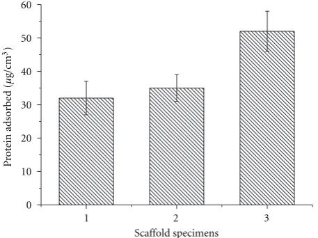
composition were incubated for 3 hrs, 22 hrs and 42 hrs in BSA/PBS solution in order to assess protein adsorption behavior. After 22 hrs, protein adsorption of all the scaffolds reached equilibrium as there was no significant change observed from 22 hrs to 42 hrs (Figure 3(b)).Figure 4shows
the effect of polymer blend composition and incorporated
nHA on the protein adsorption capacity of composite
scaffolds. The amount of adsorbed protein was calculated
from the standard curve. After 22 hr, of immersion in
BSA/PBS solution, 50/50 PHBV/PLLA scaffold adsorbed
more BSA proteins than 100/0 PHBV/PLLA scaffold and 10%
nHA incorporated 50/50 PHBV/PLLA composite scaffolds
adsorbed more BSA proteins than plain PHBV scaffold.
Protein adsorption is of importance to evaluate a scaffold for the tissue engineering application. It was observed from
this part of the study that composite scaffolds containing
nanosized HA could adsorb more protein than pure PHBV
or PHBV/PLLA blend scaffolds. Incorporation of HA can
alter the pore surface morphology of the scaffolds and may
made them more suitable for increased protein adsorption.
As HA has better affinity towards protein adsorption,
particles exposed on the pore walls in the composite scaffolds had increased capacity of protein adsorption.
(a) (b)
(c)
Figure 2: Microstructures examined by SEM of three types of scaffolds: (a) 100/0 PHBV scaffold, (b) 50/50 PHBV/PLLA scaffold, and (c)
10% HA in 50/50 PHBV/PLLA scaffold.
(a)
0 10 20 30 40 50 60
10% HA in 50/50 PHBV/PLLA scaffold 50/50 PHBV/PLLA scaffold
3 22 42
PHBV scaffold
P
rot
ein adsor
b
ed (
µ
g/cm
3)
Scaffold specimens (hr)
(b)
Figure 3: (a) General appearance of the scaffolds and (b) protein adsorption of different scaffolds at 3 hrs, 22, hrs and 42 hrs (data are
Table 4: Calculated main effects of the variables on compressive strength and modulus.
Main effects Effect on compressive yield strength (MPa)∗ Effect on modulus (MPa)∗
(A) Polymer concentration +0.25±0.03 +3.4±1.12
(B) Water phase composition −0.26±0.03 −4.0±1.12
(C) Ratio of PHBV/PLLA −0.01±0.03 −3.1±1.12
(D) The amount of HA +0.18±0.03 +6.5±1.12
(E) Molecular weight of PLLA +0.16±0.03 +1.9±1.12
∗mean±standard error.
1 2 3
0 10 20 30 40 50 60
Scaffold specimens
P
rot
ein adsor
b
ed (
µ
g/cm
3)
Figure 4: Protein adsorption of different scaffolds: (1) PHBV
scaffolds, (2) 50/50 blend scaffolds, and (3) 10% HA in 50/50 blend scaffolds; (Data are expressed as mean±SD;n=3).
arrival of protein at the interface is driven solely by diffusion process which is dependent on bulk concentration and diffusion coefficient [18,19]. It was also demonstrated that the particular surface chemistry of the protein also controls the adsorption. When protein adsorption takes place on neutral hydrophilic surfaces, it will be relatively weak; on the other hand, adsorption of proteins on hydrophobic surfaces tends to be very strong and irreversible. When adsorption takes place on charged surfaces, it tends to be a strong function of the charge character of the protein, the pH of the medium, and ionic strength. In this study, protein
adsorption onto the scaffolds was reported. In order to
study the mechanisms whether the protein is adsorbed on the scaffolds strongly or moderately, protein release study is needed.
3.4. Cell Morphology and Attachment. Both optical
micro-scopy (Figure 5) and SEM (Figure 6) were used to examine
the state and morphology of Saos-2 cells.Figure 5shows the
state and morphology of Saos-2 cells after biocompatibility test for 3 days. It was observed that the cell population and
cell morphology of all specimens of PHBV scaffolds, 50/50
PHBV/PLLA scaffolds, and 10% HA in 50/50 PHBV/PLLA
scaffolds were comparable to the control. This can indicate
that the cell viability remained high after being in contact with all three types of scaffolds.
Using SEM analysis, the morphology of Saos-2 cells on the surface of the scaffolds specimens was also studied. Cells were appeared with clear substrate attachments and cellular processes. After culturing for 1 day, flat cells were observed
by SEM to be well attached on all three types of scaffolds.
At day 7 of Saos-2 cultures on all types of scaffolds, SEM
analysis verified that the Saos-2 cells were well proliferated. The cells were more elongated as well as very well attached and spread on the surface of the scaffolds. The difference in morphology observed among the cells grown on three types
of scaffolds was not obvious. The cells were found anchored
to the surface by discrete filopodia on the composite scaffolds (Figure 6).
In this study, all three types of scaffolds were evaluated
for cell attachment (Figure 7). It was observed that after
24 hours, the number of cells attached to HA incorpo-rated composite scaffolds was significantly different (P <
0.05) than that of 100/0 PHBV/PLLA scaffolds or 50/50
PHBV/PLLA blend scaffolds. On the other hand, the number
of Saos-2 cells attached on the PHBV/PLLA scaffold was
apparently higher than PHBV scaffolds. The difference was
not statistically significant.
In our previous studies [5–7], we reported the fabri-cation, in vitro degradation and biological evaluation of
PHBV-based scaffolds using emulsion freezing/freeze-drying
process. In the present paper, a factorial design approach was used to systematically investigate the influence of
different material parameters on the compressive
mechan-ical properties of the PHBV-based scaffolds. Besides the
other requirements, the scaffolds should possess appropriate mechanical properties. In order to control the compressive stress and compressive modulus of the polymer and com-posite scaffolds, careful analysis was possible using factorial design approach. The protein adsorption and osteoblast (Saos-2) cell response were also studied in order to evaluate
the scaffolds for bone tissue engineering application.
Suc-cessful control of compressive properties is important for constructing composite scaffolds for bone tissue engineering applications.
4. Conclusions
A factorial design study was performed to assess the
main effects of different material parameters on the
com-pressive properties of the scaffolds. Polymer
concentra-tion and the amount of HA had the strongest effect to
(a) PHBV Scaffold (b) 50/50 PHBV/PLLA Scaffold
(c) 10% HA in PHBV/PLLA Scaffold (d) Control
Figure 5: Optical microscopic image of state and morphology of Saos-2 cells in contact with scaffold specimens, (the dark area in (a), (b),
(c) is the test specimen (d) control). (Magnification×10).
(a) (b)
Figure 6: SEM micrographs of 10% HA in 50/50 PHBV/PLLA composite scaffold after 7-day culture with Saos-2 cells (a, b).
PHBV/PLLA scaffolds, 50/50 PHBV/PLLA scaffolds, and
10% HA incorporated 50/50 composite scaffolds showed
satisfactory adsorption of BSA protein. Adsorption of
pro-tein of all the scaffolds reached equilibrium after 22 hrs.
The HA incorporated composite scaffolds exhibited more
affinity towards protein adsorption than pure other two
types of scaffolds. The attachment of Saos-2 cells was
also significantly higher in 10% HA incorporated 50/50 composite scaffolds.
Conflict of Interests
0 10 20 30 40 50 60 70
Control
Composite scaffold PHBV/PLLA
scaffold PHBV
scaffold
Scaffolds
Cell n
umbers
×
10
4 ∗
∗
Figure 7: The attachment of Saos-2 cells on PHBV and
PHBV-based composite scaffolds (data are expressed as mean±SD; 2=3. ∗P <0.05).
Acknowledgment
N. Sultana acknowledges The University of Hong Kong (HKU), GRF Grant (HKU 7182/05E) from the Research Grants Council of Hong Kong. Assistance provided by
tech-nical staff in the Department. of Mechanical Engineering,
Faculty of Engineering and Department. of Orthopedics and Traumatology, Li Ka Shing Faculty of Medicine, HKU, is acknowledged. N. Sultana and T. H. Khan also acknowledge the financial support provided by UTM research Grants GUP Tier 1 (Vote: 03H13), GUP Tier 2 (vote: 06J34), FRGS (vote: 4F126), and ERGS (Vote: 4L063), Ministry of Higher Education (MOHE), and RMC for financial support.
References
[1] P. X. Ma, “Scaffolds for tissue fabrication,” Materials Today, vol. 7, no. 5, pp. 30–40, 2004.
[2] P. A. Holmes, Developments in Crystalline Polymers, Edited by D. C. Bassett, Elsevier Applied Science, London, UK, 1982. [3] B. Duan, M. Wang, W. Y. Zhou, W. L. Cheung, Z. Y. Li, and
W. W. Lu, “Three-dimensional nanocomposite scaffolds fabri-cated via selective laser sintering for bone tissue engineering,”
Acta Biomaterialia, vol. 6, no. 12, pp. 4495–4505, 2010.
[4] H. W. Tong, M. Wang, Z. Y. Li, and W. W. Lu, “Electrospin-ning, characterization and in vitro biological evaluation of nanocomposite fibers containing carbonated hydroxyapatite nanoparticles,” Biomedical Materials, vol. 5, no. 5, Article ID 054111, 2010.
[5] N. Sultana and M. Wang, “PHBV/PLLA-based composite scaffolds fabricated using an emulsion freezing/freeze-drying technique for bone tissue engineering: surface modification and in vitro biological evaluation,” Biofabrication, vol. 4, Article ID 015003, 2012.
[6] N. Sultana and T. H. Khan, “In Vitro degradation of PHBV scaffolds and nHA/PHBV composite scaffolds containing hydroxyapatite nanoparticles for bone tissue engineering,”
Journal of Nanomaterials, vol. 2012, Article ID 190950, 12
pages, 2012.
[7] N. Sultana and M. Wang, “PHBV/PLLA-based composite scaffolds containing nano-sized hydroxyapatite particles for bone tissue engineering,” Journal of Experimental Nanoscience, vol. 3, no. 2, pp. 121–132, 2008.
[8] P. X. Ma, R. Zhang, G. Xiao, and R. Franceschi, “Engineering new bone tissue in vitro on highly porous poly(alpha-hydroxyl acids)/hydroxyapatite composite scaffolds,” Journal
of Biomedical Materials Research, vol. 54, pp. 284–293, 2001.
[9] H. Wang, Y. Li, Y. Zuo, J. Li, S. Ma, and L. Cheng, “Biocompatibility and osteogenesis of biomimetic nano-hydroxyapatite/polyamide composite scaffolds for bone tissue engineering,” Biomaterials, vol. 28, no. 22, pp. 3338–3348, 2007.
[10] K. Whang and K. E. Healy, “Processing of polymer scaffolds: freeze-drying,” in Methods of Tissue Engineering, A. Atala and R. P. Lanza, Eds., p. 1285, Academic Press, San Diego, Calif, USA, 2002.
[11] N. Sultana and M. Wang, “Fabrication of tissue engineering scaffolds using the emulsion freezing/freeze-drying technique and characteristics of the scaffolds,” in Integrated Biomaterials
in Tissue Engineering, pp. 63–89, John Wiley & Sons, 2012.
[12] T. J. Webster, C. Ergun, R. H. Doremus, R. W. Siegel, and R. Bizios, “Specific proteins mediate enhanced osteoblast adhesion on nanophase ceramics,” Journal of Biomedical
Materials Research, vol. 51, pp. 475–483, 2000.
[13] S. Patil, A. Sandberg, E. Heckert, W. Self, and S. Seal, “Protein adsorption and cellular uptake of cerium oxide nanoparticles as a function of zeta potential,” Biomaterials, vol. 28, no. 31, pp. 4600–4607, 2007.
[14] W. Y. Zhou, M. Wang, W. L. Cheung, B. C. Guo, and D. M. Jia, “Synthesis of carbonated hydroxyapatite nanospheres through nanoemulsion,” Journal of Materials Science: Materials
in Medicine, vol. 19, no. 1, pp. 103–110, 2008.
[15] G. E. P. Box, J. S. Hunter, and W. G. Hunter, Statistics
for Experimenters: Design, Innovation, and Discovery,
Wiley-Interscience, Hoboken, NJ, USA, 2nd ed edition, 2005. [16] N. Sultana and M. Wang, “Fabrication of HA/PHBV
com-posite scaffolds through the emulsion freezing/freeze-drying process and characterisation of the scaffolds,” Journal of
Materials Science: Materials in Medicine, vol. 19, no. 7, pp.
2555–2561, 2008.
[17] M. J. Yaszemski, R. G. Payne, W. C. Hayes, R. S. Langer, T. B. Aufdemorte, and A. G. Mikos, “The ingrowth of new bone tissue and initial mechanical properties of a degrading polymeric composite scaffold,” Tissue Engineering, vol. 1, pp. 41–52, 1995.
[18] J. D. Andrade, V. Hlady, and A. P. Wei, “Adsorption of complex proteins at interfaces,” Pure and Applied Chemistry, vol. 64, pp. 1777–1781, 1992.
[19] T. Sun, M. Wang, and W. C. Lee, “Surface characteristics, properties and in vitro biological assessment of a NiTi shape memory alloy after high temperature heat treatment or surface H2O2-oxidation: a comparative study,” Materials Chemistry
Submit your manuscripts at
http://www.hindawi.com
Scientifica
Hindawi Publishing Corporation
http://www.hindawi.com Volume 2014
Corrosion
Hindawi Publishing Corporation
http://www.hindawi.com Volume 2014
Hindawi Publishing Corporation
http://www.hindawi.com Volume 2014
Polymer Science
International Journal ofHindawi Publishing Corporation
http://www.hindawi.com Volume 2014
Ceramics
Journal ofHindawi Publishing Corporation
http://www.hindawi.com Volume 2014
Composites
Nanoparticles
Journal ofHindawi Publishing Corporation
http://www.hindawi.com Volume 2014
Hindawi Publishing Corporation
http://www.hindawi.com Volume 2014 International Journal of
Biomaterials
Hindawi Publishing Corporation
http://www.hindawi.com Volume 2014
Nanoscience
Journal ofTextiles
Hindawi Publishing Corporation
http://www.hindawi.com Volume 2014 Journal of
Nanotechnology
Hindawi Publishing Corporation
http://www.hindawi.com Volume 2014
Hindawi Publishing Corporation
http://www.hindawi.com Volume 2014
Crystallography
Journal ofThe Scientific
World Journal
Hindawi Publishing Corporationhttp://www.hindawi.com Volume 2014
Hindawi Publishing Corporation
http://www.hindawi.com Volume 2014
Coatings
Journal ofAdvances in
Materials Science and Engineering
Hindawi Publishing Corporation
http://www.hindawi.com Volume 2014
Research
Hindawi Publishing Corporation
http://www.hindawi.com Volume 2014
Hindawi Publishing Corporation
http://www.hindawi.com Volume 2014
Metallurgy
Journal ofHindawi Publishing Corporation
http://www.hindawi.com Volume 2014
Research International
Hindawi Publishing Corporation
http://www.hindawi.com Volume 2014
Materials
Journal ofN
a
no
ma
te
ria
ls
Hindawi Publishing Corporation
http://www.hindawi.com Volume 2014
Journal of