Received October 21, 1963
A
homeotic mutant alters an appendage of a segmental series from its typical form to that of some other member of the series. One mutant usually classi- fied as homeotic is aristapedia (BALKASCHINA 1929), which transforms the arista of the antenna into a tarsus-like structure (Figure 1). Most mutants that have been studied by means of genetic mosaics can be considered late acting in that they appear to alter the competence of cells to respond to preexisting inducing fields or "prepatterns" (STERN 1954; TOKUNAGA 1961 ).
Because of the drastic nature of the transformation, it has been suggested (HANNAH-ALAVA 1958) that homeotic mutants alter prepatterns. The study reported here presents evidence that the homeotic mutant, aristapedia, alters the response of antennal cells rather than altering the prepattern.MATERIALS A N D METHODS
Genetic mosaics were produced by X-ray induced somatic crossing over. An insertional trans-
location, T(1;3)05 has a small segment of the third chromosome (section 88 A-C to 92) bearing the wild-type allele of aristapedia (ss", 3-58.5) inserted close to the tip of the X chromosome which also carries the wild-type allele of yellow ( y , 1-0.0, bristles and hairs yellow). The deficient third chromosome is marked by Dichaete (D, 3 4 . 4 , wings spread). For a more com- plete description of the translocation and mutants see LEWIS (1951) and BRIDGES and BREHME
(1%).
Males of the constitution T(1;3)05, D/ss" were crossed to y / y ; ss"/ss" females. First instar larvae were exposed ta 1200r of 250 kvp X-irradiation, allowed to mature, and adult females were examined for antennal mosaicism. Somatic crossing over involving the X chromosome is usually proximal to the inserted piece of chromosome 3 and produces homozygous ss" tissue which is identifiable by the presence of yellow bristles and hairs while wild-type tissue has black bristles and hairs. I t should be noted that the chief limitation of this technique is the inability to detect small mosaic patches without bristles or hairs or to determine the precise boundary between mutant and wild-type tissue since the cuticle is yellowish in both.
RESULTS
Examination of 105 7 duplication-bearing (non-D) females disclosed 46 mosaic antennae. No mosaicism was observed on the heads of 1368 D females indicating that the heterozygous deficiency is cell lethal, at least in the head.
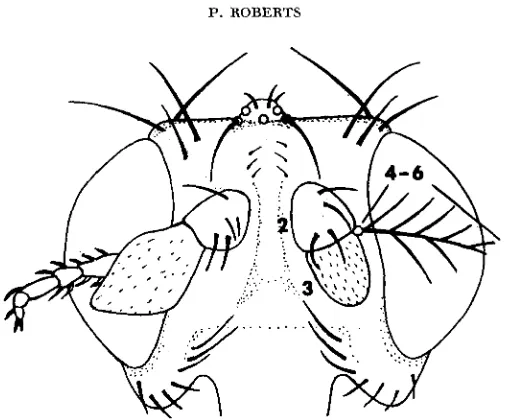
Figure 1 illustrates a mosaic head bearing a wild-type antenna with its feathery
Operated by Union Carbide Corporation for the United States Atomic Energy Commission.
594 P. ROBERTS
FIGURE 1.-A head bearing a wild-type antenna (right) and a mosaic antenna (left). In this and other figures wild-type bristles and hairs are in solid black and homozygous S S ~ bristles and
hairs that are yellow in the fly are in outline. Segment numbers are indicated on the wild-type antenna. 40 x.
arista (right) and the tarsus-like appendage which is formed when most of the distal antennal tissue is homozygous sp (left). This antenna was almost as tarsus- like as the appendage found on nonmosaic ssa homozygotes in spite of the juxtapo- sition of wild-type tissue in segment 2 and s e tissue in segments 3 to 6.
Table 1 divides the 46 recovered mosaics into five categories. The divisions are somewhat arbitrary since Types 4 and 5 can be considered variants of Type 3. The ensuing description applies only to segments 4 to 6 since no mosaicism for segment 2 was observed and segment 3, while frequently modified by mosaicism (Figures 1 to 3), shows less contrast between the two genotypes than the distal segments.
Ten of the mosaics (Type 2) were similar to Figure 1 in that they had no de- tectable wild-type tissue distal to segment 2. Segments 4 to 6 were, in general, strongly segmented but showed more variability than a sample of ten nonmosaic
ssa antennae, suggesting that there was nondetectable mosaicism.
Except for two mosaics (Type l ) , which had little tissue beyond segment 3,
the combined length of the distal segments fell within a narrower range than
TABLE 1
The frequency of occurrence of fiue types of mosaic antenna among 46 antennal mosaics
Type 1 Type 2 Type 3 Type 4 Type 5
Tarsus proximally, arista distally ~~
Little tissue Uniformly Half tarsus, Tarsus with Tissues
segment 3 (Figure 1 ) tarsus (Figure 2 ) arista (Figure 3 ) (Figure 4)
distal to tarsus-like Mostly half arista Mostly arista1 filaments intermingled
FIGURE %-A mosaic antenna which is tarsus-like proximally and arista-like distally. 90 X.
might have been expected, about twice the width of the distal part of segment 2 regardless of the proportion of mutant and wild-type tissue. The majority of mo- saics, Type 3, were tarsus-like proximally and arista-like distally. The composi- tion of these mosaics vaned from mostly tarsus to mostly arista with Figure 2, half tarsus and half arista, midway in this series.
No mosaic antennae had appreciable tarsus development distal to a thin arista (Figure
4).
When tissues of the two genotypes were intermingled to an even greater degree as in Types4
and5
(Figures 3 and 4), ssa tissue was usually thick when proximal, and when thick, strongly segmented with short bristles while wild-type tissue tended to be thinner, more chitinous, and weakly segmented withlong filaments.
DISC U S S IO N
The development of distinct tarsal segmentation in tissue that is predominantly
sa appears to depend on the degree of dilution with wild-type tissue. Where this is not apparent (Figure 1) o r slight (Figures 2 and 3, proximal portion of seg- ments 4 to 6 ) segmentation is distinct. The formation of aristal filaments, how- ever seems to depend only on the presence of a continuous band of wild-type tissue running proximal to distal in a mosaic antenna. This is apparently so in Figure 3
596 P. ROBERTS
FIGURE 4.--Segments 4 to 6 of an antenna in which tissues of the two genotypes are quite intermingled. 156 x.
but not in Figure 4, where the tip of the antenna is undoubtedly ss", a condition which has prevented the development and separation of all but the bottom fila- ment. Segmentation in the proximal part of segments 4 to 6 of this particular antenna is weak compared with the same region in Figures 1 to 3. Close examina- tion reveals traces of internal pigmentation suggesting that considerable wild- type tissue lies internally in this region. One interpretation is that owing to the diffusion of substances from one tissue to another, autonomy is not complete. A simpler explanation is suggested by a comparison of antennal development in the two genotypes.
The development of SS" and wild-type antennal disks has been described by
VOGT (1946). At 72 hours of development, the antennal disks of the two geno- types are quite similar except for the thickness of the "end knob" which is one third again as thick in the mutant as in the wild-type disk. At the time of prepupa formation the mutant end knob is more than twice the thickness of the wild-type. Subsequently, in the ss" disk, segmentation occurs within the thickened end knob while in the wild-type disk the end knob grows distally to a long, thin point out of which the arista forms. Since the two alleles control, among other things, growth rates of antennal cells, it is possible that the primary effect of dilution of
ssa tissue by wild-type tissue is to limit the thickness of the antenna. This may then weaken tarsal segmentation if segmentation is, in turn, dependent on thick- ness as is suggested by the mosaics. The studies of VOGT also suggest that the failure to recover mosaic antennae with appreciable tarsal development distal to an arista may be attributable to the necessary development of the arista from a terminal outgrowth.
If the mutant and wild-type alleles of aristapedia control antennal prepatterns, an antenna which is predominantly one type of tissue should have the correspond- ing prepattern. A small amount of tissue of the other genotype would be expected to conform to the prepattern set up by the tissue which predominates. Instead of this, the two genotypes exhibit a high degree of autonomy. Small patches of wild- type tissue form long filaments characteristic of an arista even when surrounded by ss" tissue. Conversely, sstc tissue forms tarsus-like segments with wild-type tis- sue both proximally (in segments 2 and 3 ) and distally. It is unlikely that the aristapedia alleles could be controlling antennal prepatterns, but because the an- tennal disk was wild-type until the first instar when mosaicism was induced, it
hours before they became determined. The aristapedia alleles clearly do not con- trol antennal prepatterns.
The evidence suggests that the aristapedia alleles control the competence of cells to respond to what is, in this experiment, an invariant prepattern. Not only do aristapedia and, to a lesser extent, wild-type antennae show evidence of seg- mentation, but mosaic antennae with the two types of tissue intermingled often have a periodicity of structure indicative of underlying segmentation (Figure 3 ) .
Also, the length of a mosaic antenna usually falls within definite limits and is not merely an arista added onto a tarsus (Figure 2 ) . These observations suggest that the aristapedia alleles control the competence of antennal cells to respond to a prepattern which is more general than either of the two mutant phenotypes re- sembling the primitive, unspecialized arthropod appendage, a segmented append- age of a definite length.
At this point it should be mentioned that serial homology of the eyes and an- tennae with other appendages is not supported by studies of their innervation
(FERRIS 1950). It is impossible, at this time to weigh one type of evidence against the other, but a developmental pathway which has persisted throughout the course of insect evolution, phenotypically concealed until revealed by mutation, may have been less susceptible to modification by selection than patterns of inner- vation. Mutants are known which produce limb-like appendages from segments now bearing organs which are farther from limb morphology than the antennae. Tetraltera and podoptera transform the wings into segmented appendages (GOLD-
SCHMIDT 1945), and even the eyes can be caused to bear segmented appendages
by erupt (GLASS 1944) and ophthalmopedia (BRIDGES and BREHME 1944). The normal segmentation pattern of the tarsi involves at least seven known loci (WADDINGTON 1962). Moreover, the characteristic effect of each of these genes is produced on developing leg material in any body segment (e.g., the antennal tarsus). It may be that these genes also control the response of imaginal disk cells to a prepattern which is even more general than suggested above, lacking even the characteristic of segmentation, but it is also a distinct possibility that the “ap- pendage prepattern” is polygenically controlled and that seven of the controlling genes are known.
There is additional evidence that the appendage prepattern is quite general in the recent report of LEWIS (1963). It is accepted that the Diptera are descended from four-winged ancestors, a condition which is duplicated by the mutant bi- thorax. LEWIS, using the duplication described above but a different technique of producing mosaics, has reported that bithorax is also autonomous. This can be in-
598 P. ROBERTS
podoptera. Also, KROEGER (1959) has presented evidence that the fore- and hind- wings of the moth Ephestia have identical prepatterns.
The production, by mutation, of limb-like appendages in all these body seg- ments suggests that there is a relict developmental pathway producing an append- age prepattern which is common to all the imaginal disks. The appendage pre- pattern was probably expressed with comparatively little modification by other genes in some remote ancestor of Drosophila which bore similar segmented ap- pendages on all body segments. Its expression has been modified in various ways -profoundly in the eyes, wings and antennae, to a lesser degree in the legs-by striking, gene controlled alterations of the competence of cells to respond to this prepattern during the course of insect evolution.
SUM MARY
Evidence is presented from a study of antennal mosaics that the mutant and wild-type alleles of a homeotic mutant, aristapedia, do not control “antennal” pre- patterns but control the competence of antennal cells to respond to an “appendage prepattern.”
LITERATURE CITED
BALKASCHINA, E. I., 1929 Ein Fall der Erbhomijosis (die Genovariation “Aristopedia”) bei
BRIDGES, C. B., and K. S. BREHME, 1944 The mutants of Drosophila melanogaster. Carnegie
FERRIS, G. F., 1950 External morphology of the adult. Pp. 368-418 Biology of Drosophila,
Edited by M. DEMEREC. Wiley, New York.
GLASS, H. B., 1944. The effect of X-rays upon the action of a specific gene in Drosophila melanogaster. Genetics 29 : 4 3 W 6 .
GOLDSCHMIDT, R., 19.15 The structure of podoptera, a homeostic mutant in Drosophila naelano- gaster. J. Morph. 77: 71-101.
HANNAH-ALAVA, A., 1958 Developmental genetics of the posterior legs in Drosophila melano- gaster. Genetics 43: 878-905.
KROEGER, H., 1959 Determinationsmosaike aus kombiniert implantierten Imaginalscheiben yon Ephestia Kiihniellu Zeller. Arch. Entwicklungsmech. Organ. 151 : 113-135.
LEWIS, E. B., 1951 Additions and corrections to the cytology of rearrangements. Dromsophila Inform. Serv. 25: 108-109. ~ 1963 Genes and developmental pathways. Am. Zool- ogist 3 : 33-5,6.
Drosophila melanogaster. Arch. Entwicklungsmech. Organ. 115: 44.8-463.
Inst. Wash. Publ. 552.
STERN, C., 1954
TOKUNAGA, C., 1961
VOGT, M., 1946
WADDINGTON, C. H., 1962
Two or three bristles. Am. Scientist 42: 213-247.
The differentiation of a secondary sex comb under the influence of the
Zur labilen Determination der Imaginalscheiben von Drosophila. 11. Die
pp. 199-234. N e w Paiterns in Genetics and Deuelapment. Columbia gene engrailed in Drosophila melanogaster. Genetics 46: 157-1 76.
Umwandlung prasumptiven Fiihlergewebes in Beingewebe. Biol. Zentr. 65 : 238-254.