CME
Premenopausal women and low bone density
Aliya Khan,
MD, FRCPC, FACPThis article has been peer reviewed.
Cet article a fait l’objet d’une révision par des pairs.
Can Fam Physician
2006;52:743-747.
ABSTRACT
OBJECTIVE
To review the evidence and provide an approach to management of low bone density among premenopausal women.QUALITY OF EVIDENCE
MEDLINE was searched from January 1990 to November 2004 and articles graded by level of evidence (I to III). Diagnosis and management recommendations were based on evidence from randomized controlled trials and expert consensus.MAIN MESSAGE
Bone mineral density (BMD) testing among premenopausal women should becompleted only in the presence of approved indications. Current evidence does not support screening for osteoporosis among premenopausal women. Low BMD in premenopausal women is associated with a lower fracture risk than seen in postmenopausal women. In the absence of fragility fractures, low BMD might refl ect low peak bone mass based on genetic predisposition, environment, and lifestyle factors. Clinical evaluation enables distinction between low peak bone mass and a systemic disorder resulting in low BMD and skeletal fragility. Common causes of low bone density among premenopausal women include ovulatory disturbances and low body weight.
CONCLUSION
Bone mineral density alone is insuffi cient for diagnosis of osteoporosis amongpremenopausal women in the absence of fragility fractures. Antiresorptive therapy has been evaluated and shown to benefi t premenopausal women using glucocorticoid therapy or those with primary hyperparathyroidism.
RÉSUMÉ
OBJECTIF
Vérifi er les données probantes concernant la présence d’une densité osseuse basse chez les femmes en préménopause et proposer une approche pour le traitement de cette condition.QUALITÉ DES PREUVES
On a consulté MEDLINE entre janvier 1990 et novembre 2004 et les articles retenus ont été classés selon leur niveau de preuve (I à III). Les recommandations de diagnostic et de traitement étaient fondées sur des preuves provenant d’essais cliniques randomisés et de consensus d’experts.PRINCIPAL MESSAGE
Chez les femmes préménopausique, la densité minérale osseuse (DMO) ne devrait être déterminée qu’en présence d’indications reconnues. Les données actuelles indiquent que le dépistage de l’ostéoporose n’est pas indiqué pour ces femmes préménopausique. En présence d’une faible DMO, le risque de fracture est plus faible en préménopause qu’en postménopause. En absence de fracture de fragilité, une DMO basse pourrait refl éter un pic de masse osseuse bas en lien avec le mode de vie, certains facteurs environnementaux ou une prédisposition génétique. L’évaluation clinique permet de distinguer un pic de masse osseuse bas d’une affection systémique causant une faible DMO et une fragilité squelettique. Les causes fréquentes de faible DMO chez la femme préménopausique incluent les troubles ovulatoires et un poids corporel bas.O
steoporosis is common among postmenopausal women. Younger premenopausal women who have diseases or conditions associated with pro-gressive bone loss could be at increased risk of osteopo-rosis. Some asymptomatic women ask to have bone mineral density (BMD) tested. Premenopausal women can have low bone density without fragility fractures or serious risk factors for fracture. It is thus necessary for primary care physicians to know the indications for BMD testing among premenopausal women as well as the appropriate interpretation of BMD tests and man-agement of low BMD among premenopausal women.Low bone density (T-score of less than 1 standard deviation below the mean for young adults) affects approximately 15% of young healthy women between the ages of 30 and 40.1 Bone density follows a bell curve
distribution, and approximately 0.5% of young healthy women between the ages of 30 and 40 have T-scores of –2.5 or less.2,3 Osteoporosis in premenopausal women is
diagnosed when fragility fractures are present and diag-nosis is not based solely on the results of a BMD test.1
Premenopausal women experiencing fragility fractures should be further evaluated to determine why bones fracture despite adequate estrogen levels. A few patients require a bone biopsy to evaluate the underlying pathol-ogy and to identify a possible cause for bone fragility before menopause.
This paper addresses managing low BMD among pre-menopausal women and reviews the underlying patho-physiology, diagnosis, and therapy.
Quality of evidence
MEDLINE was searched to identify all English-language abstracts evaluating low BMD among premenopausal women. The search was limited to articles published in peer-reviewed journals from January 1990 to November 2004. The term “low bone density” was cross-matched with “premenopausal women” and with the MeSH head-ings “management” and “pathophysiology.” All studies evaluating premenopausal women were included except for case reports.
Diagnosis
The World Health Organization defi nes osteoporosis as a progressive systemic disease characterized by low bone density and microarchitectural deterioration in bone that predisposes patients to increased bone fragility and fracture.1 The WHO criteria for diagnosis of
osteoporo-sis by T-scores applies only to postmenopausal women. These criteria were not intended to apply to premeno-pausal women with low BMD.1
Low BMD among premenopausal women can be either physiologic or pathologic. Bone mineral den-sity among young healthy women follows a bell curve distribution; approximately 15% of young healthy women have T-scores of less than –1, and 0.5% of young healthy women have T-scores of –2.5 or less.2
In the normal population, people at the upper or the lower end of the normal bell curve could represent the normal variation in BMD. Lower BMD might not reflect underlying disease and might not be associ-ated with increased fracture risk before menopause.1
In premenopausal women without fragility fractures or height loss, low BMD could simply refl ect an under-lying low peak bone mass. Low peak bone mass is genetically determined and also affected by envi-ronmental factors, such as inadequate exercise and dietary calcium intake, as well as smoking and excess alcohol consumption during patients’ years as teenag-ers and young adults.1
Low BMD among young women is not associated with the same increased risk of fracture as low BMD among older women. Premenopausal women, being younger, have a substantially lower risk of fracture even with falls. Premenopausal women have relatively increased muscle mass. They are estrogen replete and have lower rates of bone turnover than postmeno-pausal women have. Therefore, the risk of fracture in young women with low BMD is much lower than that seen in postmenopausal women.4-6 Age is an
indepen-dent risk factor for fracture,1 and low BMD among
post-menopausal older women is associated with higher risk of fracture.1
Bone density testing should be completed among women with identifi able causes of bone loss (Table 1).1
The decision to complete BMD testing is made on clini-cal grounds. Secondary causes of bone loss (Table 21)
can be important indications for BMD testing.
Levels of evidence
Level I:
At least one properly conducted randomizedcontrolled trial, systematic review, or meta-analysis
Level II:
Other comparison trials, non-randomized,cohort, case-control, or epidemiologic studies, and preferably more than one study
Level III
:
Expert opinion or consensus statementsDr Khan is Clinical Professor of Medicine in the Divisions of Endocrinology and Geriatrics at McMaster University in Hamilton, Ont.
Table 1
. Indications for BMD testing in premenopausal
women
Glucocorticoid therapy
Premature ovarian failure (fi rst-degree or second-degree)
Diseases and conditions associated with bone loss (Table 2)
Clinical evaluation
It is necessary to evaluate premenopausal women with low BMD to ensure that no secondary causes of bone loss have contributed to the low BMD. Clinical assess-ment includes taking a complete history, performing a physical examination of the patient, and requesting appropriate laboratory tests to exclude common condi-tions associated with bone loss.7 Patients with low BMD
in the absence of fragility fractures need to be evaluated, as do those with a history of fracture and those who have a secondary cause of bone loss likely to have con-tributed to development of low BMD levels.
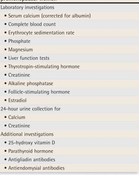
Evaluation should include assessment of thyroid, liver, and renal function (Table 3). Serum calcium (cor-rected for albumin) is elevated in hyperparathyroidism or malignancy. Low serum calcium levels are seen among people with vitamin D deficiency or malabsorption. Malabsorption of calcium and vitamin D can result in low BMD as well as osteomalacia. A low measurement for calcium in 24-hour urine assessment is an early indi-cator of inadequate calcium intake or malabsorption. If results from the 24-hour urine assessment for calcium do not return to normal when calcium supplementation is increased, the possibility of malabsorption or occult
celiac disease should be considered. A celiac panel with assessment of antiendomysial and antigliadin antibod-ies can help to diagnose celiac disease. History taking and physical examination will guide physicians consid-ering additional investigations. It is also important to ensure that young women truly are estrogen replete. Subclinical defi ciency in estrogen has been associated with low BMD among young women.8-10
Amenorrhea, as a result of estrogen deficiency, is associated with accelerated bone loss. Menstrual sta-tus is an important factor in achieving peak bone mass and in maintaining BMD among women before menopause.1,8-11 It is necessary to ensure that women
with low bone mass are not experiencing estrogen defi -ciency either clinically or subclinically.1 Elevations in
fol-licle-stimulating hormone of more than 20 mIU/L have been associated with increased bone turnover and pro-gressive bone loss in the perimenopausal period.10,12
Therefore, a detailed assessment of menstrual status is necessary, as ovulatory disturbances are frequently observed among premenopausal women with low BMD.8-10
A premenopausal woman with fragility fractures or progressive bone loss should be referred to a meta-bolic bone clinic for further assessment. A bone biopsy can provide further information regarding the under-lying pathology resulting in fractures and might be of value in the assessment. A bone biopsy is completed using a technique similar to that of a bone marrow
Table 2
. Important secondary causes of bone loss
Diseases and conditions
• Hypogonadism (primary and secondary) • Primary hyperparathyroidism
• Thyrotoxicosis • Hypercortisolism
• Growth hormone defi ciency • Osteomalacia
• Myeloproliferative disorders • Connective tissue disorders
• Malabsorptive states (eg, celiac disease) • Hepatic disorders (eg, primary biliary cirrhosis) • Infl ammatory bowel disease
• Renal disease • Hypercalciuria
• Osteogenesis imperfecta Medications
• Glucocorticoids • Thyroxine (excessive)
• Anticonvulsants (eg, phenytoin, phenobarbital) • Heparin (long-term)
• Lithium
• Cytotoxic chemotherapy
• Gonadotropin-releasing hormone agonists • Depot medroxyprogesterone acetate
Data from Khan et al.1
Table 3
. Workup for low bone mineral density in
premenopausal women
Laboratory investigations• Serum calcium (corrected for albumin) • Complete blood count
• Erythrocyte sedimentation rate • Phosphate
• Magnesium • Liver function tests
• Thyrotropin-stimulating hormone • Creatinine
• Alkaline phosphatase • Follicle-stimulating hormone • Estradiol
24-hour urine collection for • Calcium
• Creatinine
biopsy; however, the bone biopsy needle removes a core of bone tissue with both cortical and cancellous bone intact. This removal is completed while the patient receives local anesthetic, and risks are essentially lim-ited in experienced hands to local bleeding.
Intervention
There are no data evaluating the usefulness of bisphosphonates among premenopausal women with low BMD in the absence of glucocorticoid therapy. Bisphosphonates have been evaluated only among pre-menopausal women who have received glucocorticoid therapy. Glucocorticoid therapy is associated with an increase in the rate of bone turnover in addition to decreasing intestinal calcium absorption, increasing uri-nary calcium losses, and impairing osteocyte lifespan and function. Bisphosphonates lower bone turnover and, therefore, are effective among premenopausal women who have been treated with glucocorticoid therapy.13-16
Women who have not received glucocorticoid therapy and who are estrogen replete could have normal bone turnover rates. Suppressing these rates with a bisphos-phonate might not be safe or effective in improving bone density or reducing fracture risk. This patient popula-tion is thus best served by a specialized metabolic bone clinic before antiresorptive therapy is implemented.
Antiresorptive therapies. Antiresorptive therapy benefits premenopausal women with secondary causes of bone loss, such as glucocorticoid use or primary hyperparathy-roidism (level I evidence).17 Bisphosphonates have
long-term skeletal retention, and these agents can be released from the skeleton several years later, potentially in a subsequent pregnancy. The effects of bisphosphonates on the developing fetal skeleton are unknown. Cyclic etidronate, a less potent bisphosphonate, has not been evaluated among premenopausal women with osteopo-rosis. Alendronate has been evaluated among premeno-pausal women with primary hyperparathyroidism and has been shown to be effective in improving BMD in this population.17 Alendronate and risedronate are effective
in improving BMD in glucocorticoid-induced bone loss (level I evidence). Antiresorptive therapy has not been evaluated among premenopausal women with low BMD in the absence of secondary causes of osteoporosis.
Lifestyle modification. Lifestyle modification should be encouraged among premenopausal women in order to improve BMD. This would include weight-bearing exer-cises, adequate dietary calcium intake, smoking cessa-tion, limiting caffeine, and reducing excessive alcohol consumption (level II evidence).18
It is important to ensure adequate calcium and vita-min D intake. Maintenance of normal body weight, with a body mass index of 20 to 25, and a daily exercise program are of value in maintaining BMD (level II evidence).18
Estrogen supplementation. Women who are estro-gen deficient, either clinically or subclinically, benefit from estrogen supplements. Estrogen supplementa-tion, in the form of either oral contraceptive pills or 17beta-estradiol in combination with a progestin, should be considered in order to prevent progressive bone loss (level III evidence). Estrogen supplementa-tion is associated with improvements in BMD among estrogen-deficient women (level I evidence).19 Cyclic
medroxyprogesterone in women with ovulatory dis-turbance has been shown in a randomized placebo-controlled prospective study over 1 year to result in gains in BMD (level I evidence).20 Prospective data are
needed to evaluate further the effect of short luteal-phase cycles on BMD. Further evaluation of interven-tion with estrogen or progesterone supplements is also warranted. The role of estrogen supplementation for women who are not estrogen deficient is contro-versial (level III evidence).
Data regarding oral contraceptives have been conflict-ing; some studies demonstrate positive effects on BMD in premenopausal women and other studies demonstrate negative effects on BMD (level II evidence).21,22 Data from
the Canadian Multicentre Osteoporosis Study indicated that oral contraceptive users had lower BMD at the tro-chanter and spine than non-users.23 Evaluation of this
patient population indicated, however, that these women also had higher rates of smoking and alcohol use and had a higher prevalence of menstrual irregularity before initi-ating oral contraceptive use than non-users had (level II evidence).23 Thus, prospective data are required to assess
the effects of oral contraceptive use on BMD among pre-menopausal women. Depot medroxyprogesterone acetate, an injectable contraceptive, inhibits release of luteinizing hormone and follicle-stimulating hormone and results in suppression of ovarian synthesis of estradiol and proges-terone (level II evidence).24,25 Depot medroxyprogesterone
use has been associated with decreased BMD at the lum-bar spine (level II evidence).26,27 Discontinuation of depot
medroxyprogesterone is associated with rapid improve-ment in BMD and reversal of bone loss, as demonstrated in a prospective cohort study.28
Conclusion
Referral to a metabolic bone clinic is appropriate for patients with fragility fractures or progressive bone loss. Antiresorptive therapy has been evaluated only for
pre-menopausal women receiving glucocorticoid therapy or those with primary hyperparathyroidism. Only in these conditions has antiresorptive therapy been shown to improve BMD measurements. Screening for osteoporo-sis among premenopausal women is not justifi ed based on available evidence.
Acknowledgment
This article was originally published as “Management of low bone mineral density in premenopausal women.” J Obstet Gynaecol Can 2005;27(4):345-9 and is reprinted courtesy of the Society of Obstetrics and Gynaecologists of Canada.
Competing interests
None declared
Correspondence to:Dr Aliya Khan, Divisions of Endocrinology and Geriatrics, McMaster University, 331— 209 Sheddon Ave, Oakville, ON L6J 1X8; telephone 905 844-5677; fax 905 844-8966; e-mail [email protected]
References
1. Khan AA, Bachrach L, Brown JP, Hanley DA, Josse RG, Kendler DL, et al. Standards and guidelines for performing central dual-energy x-ray absorptiometry in premenopausal women, men, and children. J Clin Densitom 2004;7:51-64.
2. Kanis JA, Melton LJ III, Christiansen C, Johnston CC, Khaltaev N. The diagnosis of osteo-porosis. J Bone Miner Res 1994;9:1137-41.
3. Gullberg B, Johnell O, Kanis JA. World-wide projections for hip fracture. Osteoporos Int 1997;7:407-13.
4. Hui SL, Slemenda CW, Johnston CC. Age and bone mass as predictors of fracture in a prospective study. J Clin Invest 1988;81:1804-9.
5. Sowers M, Kshirsagar A, Crutchfi eld M, Updike S. Body composition, age and femoral bone mass of young adult women. Ann Epidemiol 1991;1:245-54.
6. Hosmer WD, Genant HK, Browner WS. Fractures before menopause: a red fl ag for phy-sicians. Osteoporos Int 2002;13:337-41.
7. Khan AA, Syed Z. Bone densitometry in premenopausal women. J Clin Densitom 2004;7:85-92.
8. Prior JC, Vigna YM, Schechter MT, Burgess AE. Spinal bone loss and ovulatory distur-bances. N Engl J Med 1990;323:1221–7.
9. Sowers M, Randolph JF Jr, Crutchfi eld M, Jannausch ML, Shapiro B, Zhang B, et al. Urinary ovarian and gonadotropin hormone levels in premenopausal women with low bone mass. J Bone Miner Res 1998;13:1191–202.
10. Sowers M, Crutchfi eld M, Bandekar R, Randolph JF, Shapiro B, Schork MA, et al. Bone mineral density and its change in pre- and perimenopausal white women: the Michigan Bone Health Study. J Bone Miner Res 1998;13:1134–40.
11. Sowers MR, Clark MK, Hollis B, Wallace RB, Jannausch M. Radial bone mineral density in pre- and perimenopausal women: a prospective study of rates and risk factors of loss. J Bone Miner Res 1992;7:647–57.
12. Warming L, Hassager C, Christiansen C. Changes in bone mineral density with age in men and women: a longitudinal study. Osteoporos Int 2002;13:105–12.
13. Saag KG, Emkey R, Schnitzer TJ, Brown JP, Hawkins F, Goemaere S, et al, for the Glucorticoid-Induced Osteoporosis Intervention Study Group. Alendronate for the preven-tion and treatment of glucocorticoid-induced osteoporosis. N Engl J Med 1998;339:292-9. 14. Adachi JD, Saag KG, Delmas PD, Liberman UA, Emkey RD, Seeman E, et al. Two-year effects of alendronate on bone mineral density and vertebral fracture in patients receiv-ing glucocorticoids: a randomized, double-blind, placebo-controlled extension trial. Arthritis Rheum 2001;44:202-11.
15. Reid DM, Hughes RA, Laan RF, Sacco-Gibson NA, Wenderoth DH, Adami S, et al. Effi cacy and safety of daily risedronate in the treatment of corticosteroid-induced osteo-porosis in men and women: a randomized trial. European Corticosteroid-Induced Osteoporosis Treatment Study. J Bone Miner Res 2000;15:1006-13.
16. Wallach S, Cohen S, Reid DM, Hughes RA, Hosking DJ, Laan RF, et al. Effects of rise-dronate treatment on bone density and vertebral fracture in patients on corticosteroid therapy. Calcif Tissue Int 2000;67:277-85.
17. Khan AA, Bilezikian JP, Kung AW, Ahmed MM, Dubois SJ, Ho AY, et al. Alendronate in primary hyperparathyroidism: a double-blind, randomized, placebo-controlled trial. J Clin Endocrinol Metab 2004;89:3319-25.
18. Brown JP, Josse RG; Scientifi c Advisory Council of the Osteoporosis Society of Canada. Clinical practice guidelines for the diagnosis and management of osteoporosis in Canada. CMAJ 2002;167(10 Suppl):S1-34.
19. Kuohung W, Borgatta L, Stubblefi eld P. Low dose oral contraceptives and bone mineral density: an evidence based analysis. Contraception 2000;61(2):77-82.
20. Prior JC, Vigna YM, Barr SI, Rexworthy C, Lentle BC. Cyclic medroxyprogesterone treat-ment increases bone density: a controlled trial in active women with menstrual cycle disturbances. Am J Med 1994;96:521-30.
21. DeCherney A. Bone-sparing properties of oral contraceptives. Am J Obstet Gynecol 1996;174:15-20.
22. Ott SM, Scholes D, LaCroix AZ, Ichikawa LE, Yoshida CK, Barlow WE. Effects of con-traceptive use on bone biochemical markers in young women. J Clin Endocrinol Metab 2001;86:179-85.
23. Prior JC, Kirkland SA, Joseph L, Kreiger N, Murray TM, Hanley DA, et al. Oral con-traceptive use and bone mineral density in premenopausal women: cross-sectional, population-based data from the Canadian Multicentre Osteoporosis Study. CMAJ 2001;165(8):1023-9.
24. Fraser IS, Weisberg E. A comprehensive review of injectable contraception with spe-cial emphasis on depot medroxyprogesterone acetate. Med J Aust 1981;24(Suppl 1):3-19. 25. Jeppsson S, Johansson EDB. Medroxyprogesterone acetate, estradiol, FSH, and LH
in peripheral blood after intramuscular administration of Depo-Provera® to women. Contraception 1976;14:461-9.
26. Paiva LC, Pinto-Neto AM, Faundes A. Bone density among long-term users of medroxyprogesterone acetate as a contraceptive. Contraception 1998;58:351-5. 27. Wanichsetakul P, Kamudhamas A, Watanaruangkovit P, Siripakarn Y, Visutakul P. Bone
mineral density at various anatomic bone sites in women receiving oral contraceptives and depot-medroxyprogesterone acetate for contraception. Contraception 2002;65:407-510. 28. Scholes D, LaCroix AZ, Ichikawa LE, Barlow WE, Ott SM. Change in bone mineral
den-sity among adolescent women using and discontinuing depot medroxyprogesterone acetate contraception. Arch Pediatr Adolesc Med 2005;159(2):139-44.
22. Ott SM, Scholes D, LaCroix AZ, Ichikawa LE, Yoshida CK, Barlow WE. Effects of
con-EDITOR’S KEY POINTS
•
Bone density follows a bell curve, so that among
premenopausal women, low bone density might
refl ect normal variation in BMD and achievement of
a lower peak bone mass owing to genetic and
envi-ronmental factors, such as poor calcium intake, lack
of exercise, smoking, or excessive alcohol intake.
•
Osteoporosis should be diagnosed in premenopausal
women only if fragility fractures are present and not
on the basis of low bone density measurements alone.
•
Low bone density should be investigated as it might
be secondary to medical conditions associated with
bone loss.
•
In this population, bisphosphonates have been
eval-uated only for treating patients who have received
glucocorticoid therapy; they have improved bone
density. Other measures to increase bone mass,
depending on the cause, include estrogen
replace-ment and lifestyle changes.
POINTS DE REPÈRE DU RÉDACTEUR
•
La densité osseuse suit une courbe en forme de
cloche, de sorte qu’une faible densité osseuse en les
femmes préménopause pourrait refl éter une
varia-tion normale de ce paramètre et l’atteinte d’un pic
de masse osseuse moins élevé en raison de facteurs
génétiques ou environnementaux, tels qu’un apport
insuffi sant de calcium, un manque d’exercice, l’usage
de tabac ou une consommation excessive d’alcool.
•
Un diagnostic d’ostéoporose chez les femmes
prémé-nopausique ne devrait être évoqué qu’en présence de
fractures de fragilité osseuse, et non en raison d’une
densitométrie osseuse trop basse seulement.
•
Une densité osseuse trop basse doit être investiguée
parce qu’elle pourrait être secondaire à une
affec-tion qui cause une lyse osseuse.
•