P R O C E E D I N G S
Open Access
Highly precise protein-protein interaction
prediction based on consensus between
template-based and
de novo
docking methods
Masahito Ohue
1,2,3, Yuri Matsuzaki
1, Takehiro Shimoda
1,2, Takashi Ishida
1, Yutaka Akiyama
1,2*From
Great Lakes Bioinformatics Conference 2013
Pittsburgh, PA, USA. 14-16 May 2013
Abstract
Background:Elucidation of protein-protein interaction (PPI) networks is important for understanding disease mechanisms and for drug discovery. Tertiary-structure-basedin silicoPPI prediction methods have been developed with two typical approaches: a method based on template matching with known protein structures and a method based onde novo protein docking. However, the template-based method has a narrow applicable range because of its use of template information, and thede novo docking based method does not have good prediction performance. In addition, both of thesein silico prediction methods have insufficient precision, and require validation of the predicted PPIs by biological experiments, leading to considerable expenditure; therefore, PPI prediction methods with greater precision are needed.
Results:We have proposed a new structure-based PPI prediction method by combining template-based prediction
andde novodocking prediction. When we applied the method to the human apoptosis signaling pathway, we obtained a precision value of 0.333, which is higher than that achieved using conventional methods (0.231 for PRISM, a template-based method, and 0.145 for MEGADOCK, a non-template-based method), while maintaining an F-measure value (0.285) comparable to that obtained using conventional methods (0.296 for PRISM, and 0.220 for MEGADOCK).
Conclusions:Our consensus method successfully predicted a PPI network with greater precision than conventional template/non-template methods, which may thus reduce the cost of validation by laboratory experiments for confirming novel PPIs from predicted PPIs. Therefore, our method may serve as an aid for promoting interactome analysis.
Introduction
Elucidation of regulatory relationships among the tens of thousands of protein species that function in a human cell is crucial for understanding the mechanisms underlying diseases and for the development of medi-cines [1]. Predicting protein-protein interaction (PPI) networks at the genome scale is one of the main topics in systems biology.
The methods used for PPI network prediction include primary-structure-based searching [2,3], evolutionary information-based methods [4], and tertiary-structure-based methods [5-7]. Tertiary-structure-tertiary-structure-based methods are attracting attention because they provide predicted protein complex structures and because they do not depend on homologous proteins. Tertiary structural information also provides powerful features for recogni-tion [8,9] and is therefore useful for predicting binding affinity [10] in protein-protein complexes.
There are two typical approaches for tertiary-struc-ture-based PPI predictions: a method based on template matching with known protein structures and another * Correspondence: [email protected]
1
Graduate School of Information Science and Engineering, Tokyo Institute of Technology, 2-12-1-W8-76 Ookayama, Meguro-ku, Tokyo 152-8550, Japan Full list of author information is available at the end of the article
method based onde novo protein docking. The tem-plate-based method is based on the hypothesis that known complex structures or interface architectures can be used to model the complex formed between two tar-get proteins. The hypothesis is logical, and this method provides good prediction performance when complex structural information is available as a template; how-ever, if the template structure information is not avail-able, performance is poor. In addition, because the interface architecture is not always similar for similar interactions, the template-based method has a narrow applicable range. In contrast, thede novodocking based method has a wide applicable range because it uses only tertiary structural information. However, because the advantage provided by existing template information is not utilized, the prediction performance is poor.
Tuncbaget al. developed a template-based PPI predic-tion method called PRISM [5], which is based on infor-mation regarding the interaction surface of crystalline complex structures. PRISM has been applied for predict-ing PPIs in a human apoptosis pathway [11] and a p53-protein-related pathway [12], and has contributed to the understanding of the structural mechanisms underlying some types of signal transduction. Ohueet al. developed a PPI prediction method called MEGADOCK [6] and Wass et al. developed a method [13] based on protein-protein docking without interaction surface information. MEGADOCK has been applied for PPI prediction for a bacterial chemotaxis pathway [7,14] and has contributed to the identification of protein pairs that may interact.
However, the prediction results of both template-based andde novodocking-based methods in these studies con-tained many false-positive predictions. PRISM obcon-tained a precision value of 0.231 when applied to a human apop-tosis pathway that consisted of 57 proteins, which was higher than the precision obtained with random predic-tion (precision value of 0.086), and MEGADOCK obtained a precision value of 0.400 when applied to a bacterial chemotaxis pathway that consisted of 13 pro-teins, which was higher than the precision obtained with random prediction (precision value of 0.253). To identify new PPIs, the prediction results need to be validated using biological experiments. For this purpose, obtaining a low number of predicted interaction candidates with high reliability is more important than obtaining a high number of predictions with low reliability. Thus, this paper aims to improve the reliability of the method used to obtain PPI predictions.
In this study, we combined two different PPI prediction methods to improve the precision of PPI prediction. Because PRISM is a template-based method, its prediction accuracy depends on the template dataset prepared. Only PPIs whose interaction surface structures are conserved are expected to be predicted. In contrast, MEGADOCK is
a non-template-based method (also calledde novo predic-tion), which has the demerit of generating false-positives for the cases in which no similar structures are seen in known complex structure databases; thus, template-based method would be ruled out from the prediction. However, in situations where template structures are not present in databases, MEGADOCK can still predict PPIs. This quali-tative difference between the two methods typically makes their output different. Thus, the combination of both pre-diction methods may improve prepre-diction accuracy, as the intersection set (AND set) of both results may contain fewer false-positives; this improvement in precision would also contribute to improvement in the prediction reliability provided by the use of just one method.
Such an approach is called a“meta”approach. Meta approaches have already been used in the field of protein tertiary structure prediction [15], and critical experiments have demonstrated improved performance of meta predic-tors when compared with the individual methods used in the meta predictors. The meta approach has also provided favorable results in protein domain prediction [16] and the prediction of disordered regions in proteins [17]. We have therefore proposed a new PPI prediction method based on the consensus between template-based andde novo dock-ing methods. Generally, a meta prediction method may have low applicability because meta approaches require applicable conditions for every method in the approach. However, if structural information is available, thede novo
docking method introduced in this study is always applic-able with or without template information. Thus, the applicability of the consensus method is not narrower than that of a template-based method.
Materials and methods Template-based PPI prediction
We used PRISM for template-based PPI prediction. PRISM uses two input datasets: the template set and the target set. The template set consists of interfaces extracted from protein pairs that are known to interact. The target set consists of protein chains whose interactions need to be predicted. The two sides of a template interface are compared with the surfaces of two target monomers by structural alignment. If regions of the target surfaces are similar to the complementary sides of the template inter-face, then these two targets are predicted to interact with each other through the template interface architecture.
template interfaces to form a complex; and (4) the Fiber-Dock [20] algorithm is used to refine the interactions to introduce flexibility, resolve steric clashes of side chains, compute the global energy of the complex, and rank the solutions according to their energies. When the computed energy of a protein pair is less than−10 kcal/mol, the pair is determined to“interact”(personal communication with Ms. Saliha Ece Acuner Ozbabacan, July 12, 2013). This prediction protocol has been described in detail in a previous study [5,11].
PPI prediction based on thede novodocking method For de novo protein docking-based PPI prediction, we used MEGADOCK version 2.6.2 [7]. MEGADOCK does not require template structures for prediction. The PPI prediction scheme used in this study consists of two steps. First, we conducted rigid-body docking calcula-tions based on a simplified energy function considering shape complementarity, electrostatics, and hydrophobic interactions for all possible binary combinations of pro-teins in the target set. Using this process, we obtained a group of high-scoring docking complexes for each pair of proteins. Next, we applied ZRANK [21] to the pre-dicted complex structures for more advanced binding energy calculation and re-ranked the docking results based on ZRANK energy scores. The deviation of the selected docking scores from the score distribution of high-ranked complexes was determined as a standar-dized score (Z-score) and was used to assess possible interactions. This prediction protocol has been described in previous studies [22,23]. Potential complexes that had no other high-scoring interactions nearby were rejected using structural differences. Thus, we considered likely binding pairs that had at least one populated area of high-scoring structures, one of which may be the true binding site.
Consensus prediction method
In this study, we proposed a new meta-prediction method by evaluating the consensus between both previously used prediction methods. The proposed method consists of two steps: (1) prediction from the same target set by PRISM and MEGADOCK and (2) consideration that the method provides a prediction regarding target protein pair interac-tion only when both PRISM and MEGADOCK predict that the target protein pair interacts. Although some true-positives may be dropped by this method, the remaining predicted pairs are expected to have higher reliability because of the consensus between two prediction methods that have different characteristics.
Dataset
In this study, we focused on the human apoptosis sig-naling pathway previously analyzed by PRISM because
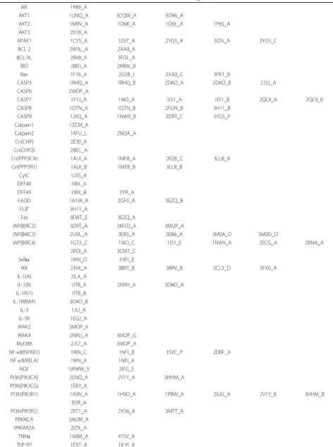
our prediction results can thus be compared directly to the results of the previous study. PRISM and MEGA-DOCK are based on three-dimensional protein struc-tures and therefore can only be applied to proteins whose tertiary structures are available. Therefore, we searched among proteins involved in the human apoptosis path-way that were present in the Protein Data Bank (PDB) (accessed on July 28, 2012). We selected several proteins that had the highest resolution for the structural group that had high sequence similarity (>0.9) with the other proteins in the dataset [11]. After filtering according to resolution and sequence similarity, we obtained 158 PDB structures that corresponded to 57 proteins in the human apoptosis pathway described in KEGG (KEGG pathway ID:hsa04210) [24]. The PDB IDs in this struc-ture dataset were the same as those used by Ozbabacan
et al.[11]. Table 1 shows the list of PDB IDs and chains of this dataset.
Known PPIs were collected from the STRING database [25]. We used only experimental data in the literature obtained from STRING with a confidence score >0.5. The number of known PPIs was 137. Because the database does not contain existing self-interactions, we did not pre-dict self-interactions. Thus, the number of target pairs was57C2= 1,596.
Evaluation of prediction performance
Here, we have defined #TP, #FP, #FN, #TN, precision, recall, and the F-measure, which we used to evaluate the prediction results: #TP is the number of predicted PPIs that were also found in STRING (true-positive), #FP is the number of predicted PPIs that were not in STRING (false-positive), #FN is the number of PPIs not predicted by the system even though the pair was found to interact in STRING (false-negative), and #TN is the number of negative predictions that were also not found in STRING (true-negative). Precision, recall, and the F-measure are represented as follows:
where the F-measure is the harmonic mean of precision and recall. To identify new PPIs in biological experiments afterin silicoscreening, precision is more important than recall to reduce the cost of validation.
Results and Discussion
Comparison of template- and non-template-based methods
Table 1 Protein and PDB ID list of human apoptosis pathway dataset
Protein Name PDB ID (_Chain)
AIF 1M6I_A
AKT1 1UNQ_A 3CQW_A 3O96_A
AKT2 1MRV_A 1O6K_A 1O6L_A 1P6S_A
AKT3 2X18_A
APAF1 1CY5_A 1Z6T_A 2YGS_A 3IZA_A 3YGS_C
BCL-2 2W3L_A 2XA0_A
BCL-XL 2B48_A 3FDL_A
BID 2BID_A 2KBW_B
Bax 1F16_A 2G5B_I 2XA0_C 3PK1_B
CASP3 1RHQ_A 1RHQ_B 2DKO_A 2DKO_B 2J32_A
CASP6 2WDP_A
CASP7 1F1J_A 1I4O_A 1I51_A 1I51_B 2QL9_A 2QL9_B
CASP8 1QTN_A 1QTN_B 2FUN_B 3H11_B
CASP9 1JXQ_A 1NW9_B 3D9T_C 3YGS_P
Calpain1 1ZCM_A
Calpain2 1KFU_L 2NQA_A
Cn(CHP) 2E30_A
Cn(CHP2) 2BEC_A
Cn(PPP3CA) 1AUI_A 1MF8_A 2R28_C 3LL8_A
Cn(PPP3R1) 1AUI_B 1MF8_B 3LL8_B
CytC 1J3S_A
DFF40 1IBX_A
DFF45 1IBX_B 1IYR_A
FADD 1A1W_A 2GF5_A 3EZQ_B
FLIP 3H11_A
Fas 3EWT_E 3EZQ_A
IAP(BIRC2) 3D9T_A 3M1D_A 3MUP_A
IAP(BIRC3) 2UVL_A 3EB5_A 3EB6_A 3M0A_D 3M0D_D
IAP(BIRC4) 1G73_C 1I4O_C 1I51_E 1NW9_A 2ECG_A 2KNA_A
2POI_A 3CM7_C
IBa 1IKN_D 1NFI_E
IKK 2JVX_A 3BRT_B 3BRV_B 3CL3_D 3FX0_A
IL-1(A) 2ILA_A
IL-1(B) 1ITB_A 2NVH_A 3O4O_A
IL-1R(1) 1ITB_B
IL-1R(RAP) 3O4O_B
IL-3 1JLI_A
IL-3R 1EGJ_A
IRAK2 3MOP_K
IRAK4 2NRU_A 3MOP_G
MyD88 2JS7_A 3MOP_A
NF-B(NFKB1) 1IKN_C 1NFI_B 1SVC_P 2DBF_A
NF-B(RELA) 1IKN_A 1NFI_A
NGF 1WWW_V 2IFG_E
PI3K(PIK3CA) 2ENQ_A 2V1Y_A 3HHM_A
PI3K(PIK3CG) 1E8Y_A
PI3K(PIK3R1) 1A0N_A 1H9O_A 1PBW_A 2IUG_A 2V1Y_B 3HHM_B
3I5R_A
PI3K(PIK3R2) 2KT1_A 2XS6_A 3MTT_A
PRKACA 3AGM_A
PRKAR2A 2IZX_A
TNFa 1A8M_A 4TSV_A
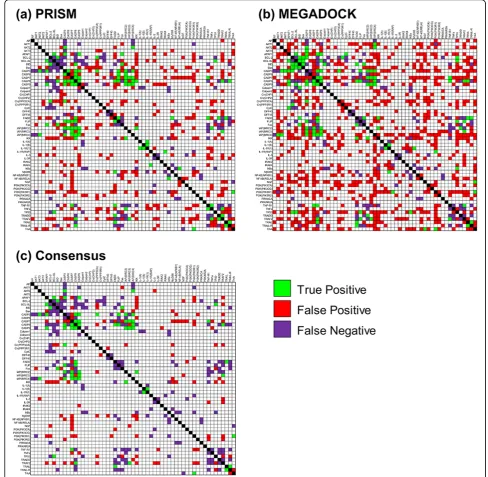
self-interactions that were not considered as prediction targets. As shown in Figure 1, PRISM was performed with fewer FPs than MEGADOCK. Table 2 shows the evalua-tion of predicevalua-tion results. With MEGADOCK, we obtained a lower value of precision and a higher value of recall relative to PRISM. When the F-measure was evalu-ated as a measure of overall performance, MEGADOCK showed lower values than PRISM. Predictions by MEGA-DOCK contained more FPs because, in contrast to PRISM, MEGADOCK does not restrict interface struc-tures to those found in template strucstruc-tures. In contrast, PRISM obtained lower recall values than MEGADOCK because it only searched interactions whose interface structures could be found in the template set.
Results of the consensus prediction
Figure 2 shows the Venn diagram of the number of TPs and FPs of the results of PRISM and MEGADOCK. A large difference was observed in the results obtained by the two methods. Thus, combining the prediction results of PRISM and MEGADOCK may provide better perfor-mance in PPI prediction. All of the predicted pairs of TPs and FPs are shown in Table S1 in Additional File 1.
Figure 1(c) shows the prediction obtained on consensus between PRISM (a) and MEGADOCK (b); notably, the number of FP samples greatly decreased. The first row of Table 2 shows that the consensus method obtained an F-measure value of 0.285, which was comparable to the PRISM result (F-measure = 0.296). The consensus predic-tion indicated a higher value of precision for the consensus method (0.333) than for PRISM (0.231). The consensus method yielded the highest precision value in the method
shown in Table 2. This method is useful when validating unknown PPI predictions using biological experiments. In contrast, OR prediction demonstrated high recall (Table 2). Thus, the OR method will be useful when prediction with high sensitivity, e.g., in the initial construction of the draft PPI network from the relevant proteins, is required.
An example of a false-positive pair and its predicted complex structure
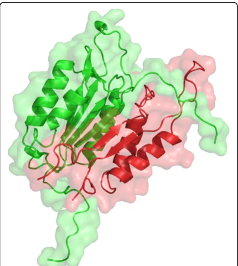
The caspase-3 and caspase-7 pair is shown as an exam-ple of FP predictions in both PRISM and MEGADOCK with a particularly high evaluation value. Both caspase-3 and caspase-7 are effector caspases, which belong to a family of cysteine proteases that play essential roles in apoptosis. Effector caspases are activated by initiator caspases (e.g., caspase-2, 8, and 9), and then induce apoptotic cell death. Although the initiator and effector caspase cascade is well known, interactions among effec-tor caspases are disputed [26].
The interaction of caspase-3 and caspase-7 was pre-dicted with a high affinity score; the PRISM energy value was less than−190 kcal/mol and the MEGADOCK dock-ing score was higher than 10,000. These values indicate a powerful affinity interaction. Figure 3 shows the predicted complex structure for caspase-3 and caspase-7. The pre-dicted complex consists of 2DKO chain A (caspase-3, p17 subunit) and 2QL9 chain B (caspase-7, p10 subunit).
Additionally, 2DKO chain B (caspase-3, p12 subunit) and 2QL9 chain B, and 2QL9 chain A (caspase-7, p20 subunit) and 2DKO chain A, respectively, have similar structures. Thus, the predicted complex with each subunit swapped, as shown in Figure 3, is similar to the original heterodimer Table 1 Protein and PDB ID list of human apoptosis pathway dataset(Continued)
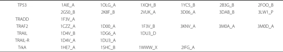
TP53 1AIE_A 1OLG_A 1XQH_B 1YC5_B 2B3G_B 2FOO_B
2GS0_B 2K8F_B 2VUK_A 3D06_A 3DAB_B 3LW1_P
TRADD 1F3V_A
TRAF2 1CZZ_A 1D00_A 1F3V_B 3KNV_A 3M0A_A 3M0D_A
TRAIL 1D4V_B 1DG6_A 1DU3_D
TRAIL-R 1D4V_A 1DU3_A
TrkA 1HE7_A 1SHC_B 1WWW_X 2IFG_A
Figure 1Apoptosis prediction by the (a) PRISM, (b) MEGADOCK, and (c) consensus methods. The green cells are true-positives, the red cells are false-positives, and the purple cells are false-negatives. The diagonal cells (black cells) have no PPI information in the STRING database and are excluded from the prediction targets.
Table 2 Accuracy of human apoptosis pathway prediction
Method #TP #FP #FN #TN Precision Recall F-measure
Consensus(AND) 34 68 103 1,391 0.333 0.248 0.285
OR 84 483 53 976 0.148 0.613 0.239
PRISM 56 186 81 1,273 0.231 0.409 0.296
and possibly predicted to occur with a high score. The interaction among effector caspases, as in this case, has not been examined by biological experiments. In fact, another PPI prediction tool based on template structure and data-base information, PrePPI [28,29] (version 1.2.0), predicted the pair of caspase-3 and caspase-7 with a high score (the final probability value was 0.99). This situation is difficult to avoid in large-scale prediction problems. However, efforts such as the Negatome project [30] will help to improve this difficulty in the future.
Relationship between the number of predicted positives and the number of structures
The structure-based PPI prediction method may generate positives with some bias regarding the type of proteins (rows and columns of Figure 1). From Table 1 and Figure 1, predictions with a large number of protein struc-tures tend to generate more positive pairs. To verify this tendency, the number of PDB chain structures used for PPI prediction and the number of positive predicted pairs containing its protein are plotted in Figure 4. The #TPs are shown in Figure 4(a) and the #FPs are shown in Figure 4(b). Pearson’s correlation coefficientRand the
P-value for the correlation coefficientt-test are shown in Table 3.
From the results of thet-tests, the number of chains and the number of positive predictions were clearly cor-related withP< 0.05 in all cases, which suggests that the structure-based PPI prediction method should address the number of used protein structures without bias. For example, in a template matching-based method such as PRISM, a protein pair with more conformations of struc-tures will have more matches in template complexes and a higher possibility of predicted interaction. In Table 3, the correlation coefficient values are particularly high in FP predictions. Therefore, for more precise prediction, we should consider one of the two ways: (i) how to gen-erate the target set without multiple conformations in each protein and (ii) develop a correction method when the target set contains multiple conformations.
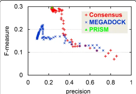
Performance evaluation with various sensitivity parameters
In this study, we used a fixed threshold value for MEGA-DOCK that provided the best F-measure value for the target dataset. Figure 5 shows a plot of precision vs. F-measure value for prediction results with various thresh-old values for MEGADOCK. Figure 5 also plots the per-formance of the consensus method with various threshold values for MEGADOCK prediction while the threshold value for PRISM prediction was fixed. When the threshold value was changed in MEGADOCK, the plotted values remained in the region of low precision (0.0-0.2), and lower F-measure values were observed in the region of higher precision because of the decreased recall value. The consensus prediction method maintained a stable F-measure value when the value of precision was approxi-mately 0.2-0.3, although the performance in the high-pre-cision region (> 0.4) was inferior to that of MEGADOCK. In this region, the consensus prediction provides a better precision value than PRISM while maintaining the same F-measure value. Figure 5 clearly shows that the perfor-mance obtained by using the consensus method is better
Figure 2 Venn diagram of apoptosis pathway prediction results. The common set (#TP = 34, #FP = 68) is denoted as “Consensus”.
over a wide range of threshold values than the prediction obtained using only MEGADOCK.
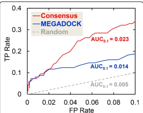
The AUC, i.e., the area under the ROC curve [31], is a more general and effective statistical measure. The ROC0.1curves, which include the ROC curves up to an
FP rate of 0.1, are shown in Figure 6. ROC curves were created by plotting the TP rate (#TP/(#TP+#FN)) against the FP rate (#FP/(#FP+#TN)). Regions with high FP rates are not useful for prediction because many FPs are gener-ated, e.g., an FP rate of 0.2 represents #FP = 292. The ROC0.1curve was thus considered to favor methods that
produce a high TP rate at low FP rates, and the asso-ciated area under the curve is referred to as AUC0.1. A
perfect prediction will produce an AUC0.1of (0.1 × 1 =)
0.1, whereas a random prediction will result in an AUC0.1
of (0.1 × 0.1/2 =) 0.005. Figure 6 shows that the consen-sus prediction (AUC0.1 = 0.023) is better than the
MEGADOCK (AUC0.1= 0.014) and random predictions
(AUC0.1= 0.005).
Conclusions
In this study, we propose a new PPI network prediction method based on the consensus between template-based
prediction and non-template-based prediction. The con-sensus method successfully predicted the PPI network more accurately than the conventional single template/ non-template method. Because such precise prediction can reduce biological screening costs, it will promote interactome analysis. For further improvement of pre-diction performance, it is necessary to further improve the combination of the two techniques, e.g., by using a strategy other than taking a simple AND/OR consensus. For example, biological information such as biochemical function and subcellular localization information could be used.
Figure 4Number of PDB chains vs. positive predictions. (a) Shows the number of true-positives and (b) shows the number of false-positives. The horizontal axis is the number of PDB chains used in the interaction prediction, and the vertical axis is the number of positives predicted by using protein structures.
Table 3 Correlation coefficientRandP-value of correlation test on Figure 4
Method (a) #TPs (b) #FPs
R P-value R P-value
Consensus 0.477 1.784 × 10⁻⁴ 0.594 1.121 × 10⁻⁶
PRISM 0.342 9.259 × 10⁻³ 0.415 1.316 × 10⁻³
MEGADOCK 0.488 1.167 × 10⁻⁴ 0.864 4.602 × 10⁻¹⁸
Additional material
Additional file 1: Supplementary table for predicted list. Table S1: The list of all true-positive pairs and false-positive pairs predicted by the PRISM, MEGADOCK, and consensus methods; (a) the true-positive list of PRISM predictions, (b) the false-positive list of PRISM predictions, (c) the true-positive list of MEGADOCK predictions, (d) the false-positive list of MEGADOCK predictions, (e) the true-positive list of consensus predictions, and (f) the false-positive list of consensus predictions.
List of abbreviations
PPI: protein-protein interaction; PDB: protein data bank; KEGG: Kyoto encyclopedia of genes and genomes; TP: true-positive; FP: false-positive; FN: false-negative; TN: true-negative; ROC: receiver operating characteristic; AUC: area under the (ROC) curve.
Competing interests
The authors declare that they have no competing interests.
Authors’contributions
MO developed the consensus interaction prediction method, designed the human apoptosis pathway problem, and wrote the manuscript. MO and YM performed the computational experiments and validated the results. TS performed the PRISM experiments. TI assisted with the method design. YA supervised and directed the entire study. All authors read and approved the final manuscript.
Acknowledgements
The authors gratefully acknowledge Saliha Ece Ozbabacan for explaining the PRISM protocols. Some of the results were obtained by using the K-computer at the RIKEN Advanced Institute for Computational Science (AICS) through early access and access granted as a High Performance Computing Infrastructure (HPCI) Systems Research Program (proposal number hp120131).
Declarations
The publication fee of this article was funded by Tokyo Institute of Technology. This work was supported in part by a Grant-in-Aid for JSPS Fellows (238750), a Grant-in-Aid for Research and Development of The Next-Generation Integrated Simulation of Living Matter (ISLiM), and by the Education Academy of Computational Life Sciences (ACLS) at the Tokyo
Institute of Technology, all of which were from the Ministry of Education, Culture, Sports, Science, and Technology of Japan (MEXT).
This article has been published as part ofBMC ProceedingsVolume 7 Supplement 7, 2013: Proceedings of the Great Lakes Bioinformatics Conference 2013. The full contents of the supplement are available online at http://www.biomedcentral.com/bmcproc/supplements/7/S7.
Authors’details
1
Graduate School of Information Science and Engineering, Tokyo Institute of Technology, 2-12-1-W8-76 Ookayama, Meguro-ku, Tokyo 152-8550, Japan.
2Education Academy of Computational Life Sciences, Tokyo Institute of
Technology, 2-12-1 Ookayama, Meguro-ku, Tokyo 152-8550, Japan.3Research
Fellow of the Japan Society for the Promotion of Science.
Published: 20 December 2013
References
1. Wass MN, David A, Sternberg MJE:Challenges for the prediction of macromolecular interactions.Curr Opin Struct Biol2011,21:382-390. 2. Higurashi M, Ishida T, Kinoshita K:Identification of transient hub proteins
and the possible structural basis for their multiple interactions.Protein Sci2008,17:72-78.
3. Shen J, Zhang J, Luo X, Zhu W, Yu K, Chen K, Li Y, Jiang H:Predicting protein-protein interactions based only on sequences information.Proc Natl Acad Sci USA2007,104:4337-4341.
4. Valencia A, Pazos F:Prediction of protein-protein interactions from evolutionary information.Structural Bioinformatics.second edition. Wiley and Sons: New York; 2009, 617-634.
5. Tuncbag N, Gursoy A, Nussinov R, Keskin O:Predicting protein-protein interactions on a proteome scale by matching evolutionary and structural similarities at interfaces using PRISM.Nature Protocols2011,
6:1341-1354.
6. Ohue M, Matsuzaki Y, Uchikoga N, Ishida T, Akiyama Y:MEGADOCK: An all-to-all protein-protein interaction prediction system using tertiary structure data.Protein Pept Lett, In press.
7. Ohue M, Matsuzaki Y, Ishida T, Akiyama Y:Improvement of the protein-protein docking prediction by introducing a simple hydrophobic interaction model: an application to interaction pathway analysis.Lecture Notes in Bioinformatics2012,7632:178-187.
8. Gromiha MM, Yokota K, Fukui K:Energy based approach for understanding the recognition mechanism in protein-protein complexes.Mol Biosyst2009,5:1779-1786.
9. La D, Kihara D:A novel method for protein-protein interaction site prediction using phylogenetic substitution models.Proteins2012,
80:126-141.
10. La D, Kong M, Hoffman W, Choi YI, Kihara D:Predicting permanent and transient protein-protein interfaces.Proteins2013,81:805-818. 11. Acuner Ozbabacan SE, Keskin O, Nussinov R, Gursoy A:Enriching the
human apoptosis pathway by predicting the structures of protein-protein complexes.J Struct Biol2012,179:338-346.
12. Tuncbag N, Kar G, Gursoy A, Keskin O, Nussinov R:Towards inferring time dimensionality in protein-protein interaction networks by integrating structures: the p53 example.Mol Biosyst2009,5:1770-1778.
13. Wass MN, Fuentes G, Pons C, Pazos F, Valencia A:Towards the prediction of protein interaction partners using physical docking.Mol Syst Biol2011,
7:469.
14. Matsuzaki Y, Ohue M, Uchikoga N, Akiyama Y:Protein-protein interaction network prediction by using rigid-body docking tools: application to bacterial chemotaxis.Protein Pept Lett, In press.
15. Zhou H, Pandit SB, Skolnick J:Performance of the Pro-sp3-TASSER server in CASP8.Proteins2009,77:123-127.
16. Saini HK, Fischer D:Meta-DP: domain prediction meta-server.
Bioinformatics2005,21:2917-2920.
17. Ishida T, Kinoshita K:Prediction of disordered regions in proteins based on the meta approach.Bioinformatics2008,24:1344-1348.
18. Hubbard SJ, Thornton JM:NaccessDepartment of Biochemistry and Molecular Biology, University College London; 1993.
19. Shatsky M, Nussinov R, Wolfson HJ:A method for simultaneous alignment of multiple protein structures.Proteins2004,56:143-156.
20. Mashiach E, Nussinov R, Wolfson HJ:FiberDock: Flexible induced-fit backbone refinement in molecular docking.Proteins2010,78:1503-1519.
Figure 6ROC0.1curves obtained when the MEGADOCK threshold parameter is changed in the apoptosis pathway prediction. AUC0.1is the area under the ROC0.1curve. For the 0-0.1 FP rate range
21. Pierce B, Weng Z:ZRANK: reranking protein docking predictions with an optimized energy function.Proteins2007,67:1078-1086.
22. Matsuzaki Y, Matsuzaki Y, Sato T, Akiyama Y:In silicoscreening of protein-protein interactions with all-to-all rigid docking and clustering: an application to pathway analysis.J Bioinform Comput Biol2009,7:991-1012. 23. Ohue M, Matsuzaki Y, Akiyama Y:Docking-calculation-based method for
predicting protein-RNA interactions.Genome Informatics2011,25:25-39. 24. Kanehisa M, Goto S:KEGG: kyoto encyclopedia of genes and genomes.
Nucleic Acids Res2000,28:27-30.
25. Szklarczyk D, Franceschini A, Kuhn M, Simonovic M, Roth A, Minguez P, Doerks T, Stark M, Muller J, Bork P, Jensen LJ, von Mering C:The STRING database in 2011: functional interaction networks of proteins, globally integrated and scored.Nucleic Acids Res2011,39:D561-568.
26. Edgington LE, van Raam BJ, Verdoes M, Wierschem C, Salvesen GS, Bogyo M:An optimized activity-based probe for the study of caspase-6 activation.Chem Biol2012,19:340-352.
27. DeLano WL:The PyMOL molecular graphics system.DeLano Scientific 2002 [http://www.pymol.org].
28. Zhang QC, Petrey D, Garzón JI, Deng L, Honig B:PrePPI: a structure-informed database of protein-protein interactions.Nucleic Acids Res2013,
41:D828-833.
29. Zhang QC, Petrey D, Deng L, Qiang L, Shi Y, Thu CA, Bisikirska B, Lefebvre C, Accili D, Hunter T, Maniatis T, Califano A, Honig B: Structure-based prediction of protein-protein interactions on a genome-wide scale.Nature2012,490:556-560.
30. Smialowski P, Pagel P, Wong P, Brauner B, Dunger I, Fobo G, Frishman G, Montrone C, Rattei T, Frishman D, Ruepp A:The Negatome database: a reference set of non-interacting protein pairs.Nucleic Acids Res2010,38: D540-544.
31. Zweig MH, Campbell G:Receiver-operating characteristic (ROC) plots: a fundamental evaluation tool in clinical medicine.Clin Chem1993,
39:561-577.
doi:10.1186/1753-6561-7-S7-S6
Cite this article as:Ohueet al.:Highly precise protein-protein interaction prediction based on consensus between template-based andde novodocking methods.BMC Proceedings20137(Suppl 7):S6.
Submit your next manuscript to BioMed Central and take full advantage of:
• Convenient online submission
• Thorough peer review
• No space constraints or color figure charges
• Immediate publication on acceptance
• Inclusion in PubMed, CAS, Scopus and Google Scholar
• Research which is freely available for redistribution