C A S E R E P O R T
Open Access
Impact of middle and lower jugular neck
dissection on supraclavicular lymph node
metastasis from endometrial carcinoma
Masataka Kojima, Junkichi Yokoyama
*, Shin Ito, Shinichi Ohba, Mitsuhisa Fujimaki and Katsuhisa Ikeda
Abstract
Supraclavicular lymph node metastasis from endometrial carcinoma is considerably rarer than metastasis from uterine cervical cancer. To date, there have been no reported cases regarding systematic neck dissection as a salvage treatment. In this report, we describe the neck dissection procedure carried out on a 74-year-old woman with supraclavicular lymph node metastasis. Our objective was to histologically determine the origin of the metastasis while simultaneously providing appropriate treatment. The patient’s past medical history included two prior cases of cancer: rectal cancer 7 years earlier and endometrial adenocarcinoma 4 years earlier. We determined that middle and lower jugular neck dissection was appropriate in treating this case based on the results of our preoperative FDG-PET and tumor markers. This surgery provided histological evidence that metastasis occurred from endometrial carcinoma. Middle and lower jugular neck dissection was expected to improve the patient’s prognosis without impacting the patient’s active daily life. We have continued to monitor the patient closely over an extended period.
Keywords:Neck dissection, Thoracic duct, Cervical lymph node, Endometrial adenocarcinoma, Chyle fistula
Background
Supraclavicular lymph node metastasis from uterine car-cinoma is rare and has been shown to negatively affect a patient’s prognosis. If supraclavicular lymph node metas-tases is detected from cervical cancer of the uterine, there is the possibility of further distant metastasis oc-curring. Therefore, the prognosis of supraclavicular lymph node metastasis from uterine cervical cancer is considered significant. In such cases, palliative radiother-apy is the recommended treatment for relief of symp-toms and improvement of the patient’s quality of life.
Metastases of uterine carcinoma to the neck is reported to spread by way of the lymph flow from the pelvis up to the para-aortic nodes into the mediastinum, then into the thoracic duct [1]. There have been no cases reported regarding the use of systematic neck dis-section for the treatment of left supraclavicular lymph nodes suspected of being endometrial adenocarcinoma
metastasis before surgery. Neck dissection for the treat-ment of metastasis from endometrial adenocarcinomas is not an established procedure.
The objective of our report is to consider the most ef-fective method of neck dissection for the treatment of supraclavicular lymph nodes from endometrial carcin-oma in relation to our patient.
Case presentation
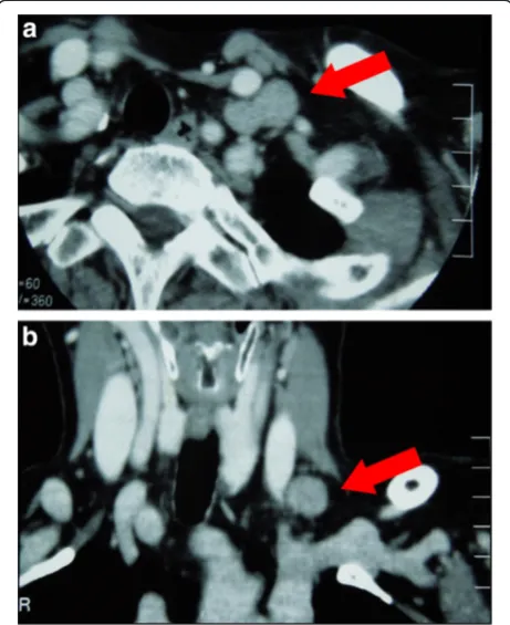
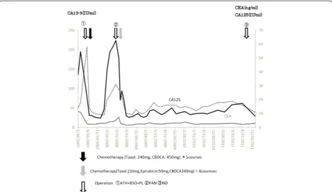
A 74-year-old woman underwent computed tomography (CT) as a follow-up procedure following rectal cancer. In the findings, we detected a left supraclavicular lymph node suspected of metastasis (Figure 1). The patient’s past medical history includes the following: 7 years earl-ier she underwent rectal cancer treatment with low an-terior resection and postoperative chemotherapy; 4 years earlier she underwent a total hysterectomy, bilateral salpingo-oophorectomy and pelvic lymphadenectomy for a stage IIIc endometrial adenocarcinoma (pT1cN1M0). Figure 2 shows the changes of tumor markers (CA19-9, CA125, and CEA) from the time of diagnosis to the present. Subsequent to the operation, the patient * Correspondence:jyokoya@juntendo.ac.jp
Department of Otorhinolaryngology Head and Neck Surgery, Juntendo University Faculty of Medicine, 2-1-1, Hongo, Bunkyo-ku, Tokyo, Japan113-8421
received adjuvant chemotherapy. Approximately 1 year later, the patient underwent para-aortic lymphadenect-omy and additional chemotherapy for the recurrence in the para-aortic lymph node of endometrial adenocarcin-oma. There was no clinical evidence of local or distant recurrence from either of the two malignancies at follow-up.
Fine needle aspiration biopsy of the left supraclavicu-lar lymph node showed adenocarcinoma metastasis. Positron emission tomography-computed tomography (PET-CT) revealed high uptake (SUVmax 6.0) in the left supraclavicular lymph node but no other metastatic lesions elsewhere (Figure 3). Middle and lower jugular neck dissection was performed in 2011 on the grounds that volume reduction would contribute to the improve-ment of prognosis, and residual metastasis was only iden-tified in the left supraclavicular lymph node. A skin incision was made 2 cm above the left clavicle. Some solid lymph nodes could be palpated between the left sternocleidomastoid muscle’s sternal head and clavicular head. The thoracic duct was also resected with the supra-clavicular metastatic lymph nodes (Figure 4a). There was no lymphorrhea from the juglosubclavian angle. To pre-vent chyle fistula, the inferior belly of the omohyoid muscle was cut distally and tightly sutured on the venous angle (Figure 4b). The sternocleidomastoid muscle’s
sternal head was obliterated into the dead space of the venous angle with absorbable sutures. A continuous suc-tion drainage tube was inserted into the supraclavicular space. Surgical time was 49 min and blood loss during surgery was 10 mL. Following the operation the amount of drainage was slight and the drainage tube was removed on the second postoperative day. The patient left hospital on the fifth postoperative day without any complications such as dysfunction of the upper limbs or shoulders.
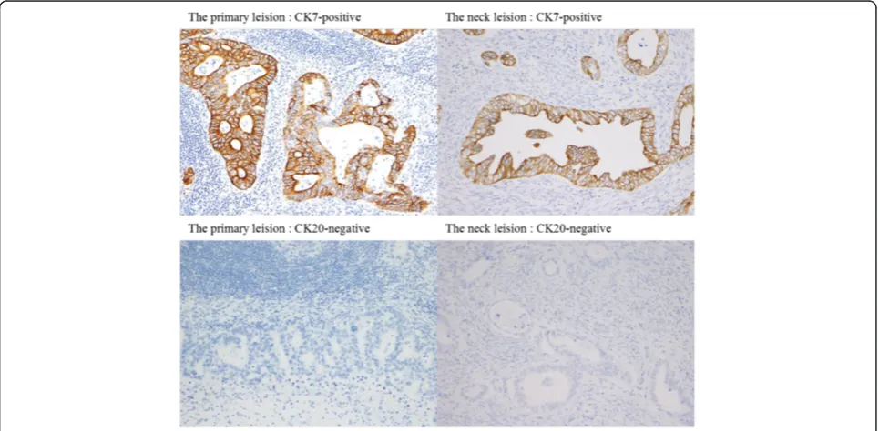
A histological examination showed that there was no metastasis among the six dissected lymph nodes at level III, and only one metastasis was detected among the 11 dissected lymph nodes at level IV. An immunohisto-chemical staining examination using cytokeratin 7 and cytokeratin 20 was performed in order to diagnose the primary disease and determine whether it was rectal cancer or endometrial cancer. The immunohistochem-ical staining examination indicated that the neck metas-tasis originated from endometrial adenocarcinoma [2,3] (Figure 5).
Extracapsular spread was not detected pathologically and consequently the patient received no additional ad-juvant treatment. The patient now lives a normal life without recurrence.
Discussion
Cervical lymph node metastases from genito-urinary neoplasms are rare. Left-sided supraclavicular metastases are predominant because of the anatomy of the lymph-atic system [1,4]. Oosakiet al. reported that there were only five patients (0.15%) with neck metastasis of endo-metrial cancer in a total number of 3,230 patients with neck metastasis of gynecological malignancy [5]. In cer-vical cancer of the uterine, however, supraclavicular lymph nodes sampling has been performed as part of the pretreatment evaluation, especially in cases in which extended field radiation was planned. The overall inci-dence of occult metastasis in the supraclavicular nodes from cervical cancer of uterine was 15.5% (55/359) [6]. When the supraclavicular lymph nodes are involved with metastasis from cervical cancer of the uterine, there is a possibility of metastasis to the para-aortic lymph nodes, lungs, liver, and peritoneal cavity [1,6,7]. The prognosis of supraclavicular lymph nodes metastasis in cervical cancer of the uterine is considered very severe. The 3-year and 5-3-year overall survival rates indicated within several studies were recorded between 0% to 28% [1,7,8]. Once supraclavicular node metastasis is detected, cure is not the objective of treatment, but rather palliative mea-sures for relief of symptoms and improving the patients’ quality of life [1,7,8].
However, there have been very few case reports doc-umenting neck dissection for the treatment of neck Figure 1Preoperative CT findings.The CT showed a single left
metastasis of endometrial cancer. Yoshitake et al. reported supraclavicular lymph node metastasis originat-ing from cancer of uterine body incidentally detected fol-lowing neck dissection carried out during the treatment of a patient with upper gingival cancer [9]. Siddiqet al. also reported on a patient with neck metastasis from la-ryngeal carcinoma, breast carcinoma, and endometrial
carcinoma. A pathological examination from a resected specimen demonstrated a clear cell endometrial carcin-oma originated from endometrial carcincarcin-oma [10]. How-ever, there has been no report of systematic neck dissection for the treatment of neck lesion from endo-metrial cancer before a preoperative diagnosis. Volume reduction surgery for the treatment of recurrent Figure 2Changes of tumor markers (CA19-9, CA125, and CEA).The course of the treatment for endometrial carcinoma and the changes of tumor markers.
endometrial cancer has been considered to be effective. Andersonet al. reported that if the metastasis was loca-lized in the lung, the resection of the pulmonary metasta-sis would improve the prognometasta-sis [11].
In our case, we chose middle and lower jugular neck dissection as a treatment and diagnosis for the supracla-vicular metastasis of endometrial carcinoma after first considering resectability, patient’s age, performance sta-tus, treatment quality of life, hospitalization, post-operative shoulder function, radiation resistibility [12], and drug resistibility [13,14].
A neck dissection method for treating endometrial adenocarcinoma metastasis to the neck has not yet been
established. If there is no extranodal invasion in the neck lymph nodes, we consider middle and lower jugular neck dissection for the apparent metastasis level to be a beneficial therapeutic procedure. Left-sided supraclavi-cular metastasis is predominant and is transferred via the thoracic duct [4]. In our case, supraclavicular lymph nodes metastases were palpable in the single level IV, and so middle and lower jugular neck dissection was performed for levels III and IV.
Postoperative cervical chyle fistula is rare, however once it occurs it is difficult to control after neck dissec-tion. Chyle fistula causes electrolyte and protein loss, wound infection, carotid blowout, chylethorax, and fatal Figure 4Intraoperative findings.(a) Thoracic duct was resected with supraclaviclar metastatic lymph nodes. Each number shows: (1) external jugular vein; (2) resected thoracic duct; (3) internal jugular vein. (b) The inferior belly of the omohyoid muscle was cut distally and tightly sutured on the venous angle. (1) External jugular vein; (2) inferior belly of omohyoid muscle augmented to resected thoracic duct; (3) superior belly of omohyoid muscle.
anoxia. It also delays postoperative treatment such as radiotherapy and chemotherapy [15]. Consequently it is necessary to prevent the chyle fistula intraoperatively. Postoperative chyle fistula occurs at a rate of 1% to 3% in left side radical neck dissections [15]. However, tying the damaged duct closure is not always an easy proced-ure. The wall of the thoracic duct is often extremely thin and over-sewing often increases leakage conversely [16]. As a result, using muscle flaps is reported to be an effective method of preventing chyle fistula [15-17]. In addition, the benefits of surgical closure of leakage with clavicular periosteal flap, or pectoralis major muscle flap used with fibrin glue have been reported [15,16,18].
The technique using the omohyoid muscle flap is straightforward and a reliable procedure for preventing chyle fistula over a short period. By cutting the infer-ior belly of the muscle distally and transferring it to the site of the juglosubclavian angle, the vascularized pedicle muscle belly flap does not result in any nega-tive functional or esthetic consequences for the patient [15].
The most important factor influencing the radiosen-sitivity of endometrial carcinoma is the duration of menopause. The older the patient’s age, the worse the radiosensitivity for endometrial carcinoma [12]. Once platinum- and taxane-based chemotherapy consistently demonstrated the highest response in the treatment of endometrial carcinoma, however, in recurrent cases, the endometrial carcinoma is often platinum = and taxane-resistant [13,14] For this reason, we selected surgical treatment for neck metastasis rather than radiotherapy due to sound control of the disease and sound pathological diagnosis of the disease. If a single neck metastasis after chemotherapy was resectable, we recommend surgical treatment with advantages includ-ing good quality of life, and short hospitalization with-out postoperative complications. However, a standard procedure for neck dissection has not been established for neck metastasis originating from endometrial can-cer. Further clinical studies of the neck dissection for gynecological malignancy including endometrial cancer are required.
Conclusions
The best procedure for carrying out neck dissection for treating neck metastases from endometrial cancer has not been established to date. Middle and lower jugular neck dissection enables pathological diagnosis and improves the patient’s prognosis without any postopera-tive complications. Furthermore, the technique of using the omohyoid muscle flap is very useful for preventing chyle fistula.
Consent
Written informed consent was obtained from the patient for publication of this case report and all accompanying images. A copy of the written consent is available for re-view by the Editor-in-Chief of this journal.
Abbreviations
CT: Computed tomography; PET-CT: Positron emission tomography-computed tomography; SUVmax: Maximum18F-FDG standardized uptake
value.
Competing interests
The authors declare that they have no competing interests.
Acknowledgments
This study was supported in part by Grants-in Aid for Scientific Research from the Ministry of Education, Culture, Sports, and Technology (22591920) of Japan.
Authors’contributions
JY carried out the initial conception and design as well as collection of data and clinical records of the patient. MK participated in its design and helped to edit the manuscript. SI and SO made up the surgical team involved in the case and carried out the initial conception. MF and KI assisted to revise the manuscript. All authors read and approved the final manuscript.
Received: 5 March 2012 Accepted: 12 July 2012 Published: 12 July 2012
References
1. Diddle AW:Carcinoma of the cervix uteri with metastases to the neck. Cancer1972,29:453–455.
2. Oien KA:Pathologic evaluation of unknown primary cancer.Semin Oncol
2009,l36:8–37.
3. Bahrami A, Truong LD, Ro JY:Undifferentiated tumor: true identity by immunohistochemistry.Arch Pathol Lab Med2008,132:326–348. 4. Donohue JP, Zachary JM, Maynard BR:Distribution of nodal metastasis in
non-seminomatous testis cancer.J Urol1982,128:315–320.
5. Oosaki T, Hirono M, Hayashi Y, Yoshihara T, Suzuki M:The therapy for the neck lymphonodi metastasis of gynecologic malignancy.Obstetrics and Gynecology Therapy1996,73:357–360.
6. Vasilev SA, Schlaerth JB:Scalene lymph node sampling in cervical carcinoma: a reappraisal.Gynecol Oncol1990,37:120–124. 7. Qiu JT, Ho KC, Lai CH, Yen TC, Huang YT, Chao A, Chang TC:
Supraclavicular lymph node metastases in cervical cancer.Eur J. Gynaec. Oncol2007,28:33–38.
8. Tran BN, Grigsby PW, Dehdashti F, Herzog TJ, Siegel BA:Occult supraclavicular lymph node metastases identified by FDG-PET in patients with carcinoma of the uterine cervix.Gynecol Oncol2003,
90:572–576.
9. Yoshitake T, Suzuki S, Kagaya M, Shigematsu H, Kusama K, Sakashita H:Case of cervical metastasis suspected to be from cancer of uterine body detected on the neck dissection in a patient with upper gingival cancer. Japanese Journal of Oral and Maxillofacial Surgery2003,49:343–346. 10. Siddiq MA, Bhudia SK, Gana P, Patel PJ:Metastatic endometrial carcinoma
of the neck.J Laryngol Otol2000,114:229–230.
11. Anderson TM, McMahon JJ, Nwogu CE, Pombo MW, Urschel JD, Driscoll DL, Lele SB:Pulmonary resection in metastatic uterine and cervical malignancies.Gynecol Oncol2001,83:472–476.
12. De Mûelenaere GF:The radiosensitivity of endometrial carcinoma.Br J Radiol1975,48:652–655.
13. Smith JA, Gaikwad A, Ramondetta LM, Wolf JK, Brown J:Determination of the mechanism of gemcitabine modulation of cisplatin drug resistance in panel of human endometrial cancer cell lines.Gynecol Oncol2006,
103:518–522.
14. Moxley KM, McMeekin DS:Endometrial carcinoma: a review of chemotherapy, drug resistance, and the search for new agents. Oncologist2010,15:1026–1033.
16. Zhengjiang L, Sabesan T, Pingzhang T, Ilankovan V:Omohyoid muscle flap in prevention of chyle fistula.J Oral Maxillofac Surg2007,65:1430–1432. 17. Crumley RL, Smith JD:Postoperative chylous fistula-Prevention and
management.Laryngoscope1976,86:804–813.
18. Yoshimura Y, Kondoh T:Treatment of chylous fistula with fibrin glue and clavicular periosteal flap.Br J Oral Maxillofac Surg2002,40:138–139.
doi:10.1186/1477-7819-10-143
Cite this article as:Kojimaet al.:Impact of middle and lower jugular neck dissection on supraclavicular lymph node metastasis from endometrial carcinoma.World Journal of Surgical Oncology201210:143.
Submit your next manuscript to BioMed Central and take full advantage of:
• Convenient online submission
• Thorough peer review
• No space constraints or color figure charges
• Immediate publication on acceptance
• Inclusion in PubMed, CAS, Scopus and Google Scholar
• Research which is freely available for redistribution