Reprogramming cell fate to pluripotency:
the decision-making signalling pathways
DANIELA SANGES
1and MARIA-PIA COSMA*
,1,21Center for Genomic Regulation (CRG) and
2Institució Catalana de Recerca i Estudis Avançats (ICREA), Barcelona, Spain
ABSTRACT Pluripotency can be defined as the ability of individual cells to initiate all of the lineages of the mature organism in response to signals from the environment. It has long been assumed that during development, pluripotency is progressively and irreversibly lost through a mechanism that requires strict coordination of the signalling pathways involved in cell prolifera-tion, differentiation and migration. However, recent breakthroughs have highlighted evidence that terminally differentiated cells can be reprogrammed into pluripotent stem cells, prompting a re-evaluation of the reversibility of cell differentiation. Generations of pluripotent cells can arise from somatic cells following ectopic expression of specific transcription factors; however, these factors might well not be the unique essential reprogramming factors. Furthermore, they can be the end-point targets of signalling pathways. Indeed, recent evidence shows that modulation of the Wnt/-catenin, MAPK/ERK, TGF- or PI3K/Akt signalling pathways strikingly enhances somatic-cell reprogramming. Nevertheless, we still know relatively little about the underlying mechanisms by which somatic cells de-differentiate to pluripotency. In this review, we provide an overview of the signalling pathways promoting the re-acquisition and maintenance of pluripo-tency and we discuss the possible mechanisms underlying nuclear reprogramming.
KEY WORDS:
pluripotency, iPS, signalling pathway
Introduction
Pluripotency of mammalian cells is defined as the ability to generate the whole organism, excluding the extra-embryonic tissue. This pluripotent potential is specific for the inner cell mass (ICM), and it is progressively lost during development. Mouse embryonic stem cells (ESCs) derived from the ICM and propa-gated in culture provide an in-vitro model of pluripotent cells: they can self-renew and differentiate into all of the different cell lineages, except the extra-embryonic trophoblast lineage (Rossant, 2008).
It has long been assumed that the differentiation state of a cell is stable and irreversible, and that specialised cells lack the ability to change their identity. However, starting from the 1980s, studies on heterokaryons that are formed by the fusion of two different cell types have demonstrated that although the differentiated state is stable, it is not irreversible and can be swiched to onother in the presence of the appropriate combinations of trans-acting regula-tory molecules (Blau et al., 1983; Wright, 1984; Baron and Maniatis, 1986). In addition, already in 1980s was reported that
BIOLOGY
www.intjdevbiol.com*Address correspondence to: Maria Pia Cosma. Center for Genomic Regulation (CRG), c/ Dr. Aiguader 88, E-08003 Barcelona, Spain. Fax: +34-93-3160 099. e-mail: [email protected]
Final author corrected PDF published online: 14 January 2011.
ISSN: Online 1696-3547, Print 0214-6282
© 2011 UBC Press Printed in Spain
Abbreviations used in this paper: AZA, 5-aza-cytidine; DNMT, DNA methyltransferase; EMT, epithelial to mesenchimal transition; ESC, embryonic stem cell; ICM, inner cell mass; iPSC, induced pluripotent stem cell, MEFs, murine embryonic fibroblasts; MET, mesenchimal to epithelial transition; OSKM, Oct4, Sox2, Klf4 and c-Myc; SAHA, suberoylanilide hydroaxamic acid; TSA, trichostatin A; VPA, valproic acid.
established that transcription factors specific for pluripotency can revert the differentiation state of somatic cells, following a ground-breaking study of Yamanaka and Takahashi in 2006, showing that mouse somatic cells can be converted to ESC-like cells with wide developmental potential: this mechanism was named “repro-gramming to pluripotency” These reprogrammed cells are known as induced pluripotent stem cells (iPSCs), and they were pro-duced using direct transduction of a cocktail composed of only four pluripotent transcription factors: Oct4 (O), Sox2 (S), Klf4 (K) and c-Myc (M) (Takahashi and Yamanaka, 2006).
This study heralded a new fascinating era in stem-cell biology. The iPSC technology has now proven successful for several species starting from different somatic cells, which has opened the amazing prospect of autologous regenerative medicine, whereby patient-specific pluripotent cells can be derived from adult somatic cells. However, our limited knowledge of the global changes during somatic cell de-differentiation still prohibits the use of iPSCs in a clinical setting.
Thus, a better understanding of the molecular mechanisms and the signalling pathways that modulate cell reprogramming is without doubt needed for the translation of this technology to the clinic. In this review, we will discuss about new advances in the
identification of key factors that promote reprogramming, with discussion of the signalling pathways that can be modulated to enhance reprogramming efficiency.
The transcriptional network that regulates
reprogram-ming
Starting from the original discovery of Tahakashi and Yamanaka, who identified the four Oct4, Sox2, Klf4 ans c-Myc (OSKM) factors that can restore pluripotency, additional studies have demon-strated that OSKM are not strickly required and are not the only factors that can induce reprogramming.
Both mouse and human iPSCs have been obtained in the absence of Klf4 and c-Myc transduction (Yu et al., 2007; Nakagawa et al., 2008). Furthermore, reprogrammed human fibroblasts have been generated with the transduction of Oct4, Sox2, Nanog and the RNA-binding protein Lin28, indicating that these last two reprogramming factors can substitute Klf4 and c-Myc (Yu et al., 2007). In addition, the orphan nuclear receptor Esrrb can replace Klf4 in the reprogramming of mouse embryonic fibroblasts (MEFs) when co-transduced with Oct4 and Sox2 (Feng et al., 2009). Esrrb is required for maintenance of self-renewal and pluripotency of ESCs (Li et al., 2010), and most importantly, it regulates the expression of Klf4, explaining its ability to replace this transcrip-tion factor in the reprogramming processes (Feng et al., 2009) (Fig. 1).
The key role of Oct4 and Sox2 is well explained by their functions as transcriptional activators that enhance the expres-sion of the genes that maintain pluripotency, including them-selves: mouse embryos lacking either Oct4 or Sox2 do not form the epiblast (Masui et al., 2007). Moreover, Oct4 and Sox2 act also as repressors in the down-regulation of lineage-specific genes. However, it has been shown that cells with high endog-enous Sox2 levels, such as neural progenitor cells and mouse or human primary melanocytes, can be reprogrammed with just Oct4 expression(Kim, LB et al., 2009; Eminli et al., 2008; Silva et al., 2008; Utikal et al., 2009a). In addition, Sox2 is not required for reprogramming by fusion of mouse ESCs and human B lympho-cytes (Pereira et al., 2008) (Fig. 1).
Surprisingly, recently it was reported that the orphan nuclear receptor Nr5a2 (also known as Lrh-1) can replace Oct4 in the production of iPSCs from mouse somatic cells, and that Nr5a2 can also enhance reprogramming efficiency. Because Nr5a2 acts in part through the activation of Nanog, it appears that unrelated transcription factors with similar functions could indeed substitute Oct4 in the reprogramming processes (Heng et al., 2010), casting doubt on the fundamental role of Oct4 in iPSC generation (Fig. 1). Nanog is another important player in pluripotency. Although for transcription factor-induced reprogramming Nanog is initially not required, it does become essential for dedifferentiated intermedi-ates to pass on to the ground-state to pluripotency (Silva et al., 2009).
In addition to induce reprogramming to the pluripotent state a few factors were also recently showed to induce lineage repro-gramming. The expression of only one factor, C/EBPalpha, can directly convert B cells into macrophage-like cells at 100% effi-ciency (Bussmann et al., 2009). Furthermore, a recent discovery showed that the combination of only three factors, Ascl1, Brn2 (also called Pou3f2) and Mytl1, is sufficient to efficiently convert Fig. 1. Replacement of the core reprogramming factors. Over the last
fibroblasts into functional neurons in vitro (Vierbuchen et al., 2010).
Within the complex gene network that regulates cellular iden-tity, microRNAs (miRNAs) have important roles as post-transcrip-tional modulators of gene expression in the maintenance of ESCs (Ivey et al., 2008). In most cases, the functions of miRNAs in ESC physiology remain unknown, but the existence of a subset of miRNAs that are exclusively expressed in ESCs and, most importantly, the failure to generate viable ESCs from mice defi-cient for Dicer (Bernstein et al., 2006), the enzyme required for miRNA processing, suggest an important role for miRNAs in ESC self-renewal. In contrast with this study, it has also been shown that the viability of ESCs was not altered after the conditional targeting of Dicer-1 gene; even if cells showed defects in the differentiation process (Kanellopoulou et al., 2005). These results indicated that rather than a role in the manteinance of pluripotency, the miRNA machinery could have a role in the control of differen-tiation potential of ESCs.
Despite these controversial reports, regulators of miRNA bio-genesis have been shown to be among the handful of factors that can convert differentiated cells into iPSCs. This is the case for the RNA-binding protein Lin28 that can, as mentioned above, effi-ciently convert fibroblasts into iPSCs when transduced together with Oct4, Sox2 and Nanog (Yu et al., 2007). Indeed, Lin28 is a well-known negative modulator of the let-7 miRNA family (Newman et al., 2008; Viswanathan et al., 2008), which are expressed at low levels in ESCs and which are rapidly induced upon differentiation (Yu et al., 2007; Kumar et al., 2008). Moreover, Lin28 expression is induced by c-Myc in multiple human and mouse tumour models (Chang et al., 2009); all of these evidences suggest that Lin28 has a central role in blocking miRNA-mediated differentiation in stem cells, as well as in the replacement of c-Myc in the induction of pluripotency during iPSC formation.
Although the high numbers of miRNAs expressed in ESCs, to date, a role in reprogramming has been shown for only a few of these. The miRNA miR-290 cluster constitutes over 70% of the entire miRNA population in mouse ESCs, and its expression is rapidly down-regulated upon ESC differentiation (Marson et al., 2008b). Some miRNAs that belong to this cluster have been tested for roles in reprogramming. In particular, 291-3p, miR-294 and miR-295 have been shown to increase the efficiency of reprogramming by Oct4, Sox2 and Klf4 when transfected into MEFs, establishing the important role of miRNAs in these repro-gramming processes (Judson et al., 2009). MiR-302 also has a role in the complex network that regulates the acquisition of pluripotency. It is expressed most abundantly in slow-growing human ESCs, and its levels quickly decrease after cell differentia-tion; most importantly, MiR-302 can convert human cancer cell lines into ESC-like cells. These transfected cells, which are known as miRNA-induced pluripotent stem cells, have been characterized for the expression of pluripotent genes, such as Oct4, SSEA1 and others, and for the global demethylation state of the genome, which was similar to a reprogrammed zygotic genome (Lin et al., 2008).
In 2009, Xu and colleagues demonstrated an important role for miR-145 in repression of the 3'-untranslated regions of Oct4, Sox2 and Klf4. Loss of miR-145 impairs differentiation and in-creases the expression of the most important reprogramming factors, suggesting its potential for the production of iPSCs (Xu et
al., 2009). Also, it has been reported that miR-145 can target and disrupt regulators of the cell-cycle and cell-proliferation path-ways, such as CDK6, a well-known player in G1/S cell-cycle transition. Thus, the ability to re-enter the cell cycle might be important during de-differentiation of iPSCs.
One of the main barriers that have to be overcome to obtain fully reprogrammed clones is the loss of replicative potential that occurs during cell senescence. Under normal conditions, senes-cence results in an irreversible arrest during G1 transition of the cell cycle that can be elicited by replicative exhaustion or in response to stress, such as DNA damage or aberrant expression of oncogenes. This arrest is implemented primarily through acti-vation of p53 and up-regulation of the cyclin-dependent kinase (CDK) inhibitors p16INK4 and p21CIP1 (Efeyan et al., 2007). It has been reported that the first phase of the reprogramming pro-cesses triggers a stress response that has characteristics of senescence and that acts as an initial barrier to limit the efficiency of these processes (Banito et al., 2009).
The existence of such senescence barrier that limits the efficiency of a successful reprogramming process has been studied by examining the reprogramming abilities of TERT-immortalised human keratinocytes. Up-regulation of TERT, the gene that encodes the enzymatic subunit of telomerase, is impor-tant to avoid telomere shortening that is specific for cell senes-cence. TERT-keratinocytes gave rise to iPSC-like colonies about 20 times more efficiently than the early passage cultures of the primary keratinocyte line from which they were derived (Utikal et al., 2009b). Furthermore, transduction of hTERT together with Oct4 and Sox2 strongly enhanced reprogramming potential (Park et al., 2008).
P53 has been proposed to antagonise reprogramming through its ability to limit cell cycle by the induction of the cyclin-dependent kinase inhibitor p21, and through its ability to induce apoptosis after cell stress (Hoffman and Liebermann, 2008). It was shown that down-regulation of the p53 gene or reductions in down-stream factors such as p21 significantly increased the reprogram-ming efficiency of human somatic cells (Hong et al., 2009; Li et al., 2009; Kawamura et al., 2009; Utikal et al., 2009b; Marion et al., 2009). However, despite the importance of these results, the role of p53 in the reprogramming process is not yet fully understood. Previous studies had shown that p53 represses Nanog in re-sponse to DNA damage in ESCs (Lin et al., 2005); thus, it has been postulated that p53 prevents Nanog expression in somatic cells that cannot be fully reprogrammed. On the other hand, down-regulation of p53 appears to allow Oct4 and Sox2 to remodel chromatin to a threshold required for expression of sufficient Nanog to drive the subsequent events involved in iPSC generation (Kawamura et al., 2009). Furthermore, p53 promotes maturation of pre-miR-145 to mature miR-145, so p53-mediated miR-145 down-regulation might also allow transcription of endog-enous Oct4, Sox2 and Klf4, which are essential for self-renewal (Suzuki et al., 2009).
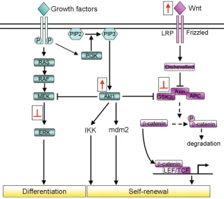
Fig. 2. Modulation of the signalling pathways to enhance iPSC technology. In ESCs, there is a complex network of signalling pathways that can respond to extracellular stimuli that strictly control the equilibrium between self-renewal and differen-tiation. These provide the positive and negative regulation pathways for ESC pluripotency. These intracellular signalling pathways can be activated (red arrows) or inhibited (red blunt arrows) in order to improve the efficiency of somatic cell reprogram-ming. For instance, the Wnt/-catenin pathway is known to have an important role in maintenance of ESC self-renewal. Interestingly, it has been demon-strated that activation of this signalling through Wnt or inhibition of GSK3 strongly enhances the repro-gramming efficiency in both direct reprorepro-gramming and in cell-fusion-mediated reprogramming. Also, stimulation of PI3K/Akt signalling is known to im-prove the reprogramming processes in MEFs after cell fusion with ESCs. In contrast, the pathways that normally stimulate ESC differentiation are usually inhibited for enhancement of reprogramming effi-ciency. For example, inhibition of the ERK pathway that normally influence ESC differentiation, pro-motes completion of the reprogramming processes in mouse somatic cell.
p21 gene is known to control the cell cycle progression. Thus, the modulation of p53 might not enhance reprogramming efficiency but might accelerate the process as cells divide more rapidly increasing the probability that some stochastic events occur earlier in time (Hanna et al., 2009).
Finally, P53-deficient iPSCs can give rise to germline-transmit-ting chimaeric mice and adult tissues when implanted into mouse embryos (Hong et al., 2009; Li et al., 2009; Kawamura et al., 2009). However, even if mice can in some cases be generated from iPSCs, they eventually develop tumours (Hong et al., 2009). Indeed, Marión and colleagues (2009) showed that p53-deficient iPSCs are genomically unstable and are not efficient for the production of mice (Marion et al., 2009). As p53 inactivation promotes genome instability and cancer, this represents a barrier for the use in a clinical setting of p53-deficient-iPSCs that have been generated. Further studies aimed at developing new meth-odologies for transient p53 inhibition using chemical antagonists or reversible approaches will be required to safely translate this p53 potential into de-differentiation of somatic cells in therapeutic strategies (Krizhanovsky and Lowe, 2009).
Controlled activation of the Wnt/-catenin signalling path-way enhances somatic-cell reprogramming
An understanding of the molecular signalling that controls pluripotency or that stimulates differentiation started when ESCs were isolated and cultured in vitro, many years before the first iPSCs were generated. Thus, studies aimed at identifying small molecules that can maintain ESCs in an undifferentiated state in culture will be useful to improve iPSC technology. Conditional medium for ESC in vitro cultures has historically supplied with the cytokine leukaemia inhibitory factor (LIF), which upon binding to
its receptor activates the Janus tyrosine kinase (JAK), and in turn, STAT3, which acts as a transcriptional factor in the sustaining of pluripotency in mouse ESCs. In human and monkey ESCs, LIF cannot promote self-renewal, because of the low expression of their signalling components and the high levels of the suppres-sors of cytokine signalling (Wei et al., 2005). However, it has also been shown that the STAT3 signalling pathway can be activated in hESCs in response to LIF treatment, but the activation level is lower as compared to that observed in mESCs and it can not maintain self-renewal (Sato et al., 2004). On the other hand, it is also reported that hESCs correspond more to pluripotent cells derived from the post-impantation epiblast of murine embryo (mEpiSCs) rather then to mESCs that are molecularly and epige-netically different (Vallier et al., 2009). This might well be the reason why hESCs respond differently to the LIF/STAT3 path-way.
Instead, it is well supported that human ESCs can be sustained in an undifferentiated state by the activation of Wnt signalling, which maintains the expression of Oct4, Rex1 and Nanog through the action of -catenin, an intracellular signalling molecule that is part of the canonical Wnt signalling pathway (Sato et al., 2004). In the absence of Wnt activation, -catenin is phosphorylated by a complex that consists of the adenomatous polyposis coli (APC) protein, Axin and glycogen synthase kinase (GSK)3, and it is rapidly degradated by the ubiquitin-proteasome system. Upon Wnt binding with the receptors Frizzled and LRP5/6, GSK3 is inhibited; as a result, -catenin accumulates in the nucleus, where it binds the lymphoid enhancer factor (LEF)/ T-cell factor (TCF) transcription factors (Hoppler and Kavanagh, 2007) (Fig. 2).
somatic-cell reprogramming after cell fusion (Lluis et al., 2008) (Table 1). The role of Wnt signalling in ESCs and reprogramming processes is puzzling since high levels of Wnt activity in GSK3 double-knockout cells were shown to lead to differentiation of ESCs (Ying et al., 2008). Cell-fusion experiments also showed a dual activity for Wnt signalling in promoting or inhibiting pluripo-tency, whereby only a modest increase in intracellular levels of -catenin in ESCs resulted in the greatest benefits for somatic-cell reprogramming after cell fusion. In contrast, ESCs with high levels of -catenin, due to its over-expression or to knock-out of GSK3, showed a block of these reprogramming events. In addition, only ESCs treated for 24 h and 96 h with Wnt3a showed an increased ability to induce reprogramming; on the contrary, Wnt3a treat-ment of ESCs for 48 or 72h did not have the same effect because
-catenin did not accumulate into the nuclei at a sufficient level (Lluis et al., 2008). These observations indicated that the timing and levels of Wnt-signalling pathway activation can result in a specific -catenin accumulation threshold that has a crucial role in the induction of reprogramming after cell fusion (Fig. 3). Interestingly, negative modulators, such as Axin2 and DKK1, are up-regulated after Wnt pathway activation, and they can promote
-catenin degradation; this further suggests controlled regulation in terms of the timing and activation levels of this pathway (Lluis et al., 2008; Lluis and Cosma, 2009).
However, the reasons why Wnt signalling stimulates repro-gramming remain unclear, and what appeared to be the most likely downstream “reprogrammer” candidates have already been excluded (Marrill, 2008). For example, c-Myc is one of the down-stream regulators of the Wnt pathway (He et al., 1998; Cole and Cowling, 2008): transduction of OSK in MEFs cultured in the presence of Wnt3a containing medium results in the increase in colony formation with respect to OSK-mediated reprogramming
without Wnt3a, which suggests that the effects of Wnt3a is in part to activate endogenous c-Myc directly, thereby substituting for exogenous c-Myc (Marson et al., 2008a).
However, in both iPSCs and cell fusion-mediated reprogram-ming experiments, Wnt3a treatment did not increase c-Myc expression, suggesting that c-Myc is not a Wnt-dependent repro-gramming factor. Also Nanog, which is a -catenin/ TCF target (Pereira et al., 2006) has been shown to enhance cell-fusion-mediated reprogramming (Silva et al., 2009), although it was not up-regulated in response to Wnt3a treatment in cell-fusion-medi-ated reprogramming experiments. Another putative “reprogrammer” activated by Wnt signalling might be Tcf3, which shows a high level of colocalisation with Oct4, Sox2 and Nanog on developmental gene promoters that regulate the balance be-tween pluripotency and differentiation (Cole and Cowling, 2008; Tam et al., 2008; Yi et al., 2008); but, if Tcf3 has a role in reprogramming remains to be elucidated. In cell-fusion and Wnt-mediated reprogramming experiments, the well-known reprogrammer genes such as Oct4, c-Myc and Nanog have never been seen to be up-regulated (Lluis et al., 2008); however, it cannot be excluded that upon activation of Wnt signalling some recruiting molecules or factors facilitate the binding of these pluripotent transcriptional factors to their target promoters, thus enhancing the global transcriptional changes that allow repro-gramming.
Inhibition of the MEK and GSK3 pathways enhances
somatic-cell reprogramming
GSK3 is an important component of Wnt signalling, and to-gether with the MEK-MAPK/ ERK pathway, is considered a fundamental player in the intracellular signalling that controls
Chemicals Function Methods Cell type Effects on reprogramming References
BIX-01294 G9a histone methyltransferase inhibitor
OK Mouse fibroblasts 5-fold increase in the efficiency Replaces S
Shi et al., 2008a
Mouse NPCs
1.5-fold increase in the efficiency Replaces S
Shi et al., 2008b
BayK8644 L-type calcium agonist OK Mouse fibroblasts 15 fold increase in the efficiency with BIX-01294 Shi et al., 2008b RG108 DNMT inhibitor OK Mouse fibroblasts 15 fold increase in the efficiency with BIX-01294 and
BayK8644
Shi et al., 2008a
AZA DNMT inhibitor OSKM Mouse fibroblasts Promotes full reprogramming with a 4-10 fold increase in the efficiency
Mikkelsen et al., 2008 Huangfu et al., 2008a
VPA HDAC inhibitor OSKM Mouse fibroblasts 100-fold increase in the efficiency Huangfu et al., 2008a OSK 50-fold increase in the efficiency
OSK Human fibroblasts 10 to 20-fold increase in the efficiency Huangfu et al., 2008b
OS Replaces K and M
TSA HDAC inhibitor OSKM Mouse fibroblasts 15-fold increase in the efficiency Huangfu et al., 2008a
SAHA HDAC inhibitor OSKM Mouse fibroblasts 2-fold increase in the efficiency Huangfu et al., 2008a PD0325901+
CHRI99021 (2i)
Inhibitors of MEK and GSK3 OK Mouse NSCs and NPCs Promote transformation of pre-iPS into fully reprogrammed iPS
Silva et al., 2008a Silva et al., 2008b
A-83-01 TGFβ inhibitor OSK Human fibroblasts and rat liver progenitors
Maintains rat iPSCs with LIF and 2i Li et al., 2009b
EMD616452 TGFβ inhibitor OSKM MEFs 5-fold increase in the efficiency Replaces K or M
Maherali et al., 2009
Wnt3a or GSK3 inhibitor (BIO)
Activation of Wnt/beta-catenin pathway
Cell fusion with ESCs Mouse NSCs 80-fold increase in the efficiency Lluis et al., 2008a
MEFs 15-fold increase in the efficiency
Wnt c.m. Activation of Wnt/beta-catenin pathway
OSK(dox) inducible Mouse fibroblasts 20-fold increase in the efficiency Marson et al., 2008b
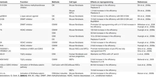
TABLE 1
CHEMICALS THAT ENHANCE REPROGRAMMING EFFICIENCY OR REPLACE REPROGRAMMING FACTORS
ESC pluripotency. Upon explant culture, the epiblast rapidly loses pluripotency and differentiates under the influence of ERK signal-ling (Buehr et al., 2003; Buehr and Smith, 2003). Furthermore, it has been reported that ERK signalling triggers differentiation of pluripotent ESCs to lineage commitment (Kunath et al., 2007) (Fig. 2). In contrast, reducing ERK activity with the inhibitor PD98059 has been shown to enhance the efficiency of ESC production by promoting retention of the Oct4-positive epiblast during the outgrowth phase (Buehr et al., 2003).
Similarly, inhibition of GSK3 activity (Sato et al., 2004) might also maintain ESC self-renewal, and inhibition of both the MEK and GSK3 pathways has been used for the production and maintenance of rat ESCs, which can form embryo bodies that contribute to chimaeras, and are germ-line competent (Buehr et al., 2008). Translating this knowledge into an improvement in iPSC technology, it was demonstrated that dual inhibition of MEK and GSK3 (2i) promoted transformation of pre-iPSCs into ground-state pluripotent cells (Silva et al., 2008) (Table 1 and Fig. 2). Indeed, combination of the MEK inhibitor PD0325901 and the GSK3 inhibitor CHIR99021 with LIF (2i/LIF) efficiently induced fully competent iPSCs from both MEFs and neural stem cell (NSC)-derived pre-iPSC clones. This study demonstrated that the 2i/LIF-medium induced transition to pluripotency, rather than selecting the rare cells that had already reached that state. It has
been postulated that this treatment induces a transcriptional and epigenetic resetting of cells that rapidly culminates in the full pluripotent status with phenotypic and functional properties that are indistinguishable from ESCs (Silva et al., 2008). However, there are some differences in the effects of 2i/LIF treatment in the MEF and NSC reprogramming efficiencies that are based on the different kinetics of the changes associated with de-differentia-tion, which appear to be faster in NSCs than in MEFs. The study indicated that NSCs might have fewer epigenetic restrictions and thus respond better to reprogramming signals than other cells. Even if the use of the 2i/LIF medium can improve iPSC technol-ogy, further studies are needed to limit the timing of the MEK inhibitor treatment: it has to be applied with care, as MEK is also required for somatic-cell survival (Roux and Blenis, 2004; Silva et al., 2008).
Role of TGF
signalling in reprogramming
The low efficiency of reprogramming technology and the ne-cessity to develop an efficient non-genetic resource of factor delivery for therapeutic use of iPSCs has prompted studies aimed at identifying more compounds that can enhance, or replace, the functions of reprogramming factors. High-content small molecule screening has helped to identify compounds that can replace, or
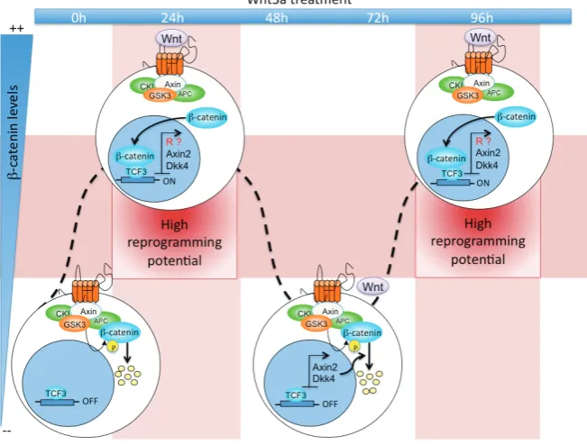
Fig. 3. Timing and level of Wnt signalling activation control somatic cell reprogram-ming. The scheme represents the periodic accumulation of -catenin in ESCs after Wnt3a
treatment. In untreated cells (0h), Wnt signalling is not activated and -catenin is rapidly degradated by the destruction complex: thus, the nuclear factor Tcf3 inhibits the expression of target genes. At 24 and 96 hours after Wnt3a treatment, the distruction complex is inactivated and -catenin can translocate into the nucleus and activates several target genes including putative reprogrammers (R) as well as Axin2 and Dkk4 that are a part of a negative feedback loop to the signal. At 48 and 72 hours, levels of Axin2 and Dkk4 become high, -catenin is degradated and reprogramming potential is inhibited. In addition, high levels of -catenin do not allow reprogramming, possibly because checkpoint control mechanisms are activated and act as negative regulators.
in some way compensate for, the activities of each of the transgenic factors. From these approaches, the important role of the highly characterized transforming growth factor
(Tgf) signalling in reprogramming processes has emerged. It is well known that the Tgf
ephitelial junctional components and morphological transforma-tion into ephitelial-like colonies, seems to be a crucial step in the reprogramming process. It was suggested that BMP signal synergized with the Yamanaka factors to induce the expression of miR-200 family, that are well known to promote MET and repro-gramming, also in absence of BMP signalling (Bracken et al., 2008; Gregory et al., 2008; Korpal et al., 2008) and to inhibit loss of pluripotency of mESCs in differentiating conditions (Lin et al., 2009).
In 2009, Maherali and colleagues showed that inhibition of Tgf receptor I kinase with an activin-like kinase 5 (Alk5) inhibitor enhanced both the efficiency and kinetics of the MEFs repro-grammed by OSKM, whereas activation of the Tgf signalling pathway blocked reprogramming (Maherali et al., 2007). As Alk5 inhibitor treatment has its strongest effects during the early stages of iPSC production, it was assumed that it acts in concert with the reprogramming factors, rather than promoting the conversion of fibroblasts to a state that is more prone to reprogramming. In support of this theory, it has been demonstrated that Alk5 inhibitor treatment can also replace the individual role of c-Myc or Sox2, even if it cannot replace them simultaneously (Maherali et al., 2007); this suggests that Alk5 acts on a pathway that completely bypasses the need for these individual reprogramming factors. On the other hand, it was demonstrated that Tgf inhibition promotes the completion of reprogramming through induction of the transcription factor Nanog (Ichida et al., 2009). Finally, be-cause Tgf is a potent inducer of ephitelial to mesenchimal transition (Zeisberg and Kalluri, 2004), it is also possible that inhibition of the pathway enhances reprogramming by promoting MET. In conclution, further studies are needed to define the role of this pathway in the enhancement of iPSC generation.
In contrast to its action on mouse ESCs, activation of the BMP pathway induces differentiation of human ESCs toward trophoectoderm (Xu et al., 2002). Indeed, an increase in effi-ciency of reprogramming was not seen in treatment of human fibroblasts, even if the use of a Tgf inhibitor (A-83-01) with the GSK3 and MEK inhibitors facilitated rat and human iPSC propa-gation and supported the mouse ESC-like phenotype (Table 1). Of note, Alk5 inhibitor treatment of mESCs did not improve reprogramming processes after ESC fusion with MEFs (Maherali and Hochedlinger, 2009). This discrepancy suggests that Tgf
inhibition acts only on direct reprogramming pathways that are already functioning in ESCs.
Finally, on the other hand, in the Tgf family signalling net-works, the Nodal signal plays an important role in the mantainance of pluripotency of hESCs (James et al., 2005). However, the role of Nodal-activin pathway in the enhancement of reprogramming mechanims has to be elucidated yet.
Activation of the PI3K/Akt signalling pathway:
repro-gramming enhancement or inhibition?
Phosphoinositide 3-kinases (PI3Ks) are lipid kinases that promote the generation of the signalling lipid phosphatidylinositol 3,4,5-trisphosphate upon activation by many different growth factor receptor tyrosine kinases, such as FGF, EGF and PDGF. This in turn regulates a complex signalling cascade. One of the main players in this pathway is Akt1, a serine/ threonine kinase that modulates the functions of numerous substrates, such as
Mdm2 and IKK, and elicits various cellular responses, including cell proliferation, adhesion, growth and death, as well as promot-ing tumorigenesis (Cantley, 2002) (Fig. 2). Activation of Akt1 signalling is sufficient to maintain the pluripotency of ESCs without LIF and feeder cells (Sun et al., 1999; Watanabe et al., 2006); on the contrary, treatment of ESCs with the PI3K inhibitor LY294002 results in a loss of ESC features even in the presence of LIF (Liu et al., 2009). Moreover, activation of PI3K signalling is crucial to the promotion of de-differentiation of embryonic germ cells from primordial germ cells (Kimura et al., 2008).
Despite our wide knowledge of the role of the PI3K pathway in ESC biology, its role in reprogramming remains elusive. Nakamura et al. (2008) showed that activation of Akt signalling stimulated reprogramming after fusion of ESCs with thymocytes or MEFs, which led to the formation of ESC-like hybrid cells. In contrast, Akt signalling significantly reduced the efficiency of reprogramming via somatic cell nuclear transfer: cloned embryos injected with the mRNA of an active form of Akt were arrested at the transition from the two-cell to eight-cell stage (Nakamura et al., 2008). The positive effects on cell-fusion-mediated reprogramming can be attributed to GSK3, the activity of which is inhibited by Akt-mediated phosphorylation (Sato et al., 2004). Furthermore, the evidence that PI3K/Akt signalling induces phosphorylation of histone methyltransferase enhancer of Zeste homolog 2 (EZH2), which reduces the levels of trimethylation of lysine 27 on histone H3, might explain how this pathway modulates global epigenetic changes during the reprogramming processes (Bredfeldt et al., 2010). However, the reasons why Akt signalling has a negative effect in reprogramming via nuclear transfer remain unclear. In contrast to the report by Nakamura and colleagues (Nakamura et al., 2008), it has been shown that microinjection of the mRNA of an active mutant of Akt into fertilised mouse eggs did not induce developmental arrest; instead, it promoted cell division and sur-vival (Feng et al., 2007). Thus, the different activities of Akt signalling in cell-fusion-mediated and nuclear-transfer-mediated cell reprogramming suggest that there are different sets of Akt downstream effectors that are involved in enhancement and inhibition of nuclear reprogramming.
Defining pluripotency in the reprogramming processes
Currently, the expression of immature-related surface anti-gens that are specific for pluripotent cells, such as alkaline phosphatase, TRA-1-60 and SSEA1 for the mouse, or SSEA3 and SSEA4 in human systems (Brambrink et al., 2008; Chan et al., 2010), is often evaluated to characterize a reprogrammed clone. However, the definition of fully reprogrammed cells based only on these early markers has promoted continuous debate due to their expression in clones that do not show reactivation of endogenous pluripotent genes.
To define an ES-like iPSC, it is also necessary to demonstrate acquisition of the same developmental potential that is specific for ESCs. In-vitro differentiation is commonly induced in cultured cells to assess the expression of different cell-type markers, although this is not considered a stringent measure of pluripotency since the set of differentiation markers used in most studies is often insufficient for the conclusion that a cell has been converted into a new state of differentiation and cellular function (Jaenisch and Young, 2008). To be considered indistinguishable from ESCs, iPSCs should generate post-natal chimaera, contribute to the germline (Maherali et al., 2007; Okita et al., 2007), and eventually generate a late-gestation embryo through tetraploid complementation (Wernig et al., 2007).
Based on these criteria, there is evidence that in all of the studies that have been performed to date to obtain iPSCs, not all of the cases have led to complete restoration of pluripotency. For example, Takahashi and Yamanaka (Takahashi and Yamanaka, 2006) reported that iPSCs selected by the reactivation of Fbx15, an Oct4 target gene, formed teratomas, but did not form chimae-ras. Molecular analysis of these clones revealed that the mainte-nance of pluripotency was in reality due to the viral expression of the transduced Oct4 and Sox2 genes, whereas these endog-enous genes were not reactivated, as demonstrated also by the high methylation state of their promoters (Masui et al., 2007). This appears to limit the potential of clones that are not fully repro-grammed in the development of therapeutic strategies; however, this evidence is fundamental to determine the still unknown molecular mechanisms that regulate somatic cell de-differentia-tion, and specifically trans-gene silencing. Indeed, the observa-tions of Takahashi and Yamanaka (Takahashi and Yamanaka, 2006) suggest that Fbx15-iPSCs were only partially reprogrammed, indicating that there exists a complex gene network along with the epigenetic events, which regulates the kinetics of the appearance of pluripotent markers and, in turn, the completion of the repro-gramming processes.
Epigenetic modulations during somatic
de-differentia-tion can overcome reprogramming barriers
Transcriptional effects induced by the OSKM reprogramming factors have been detected in transduced MEFs as early as day 4 after infection; however, only a few of the cells reactivated endogenous Nanog expression after 16 days (Mikkelsen et al., 2008). This indicates that the majority of the infected cells were trapped in a partially reprogrammed state due to their inability to overcome some reprogramming barriers. The common interme-diate state is represented by a population of partially repro-grammed cells (pre-iPSCs) that is characterized by down-regula-tion of somatic genes, incomplete reactivadown-regula-tion of pluripotent genes, maintenance of viral transgene expression and inability to
form chimaeras (Silva et al., 2008). Moreover, pre-iPSCs show incomplete genetic remodelling and persistent DNA hypermethylation (Mikkelsen et al., 2008; Sridharan et al., 2009). This suggests that as well as changes in the epigenetic state of the somatic genome, DNA methylation is an important barrier that has to be overcome to obtain fully reprogrammed colonies (Fig. 4).
Interestingly, Buthani and collegues identified an immune-system protein known as activation-induced cytidine deaminase (AID), which improves cell-fusion-mediated reprogramming through its involvement in DNA demethylation, that in turn is required for induction of Oct4 and Nanog (Bhutani et al., 2010). In the same Nature issue, Popp and collegues reported that AID is important for complete cell reprogramming in mammals, because of its ability to erase genome-wide DNA methylation in mouse primordial germ cells (Popp et al., 2010). Together, these findings have provided new insights into reprogramming processes, iden-tifying a specific role of AID in the reversion of cell fate (Deng, 2010; Agarwal and Daley, 2010).
The role of DNA demethylation in reprogramming processes was, however, already assumed from different studies that showed that treatment with the DNA methyltransferase (DNMT) inhibitor 5-aza-cytidine (AZA) converted partially reprogrammed cell lines into iPSCs through the global inhibition of DNA methylation. This thus produced fully reprogrammed clones that showed demethylation of the promoters of the pluripotent genes, and that formed teratoma. Furthermore, treatment with AZA promoted a 4-fold increase in the number of ESC-like colonies (Mikkelsen et al., 2008) (Table 1).
Histone H3 and H4 acetylation also appear to be important during iPSC generation. The use of histone deacetylase inhibi-tors, such as valproic acid (VPA), trichostatin A (TSA) and suberoylanilide hydroaxamic acid (SAHA), significantly enhanced reprogramming efficiency (Huangfu et al., 2008a) (Table 1). Not only did VPA treatment increased ESC-like colonies by more than 100-fold with three-factors (OSK) and by 50-fold with the four factors (OSKM), but treatment with both AZA and VPA induced colonies 2 days earlier than in the untreated controls, indicating that these treatments can improve both the kinetics and the efficiency of reprogramming processes (Huangfu et al., 2008b). Indeed the global transcriptional changes and histone acetylation due to the VPA treatment, allowed reprogramming of fibroblasts with two factors (OS) with the same efficiency as with three-factors (OSK) (Huangfu et al., 2008b).
damage (Palii et al., 2008); therefore, careful tests need to be performed to exclude permanent genomic or epigenomic alter-ations after transient treatments with such agents.
A model that integrates signalling pathways and
mo-lecular mechanisms that control somatic cell
repro-gramming: the possible sequence of events
Although much progress in iPSC-induction technology has been made since the Yamanaka and Takahashi landmark study in 2006, many of the molecular mechanisms that underlie repro-gramming still remain elusive. In addition, putative interconnec-tions between different players known to be involved in these processes remain obscure. As has been predicted according to an “elite model” for the de-differentiation process, it is possible that only a small number of treated somatic cells are susceptible to reprogramming (Yamanaka, 2009). However, different lines of evidence contradict this predetermined elite model. A more ac-cepted stochastic model has predicted that most differentiated cells have the potential to become iPSCs, but sequential stochas-tic events appear to be important in these processes and might contribute to the low overall efficiency of iPSC generation. One of the up-and-coming ideas is that ectopic expression of the Yamanaka cocktail of transcription factors (OSKM) triggers a sequence of epigenetic events that includes changes in DNA methylation and chromatin modifications; these could eventually result in a pluripotent state of some infected cells but not of others (Meissner et al., 2007). This suggests that the kinetics of these
processes and a large number of intracellular signalling events strongly modulate the frequency and completion of the mecha-nisms that form a fully reprogrammed clone. Virally delivered reprogramming factors might serve as triggers to initiate the processes that activate expression of other pluripotency-promot-ing genes (Loh et al., 2006) and that recruit other transcription factors and chromatin modifiers to induce more stable and global changes (Fig. 4).
The reason that some pluripotent markers, such as Fbx15, alkaline phosphatase and SSEA1, start to be expressed in the early stages of the reprogramming processes before endogenous Oct4 or Nanog are reactivated might be because of a more accessible chromatin state of the gene location, even in differen-tiated cells. Concomitant with expression of the first markers, exogenous Oct4 and Sox2, as well as Nanog, start to repress not only developmental genes (by targeting their promoters), but also increase the efficiency of the global repression, by induction of several repressive epigenetic enzymes (such as the NuRD and Setdb1 complexes) (Fig. 4).
It is well known that core transcription factors in ESCs co-regulate expression of epigenetic factors that participate in main-tenance of self-renewal and pluripotency. For example, Oct4 and Sox2 both bind genes that encode chromatin-remodelling factors, such as Smarcad1, Myst3, Jmjd2c and Jmjd1a (Loh et al., 2006; Loh et al., 2007), which leads to a general unfolding of chromatin. Interestingly, the histone H3 lysine 9 demethylase Jmjd1a has been shown to enhance the reprogramming of NSCs after fusion of cells with ESCs (Ma et al., 2008). Changes in the global
chromatin state that are necessary for full reprogramming can also arise from recruitment of the repressive Polycomb proteins, the expression of which is induced by Oct4 and Sox2. Interestingly, in this scenario, the effects of the PI3K pathway on phosphorylation of EZH2, which is the major component of Polycomb repressive complexes 2 (PRC2), might link the roles of the PI3K pathway into the global epigenetic changes of the reprogramming processes (Fig. 4).
Furthermore, it has been suggested that all of the small mol-ecules that act on the chromatin state, such as AZA and others mentioned above, function in this first stage of the reprogramming processes, where they help in the formation of pre-iPCs which are characterized by an intermediate state of the somatic genome that is more susceptible to the changes that follow (Fig. 4). Only if chromatin is open can exogenous Oct4 and Sox2 bind to their endogenous promoters and activate their expression, which is then maintained through an auto-regulatory loop. The important role the Wnt pathway in the enhancement of reprogramming efficiency after transcription-factor transduction or cell fusion might be exerted during this step. Tcf3, the transcription factor that is activated by the Wnt/-catenin canonical pathway, is known to co-regulate promoters of key pluripotency genes along with Oct4, Sox2 and Nanog (Cole and Cowling, 2008). Thus, the Wnt pathway could potentiate the effects of exogenous Oct4 and Sox2 in reactivation of the pluripotency network.
Moreover, what is the role of c-Myc and Klf4 in this scenario? Although these two factors are not essential for reprogramming, their transduction increases the efficiency and kinetics of these processes, highlighting their important roles in the molecular mechanisms that revert the loss of pluripotency.
Klf4 acts with Oct4 and Sox2 in reactivation of pluripotent gene expression through its function as a cofactor (Wei et al., 2009) (Fig. 4). Importantly, both Klf4 and c-Myc associate with several histone acetyltransferase complexes, such as p300 and CREB-binding protein (Vervoorts et al., 2003); thus, it has been proposed that both Klf4 and c-Myc contribute to the recruitment of the complex to the target genes that induces the opening of chromatin that is neces-sary for full reactivation of the pluripotent network. With regard to c-Myc function, it has been suggested that c-Myc collaborates in a cancer-like transformation of somatic cells during reprogramming processes, to induce not only cell proliferation and acceleration of the cell cycle, but also acquisition of immortality through reactiva-tion of the gene that encodes the catalytic subunit of telomerase (Dimri, 2009). The effect of c-Myc in induction of DNA replication and progression through the cell cycle appears to be crucial to the resetting of the somatic epigenome. Serial cell divisions appear to be necessary for the progressive loss of DNA methylation marks and the removal of the repressive K9me3 histone H3 at the promoters of the key pluripotent genes, which might involve inhibition of DNMT1 by unknown factors.
As c-Myc can be replaced by Tgf signalling inactivation, it is believed that this pathway has similar functions.
As described above, small regulatory miRNAs might have fundamental roles in the regulation of the cell cycle during repro-gramming processes (Fig. 4). An interesting study by Wang et al. (2008) identified 14 miRNAs that are responsible for regulation of the cell cycle in ESCs. Interestingly, miR-302, which has been reported to have a positive effect in reprogramming processes, belongs to the class of regulatory molecules that appears to act
through the Cdkn1a (or p21) target gene. Thus, miR-302 might have a function in the positive regulation of Cdk2, which is normally inhibited by Cdkn1a, which is expressed during all of the cell-cycle stages in ESCs and shows decreased expression during differen-tiation (Wang et al., 2008).
Concluding future remarks
The possibility of obtaining pluripotent stem cells from adult somatic cells promises to overcome several fundamental issues in the field of stem-cell therapy, which includes ethical concerns of using human ESCs, and the difficulty of obtaining large num-bers of adult stem cells (Belmonte et al., 2009). However, iPSC technology is far from ready to be translated into therapeutic use, at least until the safety of the pluripotent cells generated and the mechanisms of the reprogramming processes have been de-fined.
As discussed by Yamanaka and Takahashi (Takahashi and Yamanaka, 2006), the iPSC technology “is still in its infancy”. Although these studies have already provided a wealth of new information, many questions concerning iPSCs remain unan-swered today. Our progressing knowledge of the molecular mechanisms that regulate reprogramming processes will without doubt help with studies into the development of new safer proce-dures to obtain iPSCs for clinical use. At our present level of knowledge, failure of full and uniform reprogramming in iPSCs might result in resistance to differentiation and to an increase in the risk of teratoma formation, with poor control over self-renewal of pluripotency after transplantation. In the last few years, several new non-genetic reprogramming methods have been proposed, such as chemically induced pluripotent cells (Anastasia et al., 2010), which can be obtained with recombinant proteins or with small synthetic molecules; these might represent a simpler and safer approach. This will not only be useful for the improvement of iPSC technology, but it will also contribute to better character-ize the signalling pathways that operate in iPSC induction, which will itself open new avenues for mechanistic dissection of these reprogramming processes.
At the same time, the evidence that the development process can be reversed, even if it is strongly controlled by a complex network, is also shedding new light on the molecular mechanisms of tumour initiation and differentiation. Here, a fascinating idea is emerging: that when there is interference with the normal tran-scriptional and epigenetic mechanisms in charge of maintaining cellular identity, the emergence of aberrant cell lineages does appear to have a role in the development of tumours. Thus, dissection of the complex network that controls reprogramming will also contribute to a better understanding of the pathogenesis of cancer in which dysregulation of the control of cellular identity occurs.
Acknowledgments
We are grateful to Thomas Graf, Luigi Ombrato and Frederic Lluis Vinas for critical comments on the manuscript.
References
ANASTASIA, L., PELISSERO, G., VENERANDO, B., and TETTAMANTI, G. (2010). Cell reprogramming: expectations and challenges for chemistry in stem cell biology and regenerative medicine. Cell Death Differ.
BANITO, A., RASHID, S.T., ACOSTA, J.C., LI, S., PEREIRA, C.F., GETI, I., PINHO, S., SILVA, J.C., AZUARA, V., WALSH, M., et al. (2009). Senescence impairs successful reprogramming to pluripotent stem cells. Genes Dev 23: 2134-2139. BARON, M.H., and MANIATIS, T. (1986). Rapid reprogramming of globin gene
expression in transient heterokaryons. Cell 46: 591-602.
BELMONTE, J.C., ELLIS, J., HOCHEDLINGER, K., and YAMANAKA, S. (2009). Induced pluripotent stem cells and reprogramming: seeing the science through the hype. Nat Rev Genet 10: 878-883.
BERNSTEIN, B.E., MIKKELSEN, T.S., XIE, X., KAMAL, M., HUEBERT, D.J., CUFF, J., FRY, B., MEISSNER, A., WERNIG, M., PLATH, K., et al. (2006). A bivalent chromatin structure marks key developmental genes in embryonic stem cells. Cell 125: 315-326.
BHUTANI, N., BRADY, J.J., DAMIAN, M., SACCO, A., CORBEL, S.Y., and BLAU, H.M. (2010). Reprogramming towards pluripotency requires AID-dependent DNA demethylation. Nature 463: 1042-1047.
BLAU, H.M., CHIU, C.P., and WEBSTER, C. (1983). Cytoplasmic activation of human nuclear genes in stable heterocaryons. Cell 32: 1171-1180. BRACKEN, C.M., MIZERACKA, K., and MCLAUGHLIN, K.A. (2008). Patterning the
embryonic kidney: BMP signaling mediates the differentiation of the pronephric tubules and duct in Xenopus laevis. Dev Dyn 237: 132-144.
BRAMBRINK, T., FOREMAN, R., WELSTEAD, G.G., LENGNER, C.J., WERNIG, M., SUH, H., and JAENISCH, R. (2008). Sequential expression of pluripotency markers during direct reprogramming of mouse somatic cells. Cell Stem Cell 2: 151-159.
BREDFELDT, T.G., GREATHOUSE, K.L., SAFE, S.H., HUNG, M.C., BEDFORD, M.T., and WALKER, C.L. (2010). Xenoestrogen-induced regulation of EZH2 and histone methylation via estrogen receptor signaling to PI3K/AKT. Mol Endocrinol 24: 993-1006.
BROYLES, R.H. (1999). Use of somatic cell fusion to reprogram globin genes.
Semin Cell Dev Biol 10: 259-265.
BUEHR, M., MEEK, S., BLAIR, K., YANG, J., URE, J., SILVA, J., MCLAY, R., HALL, J., YING, Q.L., and SMITH, A. (2008). Capture of authentic embryonic stem cells from rat blastocysts. Cell 135: 1287-1298.
BUEHR, M., NICHOLS, J., STENHOUSE, F., MOUNTFORD, P., GREENHALGH, C.J., KANTACHUVESIRI, S., BROOKER, G., MULLINS, J., and SMITH, A.G. (2003). Rapid loss of Oct-4 and pluripotency in cultured rodent blastocysts and derivative cell lines. Biol Reprod 68: 222-229.
BUEHR, M., and SMITH, A. (2003). Genesis of embryonic stem cells. Philos Trans R Soc Lond B Biol Sci 358: 1397-1402.
CANTLEY, L.C. (2002). The phosphoinositide 3-kinase pathway. Science 296: 1655-1657.
CHAN, E.M., XU, C., MAO, A.W., HAN, G., OWEN, J.S., COHEN, B.E., and MILLIRON, D.J. (2010). Reproducible, high-throughput synthesis of colloidal nanocrystals for optimization in multidimensional parameter space. Nano Lett
10: 1874-1885.
CHANG, T.C., ZEITELS, L.R., HWANG, H.W., CHIVUKULA, R.R., WENTZEL, E.A., DEWS, M., JUNG, J., GAO, P., DANG, C.V., BEER, M.A., et al. (2009). Lin-28B transactivation is necessary for Myc-mediated let-7 repression and prolif-eration. Proc Natl Acad Sci USA 106: 3384-3389.
COLE, M.D., and COWLING, V.H. (2008). Transcription-independent functions of MYC: regulation of translation and DNA replication. Nat Rev Mol Cell Biol 9: 810-815.
DENG, W. (2010). AID in reprogramming: quick and efficient: identification of a key enzyme called AID, and its activity in DNA demethylation, may help to overcome a pivotal epigenetic barrier in reprogramming somatic cells toward pluripotency.
Bioessays 32: 385-387.
DIMRI, G.P. (2009). c-Myc and telomerase activation. Cell Cycle 8: 3075-3076. EFEYAN, A., COLLADO, M., VELASCO-MIGUEL, S., and SERRANO, M. (2007).
Genetic dissection of the role of p21Cip1/Waf1 in p53-mediated tumour sup-pression. Oncogene 26: 1645-1649.
EMINLI, S., UTIKAL, J., ARNOLD, K., JAENISCH, R., and HOCHEDLINGER, K. (2008). Reprogramming of neural progenitor cells into induced pluripotent stem
cells in the absence of exogenous Sox2 expression. Stem Cells 26: 2467-2474. EPSZTEJN-LITMAN, S., FELDMAN, N., ABU-REMAILEH, M., SHUFARO, Y., GERSON, A., UEDA, J., DEPLUS, R., FUKS, F., SHINKAI, Y., CEDAR, H., et al. (2008). De novo DNA methylation promoted by G9a prevents reprogramming of embryonically silenced genes. Nat Struct Mol Biol 15: 1176-1183. FELDMAN, N., GERSON, A., FANG, J., LI, E., ZHANG, Y., SHINKAI, Y., CEDAR,
H., and BERGMAN, Y. (2006). G9a-mediated irreversible epigenetic inactiva-tion of Oct-3/4 during early embryogenesis. Nat Cell Biol 8: 188-194. FENG, B., JIANG, J., KRAUS, P., NG, J.H., HENG, J.C., CHAN, Y.S., YAW, L.P.,
ZHANG, W., LOH, Y.H., HAN, J., et al. (2009). Reprogramming of fibroblasts into induced pluripotent stem cells with orphan nuclear receptor Esrrb. Nat Cell Biol 11: 197-203.
FENG, C., YU, A., LIU, Y., ZHANG, J., ZONG, Z., SU, W., ZHANG, Z., YU, D., SUN, Q.Y., and YU, B. (2007). Involvement of protein kinase B/AKT in early develop-ment of mouse fertilized eggs. Biol Reprod 77: 560-568.
GRAF, T. and ENVER T. (2009). Forcing cells to change lineages. Nature 462: 587-594.
GREGORY, P.A., BERT, A.G., PATERSON, E.L., BARRY, S.C., TSYKIN, A., FARSHID, G., VADAS, M.A., KHEW-GOODALL, Y., and GOODALL, G.J. (2008). The miR-200 family and miR-205 regulate epithelial to mesenchymal transition by targeting ZEB1 and SIP1. Nat Cell Biol 10: 593-601.
HANNA, J., SAHA, K., PANDO, B., VAN ZON, J., LENGNER, C.J., CREYGHTON, M.P., VAN OUDENAARDEN, A., and JAENISCH, R. (2009). Direct cell repro-gramming is a stochastic process amenable to acceleration. Nature 462: 595-601.
HE, T.C., SPARKS, A.B., RAGO, C., HERMEKING, H., ZAWEL, L., DA COSTA, L.T., MORIN, P.J., VOGELSTEIN, B., and KINZLER, K.W. (1998). Identification of c-MYC as a target of the APC pathway. Science 281: 1509-1512. HENG, J.C., FENG, B., HAN, J., JIANG, J., KRAUS, P., NG, J.H., ORLOV, Y.L.,
HUSS, M., YANG, L., LUFKIN, T., et al. (2010). The nuclear receptor Nr5a2 can replace Oct4 in the reprogramming of murine somatic cells to pluripotent cells.
Cell Stem Cell 6: 167-174.
HOFFMAN, B., and LIEBERMANN, D.A. (2008). Apoptotic signaling by c-MYC.
Oncogene 27: 6462-6472.
HONG, H., TAKAHASHI, K., ICHISAKA, T., AOI, T., KANAGAWA, O., NAKAGAWA, M., OKITA, K., and YAMANAKA, S. (2009). Suppression of induced pluripotent stem cell generation by the p53-p21 pathway. Nature 460: 1132-1135. HOPPLER, S., and KAVANAGH, C.L. (2007). Wnt signalling: variety at the core. J
Cell Sci 120: 385-393.
HUANGFU, D., MAEHR, R., GUO, W., EIJKELENBOOM, A., SNITOW, M., CHEN, A.E., and MELTON, D.A. (2008a). Induction of pluripotent stem cells by defined factors is greatly improved by small-molecule compounds. Nat Biotechnol 26: 795-797.
HUANGFU, D., OSAFUNE, K., MAEHR, R., GUO, W., EIJKELENBOOM, A., CHEN, S., MUHLESTEIN, W., and MELTON, D.A. (2008b). Induction of pluripotent stem cells from primary human fibroblasts with only Oct4 and Sox2.
Nat Biotechnol 26: 1269-1275.
ICHIDA, J.K., BLANCHARD, J., LAM, K., SON, E.Y., CHUNG, J.E., EGLI, D., LOH, K.M., CARTER, A.C., DI GIORGIO, F.P., KOSZKA, K., et al. (2009). A small-molecule inhibitor of tgf-Beta signaling replaces sox2 in reprogramming by inducing nanog. Cell Stem Cell 5: 491-503.
IVEY, K.N., MUTH, A., ARNOLD, J., KING, F.W., YEH, R.F., FISH, J.E., HSIAO, E.C., SCHWARTZ, R.J., CONKLIN, B.R., BERNSTEIN, H.S., et al. (2008). MicroRNA regulation of cell lineages in mouse and human embryonic stem cells. Cell Stem Cell 2: 219-229.
JAENISCH, R., and YOUNG, R. (2008). Stem cells, the molecular circuitry of pluripotency and nuclear reprogramming. Cell 132: 567-582.
JAMES, D., LEVINE, A.J., BESSER, D., and HEMMATI-BRIVANLOU, A. (2005). TGFbeta/activin/nodal signaling is necessary for the maintenance of pluripo-tency in human embryonic stem cells. Development 132: 1273-1282. JUDSON, R.L., BABIARZ, J.E., VENERE, M., and BLELLOCH, R. (2009).
Embry-onic stem cell-specific microRNAs promote induced pluripotency. Nat Biotechnol
27: 459-461.
centro-meric silencing. Genes Dev 19: 489-501.
KIM., JB, SEBASTIANO V., WU G., ARAÚZO-BRAVO MJ,“SASSE P, GENTILE L, KO K, RUAU D, EHRICH M, VAN DEN BOOM D, MEYER J, HÜBNER K, BERNEMANN C, ORTMEIER C, ZENKE M, FLEISCHMANN BK, ZAEHRES H, and SCHÖLER HR. (2009). Oct4-induced pluripotency in adult neural stem cells. Cell 136: 411-119.
KAWAMURA, T., SUZUKI, J., WANG, Y.V., MENENDEZ, S., MORERA, L.B., RAYA, A., WAHL, G.M., and BELMONTE, J.C. (2009). Linking the p53 tumour suppressor pathway to somatic cell reprogramming. Nature 460: 1140-1144. KIMURA, T., TOMOOKA, M., YAMANO, N., MURAYAMA, K., MATOBA, S.,
UMEHARA, H., KANAI, Y., and NAKANO, T. (2008). AKT signaling promotes derivation of embryonic germ cells from primordial germ cells. Development
135: 869-879.
KORPAL, M., LEE, E.S., HU, G., and KANG, Y. (2008). The miR-200 family inhibits epithelial-mesenchymal transition and cancer cell migration by direct targeting of E-cadherin transcriptional repressors ZEB1 and ZEB2. J Biol Chem 283: 14910-14914.
KRIZHANOVSKY, V., and LOWE, S.W. (2009). Stem cells: The promises and perils of p53. Nature 460: 1085-1086.
KUMAR, M.S., ERKELAND, S.J., PESTER, R.E., CHEN, C.Y., EBERT, M.S., SHARP, P.A., and JACKS, T. (2008). Suppression of non-small cell lung tumor development by the let-7 microRNA family. Proc Natl Acad Sci USA 105: 3903-3908.
KUNATH, T., SABA-EL-LEIL, M.K., ALMOUSAILLEAKH, M., WRAY, J., MELOCHE, S., and SMITH, A. (2007). FGF stimulation of the Erk1/2 signalling cascade triggers transition of pluripotent embryonic stem cells from self-renewal to lineage commitment. Development 134: 2895-2902.
LI, H., COLLADO, M., VILLASANTE, A., STRATI, K., ORTEGA, S., CANAMERO, M., BLASCO, M.A., and SERRANO, M. (2009). The Ink4/Arf locus is a barrier for iPS cell reprogramming. Nature 460: 1136-1139.
LI, R., LIANG, J., NI, S., ZHOU, T., QING, X., LI, H., HE, W., CHEN, J., LI, F., ZHUANG, Q., et al. (2010). A mesenchymal-to-epithelial transition initiates and is required for the nuclear reprogramming of mouse fibroblasts. Cell Stem Cell
7: 51-63.
LI, Y.Q. (2010). Master stem cell transcription factors and signaling regulation. Cell Reprogram 12: 3-13.
LIN, C.H., JACKSON, A.L., GUO, J., LINSLEY, P.S., and EISENMAN, R.N. (2009). Myc-regulated microRNAs attenuate embryonic stem cell differentiation. EMBO J 28: 3157-3170.
LIN, S.L., CHANG, D.C., CHANG-LIN, S., LIN, C.H., WU, D.T., CHEN, D.T., and YING, S.Y. (2008). Mir-302 reprograms human skin cancer cells into a pluripo-tent ES-cell-like state. RNA 14: 2115-2124.
LIN, T., CHAO, C., SAITO, S., MAZUR, S.J., MURPHY, M.E., APPELLA, E., and XU, Y. (2005). p53 induces differentiation of mouse embryonic stem cells by suppressing Nanog expression. Nat Cell Biol 7: 165-171.
LIU, N., LU, M., FENG, X.M., MA, F.X., FANG, Z.H., TIAN, X.M., REN, Q., ZHANG, L., LIU, B., HUANG, P.P., et al. (2009). Exogenous Nanog alleviates but is insufficient to reverse embryonic stem cells differentiation induced by PI3K signaling inhibition. J Cell Biochem 106: 1041-1047.
LLUIS, F., and COSMA, M.P. (2009). Somatic cell reprogramming control: signaling pathway modulation versus transcription factor activities. Cell Cycle 8: 1138-1144.
LLUIS, F., PEDONE, E., PEPE, S., and COSMA, M.P. (2008). Periodic activation of Wnt/beta-catenin signaling enhances somatic cell reprogramming mediated by cell fusion. Cell Stem Cell 3: 493-507.
LOH, Y.H., WU, Q., CHEW, J.L., VEGA, V.B., ZHANG, W., CHEN, X., BOURQUE, G., GEORGE, J., LEONG, B., LIU, J., et al. (2006). The Oct4 and Nanog transcription network regulates pluripotency in mouse embryonic stem cells.
Nat Genet 38: 431-440.
LOH, Y.H., ZHANG, W., CHEN, X., GEORGE, J., and NG, H.H. (2007). Jmjd1a and Jmjd2c histone H3 Lys 9 demethylases regulate self-renewal in embryonic stem cells. Genes Dev 21: 2545-2557.
MA, D.K., CHIANG, C.H., PONNUSAMY, K., MING, G.L., and SONG, H. (2008). G9a and Jhdm2a regulate embryonic stem cell fusion-induced reprogramming of adult neural stem cells. Stem Cells 26: 2131-2141.
MAHERALI, N., and HOCHEDLINGER, K. (2009). Tgfbeta signal inhibition
coop-erates in the induction of iPSCs and replaces Sox2 and cMyc. Curr Biol 19: 1718-1723.
MAHERALI, N., SRIDHARAN, R., XIE, W., UTIKAL, J., EMINLI, S., ARNOLD, K., STADTFELD, M., YACHECHKO, R., TCHIEU, J., JAENISCH, R., et al. (2007). Directly reprogrammed fibroblasts show global epigenetic remodeling and widespread tissue contribution. Cell Stem Cell 1: 55-70.
MARION, R.M., STRATI, K., LI, H., MURGA, M., BLANCO, R., ORTEGA, S., FERNANDEZ-CAPETILLO, O., SERRANO, M., and BLASCO, M.A. (2009). A p53-mediated DNA damage response limits reprogramming to ensure iPS cell genomic integrity. Nature 460: 1149-1153.
MARSON, A., FOREMAN, R., CHEVALIER, B., BILODEAU, S., KAHN, M., YOUNG, R.A., and JAENISCH, R. (2008a). Wnt signaling promotes reprogramming of somatic cells to pluripotency. Cell Stem Cell 3: 132-135.
MARSON, A., LEVINE, S.S., COLE, M.F., FRAMPTON, G.M., BRAMBRINK, T., JOHNSTONE, S., GUENTHER, M.G., JOHNSTON, W.K., WERNIG, M., NEWMAN, J., et al. (2008b). Connecting microRNA genes to the core transcrip-tional regulatory circuitry of embryonic stem cells. Cell 134: 521-533. MASUI, S., NAKATAKE, Y., TOYOOKA, Y., SHIMOSATO, D., YAGI, R.,
TAKAHASHI, K., OKOCHI, H., OKUDA, A., MATOBA, R., SHAROV, A.A., et al.
(2007). Pluripotency governed by Sox2 via regulation of Oct3/4 expression in mouse embryonic stem cells. Nat Cell Biol 9: 625-635.
MEISSNER, A., WERNIG, M., and JAENISCH, R. (2007). Direct reprogramming of genetically unmodified fibroblasts into pluripotent stem cells. Nat Biotechnol 25: 1177-1181.
MERRILL, B.J. (2008). Develop-WNTs in somatic cell reprogramming. Cell Stem Cell 3: 465-466.
MIKKELSEN, T.S., HANNA, J., ZHANG, X., KU, M., WERNIG, M., SCHORDERET, P., BERNSTEIN, B.E., JAENISCH, R., LANDER, E.S., and MEISSNER, A. (2008). Dissecting direct reprogramming through integrative genomic analysis.
Nature 454: 49-55.
NAKAGAWA, M., KOYANAGI, M., TANABE, K., TAKAHASHI, K., ICHISAKA, T., AOI, T., OKITA, K., MOCHIDUKI, Y., TAKIZAWA, N., and YAMANAKA, S. (2008). Generation of induced pluripotent stem cells without Myc from mouse and human fibroblasts. Nat Biotechnol 26: 101-106.
NAKAMURA, T., INOUE, K., OGAWA, S., UMEHARA, H., OGONUKI, N., MIKI, H., KIMURA, T., OGURA, A., and NAKANO, T. (2008). Effects of Akt signaling on nuclear reprogramming. Genes Cells 13: 1269-1277.
NEWMAN, M.A., THOMSON, J.M., and HAMMOND, S.M. (2008). Lin-28 interac-tion with the Let-7 precursor loop mediates regulated microRNA processing.
RNA 14: 1539-1549.
OKITA, K., ICHISAKA, T., and YAMANAKA, S. (2007). Generation of germline-competent induced pluripotent stem cells. Nature 448: 313-317.
PALII, S.S., VAN EMBURGH, B.O., SANKPAL, U.T., BROWN, K.D., and ROBERTSON, K.D. (2008). DNA methylation inhibitor 5-Aza-2'-deoxycytidine induces reversible genome-wide DNA damage that is distinctly influenced by DNA methyltransferases 1 and 3B. Mol Cell Biol 28: 752-771.
PARK, I.H., ZHAO, R., WEST, J.A., YABUUCHI, A., HUO, H., INCE, T.A., LEROU, P.H., LENSCH, M.W., and DALEY, G.Q. (2008). Reprogramming of human somatic cells to pluripotency with defined factors. Nature 451: 141-146. PEREIRA, C.F., TERRANOVA, R., RYAN, N.K., SANTOS, J., MORRIS, K.J., CUI,
W., MERKENSCHLAGER, M., and FISHER, A.G. (2008). Heterokaryon-based reprogramming of human B lymphocytes for pluripotency requires Oct4 but not Sox2. PLoSGenet 4: e1000170.
PEREIRA, L., YI, F., and MERRILL, B.J. (2006). Repression of Nanog gene transcription by Tcf3 limits embryonic stem cell self-renewal. Mol Cell Biol 26: 7479-7491.
POPP, C., DEAN, W., FENG, S., COKUS, S.J., ANDREWS, S., PELLEGRINI, M., JACOBSEN, S.E., and REIK, W. (2010). Genome-wide erasure of DNA methy-lation in mouse primordial germ cells is affected by AID deficiency. Nature 463: 1101-1105.
ROSSANT, J. (2008). Stem cells and early lineage development. Cell 132: 527-531. ROUX, P.P., and BLENIS, J. (2004). ERK and p38 MAPK-activated protein kinases: a family of protein kinases with diverse biological functions. Microbiol Mol Biol Rev 68: 320-344.